ABSTRACT
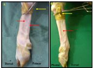
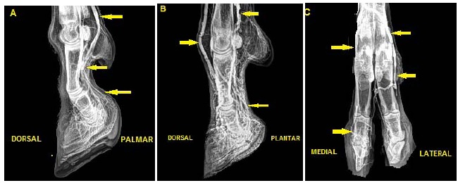
Ten forelimbs and hindlimbs of healthy sheep and goats, of varied breeds and gender with ages ranging between two and four years and an average body weight of 53kg were used in the study. The forelimbs and hindlimbs underwent a contrasted venography of the distal region. No numerical differences were observed in relation to veins between males and females and between the left and right members of the same species. Sheep had more veins than goats. The antiretrograde venography technique of both limbs in sheep and goats was proved to be applicable, showing the vasculazation of the distal region of the foot, the communication between the vessels and the quantity of vessels.
hoof; laminitis; small ruminants; venogram

 Contrast venography technique in vivo of the digits of sheep and goat
 Thumbnail
 Thumbnail
 Thumbnail
 Thumbnail
 Thumbnail
 Thumbnail


