 Digital intraoral scanning (IOS 3D) as a digital transformation on equine dentistry: in vivo evaluation
ABSTRACT
Equine dentistry is an essential healthcare practice for horses, deserving significant time and financial investment. To date, there have been no scientific reports on 3D digital intraoral scanning in equine dentistry. The objective of this pioneering study is to evaluate the safety, feasibility, dimensional compatibility, scanning patterns, and operational procedures of intraoral scanning in live horses under simple sedation. The scanning device’s wire connected to a notebook was used on 10 mixed breed adult horses, sedated with detomidine IV or detomidine associated with diazepam IV and restrained with oral speculum. The digital models obtained were exported in stl format to Exocad Dental DB® 3D imaging software. The sedation protocols were sufficient to do the IOS without equipment damage and the oral scan equipment used was compatible with the equine oral space of all the animals in this study. The digital models allowed three-dimensionally planes of views, plan section analysis and millimetric precise measurements. The occlusal colorimetric map revealed the contact and pressure areas during neutral occlusion. Intraoral 3D scanning is a feasible and useful technique in horses. More studies in live animals with different sizes and dental alterations, as well as employing different scanner devices, are needed.
Keywords:
advanced equine dentistry; intraoral scanning patterns; occlusal adjustment; veterinary dental imaging; digital equine dentistry
RESUMO
A odontologia equina é uma prática essencial para a saúde dos cavalos, merecendo investimento significativo de tempo e recursos financeiros. Até o momento, não existem relatos científicos sobre escaneamento intraoral digital 3D em odontologia equina. O objetivo deste estudo pioneiro é avaliar a viabilidade, a compatibilidade dimensional, os padrões de escaneamento e os procedimentos operacionais do escaneamento intraoral em cavalos vivos sob sedação simples. Os dispositivos de escaneamento, conectados por fio a um notebook, foram utilizados em 10 cavalos adultos de raças mistas, sedados com detomidina intravenosa ou detomidina associada a diazepam intravenoso e contidos com espéculo oral. Os modelos digitais obtidos foram exportados em formato STL para o software de imagem 3D Exocad Dental DB®. Os protocolos de sedação foram suficientes para realizar o escaneamento intraoral sem danos ao equipamento, e o equipamento de escaneamento oral utilizado foi compatível com o espaço oral de todos os animais deste estudo. Os modelos digitais permitiram visualização tridimensional dos planos de visão, análise da seção transversal e medições com precisão milimétrica. O mapa colorimétrico oclusal revelou as áreas de contato e pressão durante a oclusão neutra. A digitalização intraoral em 3D é uma técnica viável e útil em cavalos. São necessários mais estudos em animais vivos, com diferentes tamanhos e alterações dentárias, bem como com o uso de diferentes dispositivos de digitalização.
Palavras-chave:
odontologia equina avançada; padrões de escaneamento intraoral; ajuste oclusal; diagnóstico por imagem odontológico veterinário; odontologia equina digital
INTRODUCTION
Equine dentistry is increasingly valued as a continuous and essential healthcare, evidenced by the scale of investments aimed at improving power tools and diagnostic imaging techniques. Equine dentistry is a specialized area with the responsibility to promote and apply the results of evidence-based research rather than propagate claims regarding supposed intervention benefits without scientific evidence. (Carmalt, 2007; Rowlands, 2015). Despite these advances, empirical beliefs still persist among professionals and clients.
Digitization, Digitalization, Digital Transformation, Mobile Technologies, Blockchain, Big Data, Artificial Intelligence (AI) and Industry 4.0 are themes of major importance in society nowadays (Elenko et al., 2015; Bosio et al., 2017; Faddis, 2018; Konttila et al., 2019; El Idrissi et al., 2021). Digital health globally exponential growth is being accelerated by the COVID 19 pandemic and will continue to expand even after the end of this one (Calton et al., 2020; Veiga et al., 2021). Advancement in humane digital dentistry promote rapid and automated prototyping, and three-dimensional (3D) printing of dental biomaterials uses computer-aided design/computer-aided manufacturing (CAD/CAM) system in virtual occlusal records, full-arcade reconstruction, orthodontics, functional dentistry orthopedics, prosthesis fabrication, fixed partial dentures, orthodontic aligners, surgical guides, and implant abutments (Ender e Mehl., 2013; Flügge et al., 2013; Gan et al., 2016; Amornvit et al., 2020).
The aim of this study was to evaluate intraoral 3D scanning in horses as an objective method of digital transformation capable of supporting evidence-based precision equine dentistry and future artificial intelligence (AI) applications. The specific objectives were to verify the safety and feasibility of equine IOS, the compatibility between the equipment and equine oral anatomy, and aspects related to digital model reconstruction from live patients.
ETHICAL ASPECTS
The research was submitted to the Ethics Committee on Animal Use of the Universidade Federal de Viçosa, and approved under the number 50/2021”
MATERIAL AND METHODS
Ten horses, mixed breed and aged two to twenty years, from the clinical routine of dental care were submitted to the usual sedation protocols with detomidine IV (0.02mg/kg), repeated (0,01mg/kg) when needed or associated to diazepam IV (0.02 - 0.04mg/kg), and supported at an adjustable equine head stand. An autostatic mouth speculum with rubber coated bite plates promoted a full mouth access.
The scanning equipment used was the CS 3500® (CareStream®) and a notebook with specific manufacturer software installed, handled by two operators with different levels of experience. The images obtained, termed digital impressions, were processed and exported to the Exocad Dental DB® 3D imaging software.
Scanning began at the maxillary incisors followed by the mandibular incisors and then the bite registration. After that, scanning was made at the first quadrant premolars and molars, then the fourth quadrant premolars and molars and registration of the bite was obtained. Finally, the same was conducted at the second and third quadrants. Different patterns were used to better achieve the digital model image formation.
RESULTS AND DISCUSSION
Digital partial-arch models were successfully obtained. It was possible to include all incisor teeth in a single set and two additional sets containing premolars and molars on each side, as well as to record their occlusion. The interactive graphical interface provided a highly satisfactory and collaborative experience for intraoral examination. The digital models could be rotated, enlarged, and reduced for analysis from all angles of each dental arcade, separately or in occlusion. Cross-sectional plane analysis allowed millimetric measurements and comprehensive visualization of tooth surface contours.
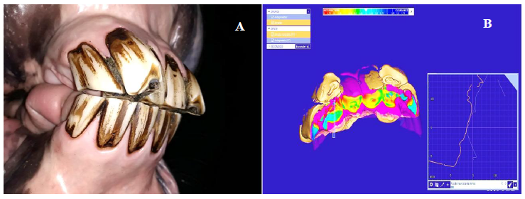
The figures below illustrate the three portions of the dental arches digitized separately for the construction of partial-arch digital models, demonstrating alterations such as horizontal incisor overjet, occlusal contact colorimetric mapping, and millimetric precision measurements. (Fig. 1), supernumerary incisors and occlusal contact colorimetric map (Fig. 2. A and B) and upper and lower molars with respective occlusal contact colorimetric mapping (Fig. 3. A, B, C and D).
Seven horses received a sedation protocol based on a bolus of detomidine, repeated two or three times when necessary, which was sufficient to maintain adequate restraint for IOS according to clinical signs. (Mama et al., 2010), although the moments of less quality or minor degree of sedation prolonged the time of procedure and increases concern about equipment damage. The continuous infusion of detomidine could be an alternative protocol to be used. Three horses received the sedation protocol with detomidine associated with diazepam and it offers better quality of restraint although also needed to be repeated two or three times too. The ataxia increased by diazepam promoted some sudden movements that limited the scan flow in a minor way because there was adequate support for the head. An important recommendation is to use low doses of diazepam due to the risk of horses recumbency or falling (Muir et al., 1982). The association of detomidine and butorfanol may be a good protocol too.
In our previous evaluation using skulls, two different commercial equipment fitted and worked effectively (Lang, 2022). In live animals, the region of greatest concern to be reached was the interocclusal space between the third molars, because of narrowing and the “Spee curvature”, followed by the lingual surface of the mandibular teeth, because of the tongue movements. The dimensions of oral scan equipment used and ergonomics was compatible with the equine oral space of all the animals in this study. Not only were the incisor teeth easily scanned, but the premolars and molars also had the entire surface digitized as the scanner works in direct contact with the teeth surface. The reach and freedom of movement without risk of damage to the equipment are highly dependent on adequate sedation and the oral speculum in horses. In humans, the size of the tip plays a role as well, especially in the case of second and third molars (Zimmermann et al., 2015; Goracci et al., 2016). The tip is removable, replaceable and can be autoclaved. The shape and length of the scanner wand and scanner tip are different among the commercial products, and its configuration can be an important desirable feature (Imburgia et al., 2017; Mangano et al., 2017).

Digital model of incisors occlusion. Occlusal colorimetric map and a detail of a cross-section plane analysis of the horizontal incisor overjet measuring 6,647 millimeters, right below. (ExocadDental BD Software).

A) Oblique rostral view of a superior supranumerary incisor tooth from 11 year old mixed breed mare. B) Occlusal colormap of the incisor superior supranumerary clinical case. Note the contact of antagonist with most caudal 102 teeth, cross-sectional plane detail right below. (ExocadDental BD Software).
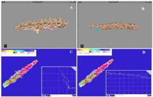
A) Digital model of maxillary premolars and molars, occlusal surface. from 14 years old mixed breed mare. Note periodontal disease (yellow arrow) between 108 and 109 teeth. (CareStream software). B) Digital model of mandibular premolars and molars, occlusal surface. from 14 years old mixed breed mare. Note the partial image loss (blue arrow) of 411 oclusal surface. (CareStream software). C) Digital model of premolars and molars occlusal colorimetric map and transverse cross-section plane (right below) detailed design of occlusion contact areas and D) longitudinal cross-section plane (right below) detailed design of occlusion contact areas. (ExocadDental BD Software).
The time to obtain the digital model for each area separately ranged from 15 to 25 minutes. Therefore, to assemble a digital model with upper and lower incisors and their occlusion, it took 30 to 50 minutes. The same time was needed for the premolars and molars on each side. Besides the movements of the horses and the interval of sedative latency according to protocol used, already discussed, the time consuming found in this study can be explained by the speed of action of the equipment and by the scan steam. The image capture speed is an inherent characteristic of the equipment and differs between brands and models on the market (Mangano et al., 2017). The equipment used in this study is not the latest model equipment of the brand, already receiving two updates to the present day that are still not yet among the fastest available on the market. This characteristic does not limit the work on human dentistry, mainly in the scans of small areas, but for the large teeth and large dental arcades of horses it is quite time-consuming. A previous skull evaluation showed that the 3Shape® Trios3® scanner was at least twice as fast as the CareStream® CS 3500® scanner. (Lang, 2022).
The scan steam to construct the digital model is based on the partial overlapping of images, while gradually sliding the tip of scanner at small distances. The patterns of scanning are the focus of a series of studies in human dentistry (Bosio et al., 2017; Mennito et al., 2018; Latham et al., 2020). When slide a little bit longer distance, there is a risk of losing image references and the construction of the digital model is interrupted. To resume the model construction, it is necessary to return to an area that has already been scanned and then proceed. Sometimes there is an error in the construction and the images are superimposed in wrong places. A greater number of errors were observed mainly in the acquisition of images of the labial surface of incisor teeth and, less frequently, in the buccal aspect of upper premolars and molars. The similarity of large areas between different teeth made it difficult to the software to fit the images of the digital model in the correct place. To avoid mistakes, some colored marks were made with pen brushes, and it can be useful as long as they are not eventually erased during the procedure. It is possible to delete some mistaken images and return to construct the digital model, but it is another time-consuming interference. Differences of scanning patterns are to be evaluated in horses’ dental arcade in the future.
A frequent topic in scientific publications regarding the quality of intraoral scanning equipment is the accuracy, or proximity of measurements between real and digital models, which is determined by the sum of trueness and precision (Kravitz et al., 2014; Kustrzycka et al., 2020). The trueness of an IOS is established by the overlapping of a reference scan obtained with a powerful industrial machine, what is currently not possible in vivo, and some digital models obtained through IOS, then a reverse-engineering software can be used to generate colorimetric maps displaying the distances/differences between the surfaces of the IOS and the reference model at micrometric level. Precision can be established simply by overlapping different digital models taken with the same equipment and evaluating the differences at micrometric level. Trueness and precision mainly depend on the scanner acquisition/processing software, but the pattern of scanning, the amount of area to be scanned and expertise of operator may influence in a negative way. However, the latest-generation scanners are characterized by very low errors in digital impressions (Imburgia et al., 2017). The data in the literature must be interpreted critically, according to the equipment used as it is possible that different machines, using different scanning strategies, would produce different results as they are in human dentistry (Goracci et al., 2016; Kustrzycka et al., 2020). Furthermore, it is just the beginning of testing in equine dentistry.
The equipment and software are still costly for routine use in equine practice; however, the availability of these technologies on the market is increasing, as competition among manufacturers promotes improvements in software quality as well as reductions in acquisition and operating costs. (Kravitz et al., 2014). Portable and wireless equipment connected to a notebook is gradually replacing large systems mounted on wheeled cabinets and monitors, as the use of surface powder has become unnecessary and image quality has evolved to provide well-defined color images, although still limited in some deep anatomical regions, such as equine dental infundibulae and narrow interdental spaces, as observed in this study. Software development is continuous; however, some systems still present limited analysis tools. (Schmidt et al., 2021). Open systems can export in different formats like stl to more feature-rich software like Exocad Dental DB®3D as other software is still expensive, requires payment of annual fees and periodic updates. The development of software for equine dentistry can contribute to maximizing the use of the technique
Many applications and benefits of IOS 3D in horses can be glimpsed in an affordable way and in a short time, such as for example the collection of anatomy images, clinical cases and pathological findings, to educational purpose archives. The coupled fluorescent technology enables the early identification of cariogenic bacterial activity (Stookey e González-Cabezas, 2001; Gimenez et al., 2013; Moreira et al., 2020). Precise millimetric measures of reference and variation within different stages of disorder like excessive enamel points, diastemas, gingival recession, gingival pouch and EOTRH can be standardized and measurements to define when orthodontics are indicated or not and the treatments follow up. Access to standard digital models showing odontoplasty and occlusal adjustment defined by world-renowned professionals will be available to professionals in the specialization process. Digital models can be 3D printed and used for training, what shortens the learning curve, reduces time to do procedures and reduces risks to patients. It can encourage the development of underrated areas such as implant dentistry in equine practice, since it is widely used in implant process for human dentistry. A market for products and operating models can be created or enhanced to online apps with graphic charts platforms and patient/customer files including archives of digital models obtained from IOS 3D and Blockchains of specialized professionals.
CONCLUSION
The IOS 3D technique can be safely performed under routine sedation protocols, and the tested commercial scanner configuration fitted properly within the equine oral cavity of live animals. This technology can be incorporated into the digital transformation of various areas of equine dentistry, including research, education, evidence-based clinical practice, client communication, and marketing, mainly benefiting from data storage, precise tooth morphology measurements, and objective assessment of occlusal contact. Aspects that still require improvement for broader application of this technology in equine dentistry include software and equipment design, image acquisition and processing speed, and equipment cost.
REFERENCES
- AMORNVIT, P.; SANOHKAN, S.; PEAMPRING, C. Studying the optical 3D accuracy of intraoral scans: an in vitro study. J. Healthc Eng, v.14, p.5739312, 2020.
- BOSIO, J.A.; DEL SANTO, M.; JACOB, H.B. Odontologia digital contemporânea - scanners intraorais digitais. Orthod. Sci. Pract, v.10, p.355-362, 2017.
- CALTON, B.; ABEDINI, N.; FRATKIN M, Telemedicine in the Time of Coronavirus, J Pain Symptom Manage, v.60, p.e12-e14, 2020.
- CARMALT, J.L. Evidence-based equine dentistry: preventive medicine. Vet. Clin. North Am. Equine Pract, v.23, p.519-524, 2007.
- EL IDRISSI, A.H.; LARFAOUI, F.; DHINGRA, M. et al Digital technologies and implications for veterinary services. Rev. Sci Tech, v.40, p.455-468, 2021.
- ELENKO, E.; UNDERWOOD, L.; ZOHAR, D. Defining digital medicine. Nat. Biotechnol., v.33, p.456-461, 2015.
- ENDER, A.; MEHL, A. Accuracy of complete-arch dental impressions: a new method of measuring trueness and precision. J. Prosthet. Dent, v.109, p.121-128, 2013.
- FADDIS, A. The digital transformation of healthcare technology management. Biomed. Instrum. Technol, v.52, p.34-38, 2018.
- FLÜGGE, T.V., SCHLAGER, S.; NELSON, K. et al Precision of intraoral digital dental impressions with iTero and extraoral digitization with the iTero and a model scanner. Am. J. Orthod. Dentofacial Orthop, v.144, p.471-478, 2013.
- GAN, N.; XIONG, Y.; JIAO, T. Accuracy of intraoral digital impressions for whole upper jaws, including full dentitions and palatal soft tissues. PLoS One, v.11, p.e0158800, 2016.
- GIMENEZ, T.; BRAGA, M.M.; RAGGIO, D.P. et al Fluorescence-based methods for detecting caries lesions: systematic review, meta-analysis and sources of heterogeneity. PLoS One, v.8, p.e60421, 2013.
- GORACCI, C.; FRANCHI, L.; VICHI, A.; FERRARI, M. Accuracy, reliability, and efficiency of intraoral scanners for full-arch impressions: a systematic review of the clinical evidence. Eur. J. Orthod, v.38, p.422-428, 2016.
- IMBURGIA, M.; LOGOZZO, S.; HAUSCHILD, U. et al Accuracy of four intraoral scanners in oral implantology: a comparative in vitro study. BMC Oral Health, 2017;v.17, p.92, 2017.
- KONTTILA, J.; SIIRA, H.; KYNGÄS, H. et al Healthcare professionals’ competence in digitalization: a systematic review. J. Clin. Nurs, v.;28, p.745-761, 2019.
- KRAVITZ, N.D.; GROTH, C.; JONES, P.E. et al Intraoral digital scanners. J. Clin. Orthod, v.48, p.337-347, 2014.
- KUSTRZYCKA, D.; MARSCHANG, T.; MIKULEWICZ, M.; GRZEBIELUCH, W. Comparison of the accuracy of 3D images obtained from different types of scanners: a systematic review. J. Healthc Eng, v.2020, p.8854204, 2020.
-
LANG, A. Escaneamento intraoral digital 3D em odontologia equina 2022. 86f. Tese (Doutorado em Medicina Veterinária) - Universidade Federal de Viçosa, Viçosa, MG. Available from: https://www.locus.ufv.br/. Accessed on: October 20, 2025.
» https://www.locus.ufv.br - LATHAM J, MARK L, MENNITO A, KELLY A, et al Effect of scan pattern on complete-arch scans with 4 digital scanners J Prosthet Dent, v.123, p.85-95, 2020.
- MAMA, K.R.; GRIMSRUD, K.; SNELL, T.; STANLEY, S. Plasma concentrations, behavioural and physiological effects following intravenous and intramuscular detomidine in horses. Equine Vet. J, v.41, p.772-777, 2010.
- MANGANO, F.; GANDOLFI, A.; LUONGO, G. et al Intraoral scanners in dentistry: a review of the current literature. BMC Oral Health, v.17, p.149, 2017.
- MENNITO, A.S.; EVANS, Z.P.; LUDLOW, M.; RENNE, W.G. Evaluation of the effect scan pattern has on the trueness and precision of six intraoral digital impression systems. J. Esthet Restor. Dent, v.30, p.113-118, 2018.
- MOREIRA, L.M.Y.A.; AMARANTE, B.C.; MAROTTA, B.M. et al Laser fluorescence as an alternative for digital radiography in detecting caries lesions. Clin. Lab. Res. Dent, v.1, p.1-10, 2020.
- MUIR, W.W.; SAMS, R.A.; HUFFMAN, R.H.; NOONAN, J.S. Pharmacodynamic and pharmacokinetic properties of diazepam in horses Am J. Vet. Res, v.43, p.1756-1762, 1982.
-
ROWLANDS, V. Evolutions in equine dentistry. Vet Times, 2015. Available in: https://www.vettimes.co.uk Accessed on: October 20, 2025.
» https://www.vettimes.co.uk - SCHMIDT, A.; SCHLENZ, M.A.; LIU, H.; KÄMPE, H.S.; WÖSTMANN, B. The influence of hard- and software improvement of intraoral scanners on the implant transfer accuracy from 2012 to 2021: an in vitro study. Appl. Sci, v.11, p.7166, 2021.
- STOOKEY, G.K.; GONZÁLEZ-CABEZAS, C. Emerging methods of caries diagnosis. J. Dent. Educ, v.65, p.1001-1006, 2001.
- VEIGA, R.A.S.; RODRIGUES, M.V.P.; SANTOS, A.R. et al Telemedicina e Covid-19: uma revisão de literatura. Rev. Bioética Cremego, v.3, p.40-48, 2021.
- ZIMMERMANN, M.; MEHL, A.; MÖRMANN, W.H.; REICH, S. Intraoral scanning systems - a current overview. Int. J. Comput Dent, v.18, p.10-129, 2015.