Abstracts
Myxobolus platanus n. sp. infecting the spleen of Mugil platanus Günther, 1880 (Osteichthyes, Mugilidae) from Lagoa dos Patos, Brazil is described The parasites formed round or slightly oval whitish plasmodia (about 0.05-0.1mm in diameter) on the surface of the organ. The spores were round in frontal view and oval in lateral view, 10.7µm (10-11) long, 10.8µm (10-11) wide and 5µm thick, and presented four sutural marks along the sutural edge. The polar capsules, equal in size, were prominent, surpassing the mid-length of the spore, and were oval with the posterior extremity rounded, and converging with their anteriorly tapered ends. They were 7.7µm (7-8) long and 3.8µm (3.5-4) wide. A small intercapsular appendix was present. The polar filament formed five to six coils obliquely placed to the axis of the polar capsule. No mucous envelope or distinct iodinophilous vacuole were found.
Myxozoa; Myxosporea; Myxobolus platanus n. sp.; Mugil platanus; Lagoa dos Patos, Brazil
Descreve-se Myxobolus platanus n. sp. infectando o baço de Mugil platanus Günther, 1880 (Osteichthyes, Mugilidae) da Lagoa dos Patos, Brasil. Os parasitas formavam plasmódios brancos redondos ou ligeiramente ovais (diâmetro de cerca de 0,05-0,1mm) à superfície do órgão. Os esporos eram circulares em observação frontal e ovais em obervação lateral, medindo, em média, 10,7µm (10-11) de comprimento, 10,8µm (10-11) de largura e 5µm de espessura, e tinham quatro marcas suturais ao longo da linha de sutura. As cápsulas polares eram grandes e do mesmo tamanho ultrapassando a zona média do esporo. Eram de forma oval, tendo a extremidade posterior arredondada, e convergiam pelas extremidades anteriores afiladas, medindo 7,7µm (7-8) de comprimento por 3,8µm (3,5-4) de largura. Um pequeno apêndice intercapsular estava presente. O filamento polar formava cinco a seis dobras colocadas obliquamente em relação ao eixo da cápsula. Não havia envelope mucígeno nem vacúolo iodofílico.
Myxozoa; Myxosporea; Myxobolus platanus n. sp.; Mugil platanus; Lagoa dos Patos, Brasil
VETERINARY MEDICINE
Myxobolus platanus n. sp. (Myxosporea, Myxobolidae), a parasite of Mugil platanus Günther, 1880 (Osteichthyes, Mugilidae) from Lagoa dos Patos, RS, Brazil
Myxobolus platanus n. sp. (Myxosporea, Myxobolidae), parasita de Mugil platanus Günther, 1880 (Osteichthyes, Mugilidae) da Lagoa dos Patos, RS
J.C. EirasI; P.C. AbreuII; R. RobaldoII; J. Pereira JúniorII
IFaculdade de Ciências - Universidade do Porto 4099-002 - Porto, Portugal
IIFundação Universidade Federal do Rio Grande - Rio Grande, RS
ABSTRACT
Myxobolus platanus n. sp. infecting the spleen of Mugil platanus Günther, 1880 (Osteichthyes, Mugilidae) from Lagoa dos Patos, Brazil is described The parasites formed round or slightly oval whitish plasmodia (about 0.05-0.1mm in diameter) on the surface of the organ. The spores were round in frontal view and oval in lateral view, 10.7µm (10-11) long, 10.8µm (10-11) wide and 5µm thick, and presented four sutural marks along the sutural edge. The polar capsules, equal in size, were prominent, surpassing the mid-length of the spore, and were oval with the posterior extremity rounded, and converging with their anteriorly tapered ends. They were 7.7µm (7-8) long and 3.8µm (3.5-4) wide. A small intercapsular appendix was present. The polar filament formed five to six coils obliquely placed to the axis of the polar capsule. No mucous envelope or distinct iodinophilous vacuole were found.
Keywords: Myxozoa, Myxosporea, Myxobolus platanus n. sp., Mugil platanus, Lagoa dos Patos, Brazil
RESUMO
Descreve-se Myxobolus platanus n. sp. infectando o baço de Mugil platanus Günther, 1880 (Osteichthyes, Mugilidae) da Lagoa dos Patos, Brasil. Os parasitas formavam plasmódios brancos redondos ou ligeiramente ovais (diâmetro de cerca de 0,05-0,1mm) à superfície do órgão. Os esporos eram circulares em observação frontal e ovais em obervação lateral, medindo, em média, 10,7µm (10-11) de comprimento, 10,8µm (10-11) de largura e 5µm de espessura, e tinham quatro marcas suturais ao longo da linha de sutura. As cápsulas polares eram grandes e do mesmo tamanho ultrapassando a zona média do esporo. Eram de forma oval, tendo a extremidade posterior arredondada, e convergiam pelas extremidades anteriores afiladas, medindo 7,7µm (7-8) de comprimento por 3,8µm (3,5-4) de largura. Um pequeno apêndice intercapsular estava presente. O filamento polar formava cinco a seis dobras colocadas obliquamente em relação ao eixo da cápsula. Não havia envelope mucígeno nem vacúolo iodofílico.
Palavras-chave: Myxozoa, Myxosporea, Myxobolus platanus n. sp., Mugil platanus, Lagoa dos Patos, Brasil
INTRODUCTION
Myxobolus spp. are the most common Myxozoan fish parasites having a wide geographical distribution and comprising a great number of species infecting both marine and freshwater fish (Landsberg and Lom, 1991; Lom and Dyková, 1992; Eiras et al., 2005).
For Mugil spp. there are 26 species of Myxobolus, most of them (18) infecting M. cephalus from different geographical areas. These parasites can be pathogenic and this may be particularly relevant for the regions where mullets are important for aquaculture, as in Israel, Italy, Egypt and Tunisia (Bahari and Marques, 1996).
The Lagoa dos Patos, in Rio Grande do Sul State, Southern Brazil, is the biggest freshwater lagoon in the world. It is about 240km in length, and up to 48km in width. A wide sand bar separates it from the Atlantic Ocean. The lagoon is an important fishing ground and the fish species diversity is quite high (Haimovici et al., 1997; Vieira and Castello 1997). Mugil platanus is one of the most important fish species of the Lagoa dos Patos. However, its parasitology is practically unknown. Taking into account the importance of Mugil spp. for fish farming, the study of the parasites of feral specimens is highly important.
Among the 744 species of Myxobolus, nearly all described so far (Eiras et al., 2005), only 20 species were described from Brazilian hosts, while Brazilian fishes represent about 24% of all fish species (Cellere et al., 2002). The objective of this study was to describe Myxobolus platanus n. sp., a parasite of Mugil platanus Günther, 1880 (Osteichthyes, Mugilidae) from the Lagoa dos Patos.
MATERIALS AND METHODS
Forty-six specimens of Mugil platanus (total length: 19.9-35.5 cm) were captured at the Lagoa dos Patos, Rio Grande do Sul State, Brazil. Fish were brought alive to the laboratory, and all the organs were carefully inspected for parasites. Spore measurements were made from 30 fresh spores. For observation of the presence of iodinophilous vacuole, fresh spores were treated with Lugol's iodine solution. Spores were also stained with India ink for revealing any mucous envelope (Lom and Vávra, 1961).
RESULTS AND DISCUSSION
Four out of 46 M. platanus (total length: 22.1-34.6cm) had the spleen infected with Myxobolus. The parasites formed round or slightly oval whitish plasmodia (about 0.05-0.1mm in diameter) on the surface of the organ.
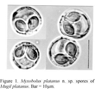
The spores (Fig. 1, 3 ) were round in frontal view and oval in lateral view. The spore valves were relatively thin, symmetrical and smooth. Spores were 10.7µm (10-11) long, 10.8µm (10-11) wide and 5µm thick and presented 4 sutural marks along the sutural edge. The polar capsules, equal in size, were prominent, surpassing the mid-length of the spore. They were oval with the posterior extremity rounded, and tapering anterior end. They were 7.7µm (7-8) long, 3.8µm (3.5-4) wide and the polar filament formed five to six coils obliquely placed to the axis of the polar capsule. A small intercapsular appendix was present. There was no mucous envelope or distinct iodinophilous vacuole.
The specific name derives from the name of the host species.
Specimens deposition: the syntipes are deposited at the Section of Animal Pathology, Department of Zoology and Anthropology from the Faculty of Sciences of Porto, Portugal, and at the Museum of Natural History from the Faculty of Sciences of Porto, Portugal.
The specimens were first compared with all the Myxobolus species described for Mugil spp. (Mugil cephalus, M. soiyu, M. chelo, M. waigensis, M. saliens, M. tade and M. curema) comprising a total of 26 species.
The Myxobolus species which have round or almost round spores are M._chiungchowensis (Chen, 1998), M. parenzani (Myxobolus branchialis Parenzan, 1966) (Landsberg and Lom, 1991), M. mugilis (Negm-Eldin et al., 1999), M. hani (Faye et al., 1999), M. hannensis (Fall et al., 1997), M. goreensis (Fall et al., 1997), and M. bizerti (Bahari and Marques, 1996).
M. mugilis, M. parenzani and M. hani could not be identified with the studied specimens once the spores were much smaller (length x width dimensions: 7.4x7.3µm, 5.4x5.4 µm and 7-9x 7.8µm, respectively). On the contrary, M. hannensis, M. goreensis and M. bizerti have larger spores (13-15x13-15µm, 10-13x10-13µm and 14-14.5x14-14.5µm, respectively) besides being different also in other characteristics as the length and width of the polar capsules or the number of coils of the polar filament. M. chiungchowensis has spores slightly longer compared with the studied specimens (10.2-11.8 x 9.6-11µm), the thickness of the spore is higher (6-6.6µm), the polar capsules are smaller and the number of coils of the polar filament is higher.
The other species infecting Mugil spp. could not be identified with the studied specimens because they had not round spores and were different in other features as the size of the spore, the number of coils of the polar filament, or the size of the polar capsules.
The present material was also compared with the full characteristics of 744 species of Myxobolus representing nearly all the species described so far (Eiras et al., 2005). Concerning the forms with rounded spores, the most similar species are M. bartai from the body wall muscles of Notropis cornutus (Salim and Desser, 2000), M. lanfyongi infecting the wall of the intestine of Spinibarbichthys denticulatus (Ha, 1971), M. nephroides parasitizing the kidney, spleen and gall-bladder of Hypophthalmichthys molitrix (Li and Nie, 1973, quoted from Chen and Ma, 1998), and M. paralintoni described from the heart of Lepomis gibbosus (Li and Desser, 1985).
M. bartai, M. nephroides and M. paralintoni despite having rounded spores with dimensions similar to the spores found in the present study, are quite different in having unequal polar capsules. Besides, the polar capsules are smaller for M. bartai and M. nephroides, and larger in the case of M. paralintoni. M. lanfyongi has round spores slightly larger than the present material (10.8-11.7µm x 10.8-11.7µm) but the polar capsules are much more smaller (4.5-5.4µm x 2.7-3.6µm).
Thus, none of the species of Myxobolus described so far fits to the characteristics of the studied material. Therefore it may be considered as a new species, Myxobolus platanus n. sp.
Recebido em 1 de agosto de 2005
Aceito em 4 de abril de 2007
E-mail: jceiras@fc.up.pt
- BAHARI, S.; MARQUES, A. Myxosporean parasites of the genus Myxobolus from Mugil cephalus in Ichkeul lagoon, Tunisia: description of two new species. Dis. Aquat. Org, v.27, p.115-122, 1996.
- CELLERE, E.F.; CORDEIRO, N.; ADRIANO, E.S. Myxobolus absonus sp. n. (Myxozoa: Myxosporea) parasitizing Pimelodus maculatus (Siluriformes: Pimelodidae), a South American freshwater fish. Mem. Inst. Oswaldo Cruz, v.97, p.79-80, 2002.
- CHEN, Q.L.; MA, C.L. Myxozoa, Myxosporea. Fauna Sinica Beijing: Science Press, 292-528, 1998 p. 292-528
- EIRAS, J.C.; MOLNÁR, K.; LU Y.S. Synopsis of the species of the genus Myxobolus Bütschli, 1882 (Myxozoa, Myxosporea, Myxobolidae). Syst. Parasitol, v.61, p.1-46, 2005.
- FALL, M.; KPATCHA, T.K.; DIEBAKATE, C. et al. Observations sur des myxosporidies (Myxozoa) du genre Myxobolus parasites de Mugil cephalus (Poisson, Téléostéen) du Sénégal. Parasite, v.2, p.173-180, 1977.
- FAYE, N.; KPATCHA, T.K.; DIEBAKATE, C. et al. Gill infections due to myxosporean (Myxozoa) parasites in fishes from Senegal with description of Myxobolus hani sp. n. Bull. Europ. Assoc. Fish. Pathol, v.19, p.14-16, 1999.
- HA, K.Y. Some myxosporidians of freshwater fish of the North Vietnam. Acta Protozool, v.8, p.283-298, 1971.
- HAIMOVICI, M.; CASTELLO, J.P.; VOOEN, C.M. Relationships and function of coastal and marine environments Fisheries. In: Seeliger U.; Odebrecht C.; Castello, J.P. (Eds). Subtropical convergence environment. The coast and sea in the Southwestern Atlantic. Berlin: Springer Verlag, 1997. p.183-197.
- LANDSBERG, J.H.; LOM, J. Taxonomy of the genera of the Myxobolus/Myxosoma group (Myxobolidae: Myxosporea), current listing of species and revision of synonyms. Syst. Parasitol, v.18, p165-186,1991.
- LI, L.; DESSER, S.S. The protozoan parasites of fish from two lakes in Algonquin Park, Ontario. Can. J. Zool, v.63, p.1846-1858, 1985.
- LOM, J.; VÁVRA, J. Mucous envelopes of spores of the subphylum Cnidospora (Doflein, 1901). Vestnik. Ceskosl. Spolec. Zoolog, v.27, p.4-6, 1961.
- LOM, J; DYKOVÁ, I. Protozoan parasites of fishes. Develop. Aquacult. Fish. Sci, v.26, p.315, 1992.
- NEGM-ELDIN, N.M.; GOVEDICH, F.R.; DAVIES, R.W. Gill myxosporeans on some Egyptian freshwater fish. Deutsche Tierarzt. Wochensch, v.106, p.459-465, 1999.
- SALIM, K.Y.; DESSER, S.S. Descriptions and phylogenetics systematics of Myxobolus_spp. from cyprinids in Algonquin Park, Ontario. J. Eukaryot. Microbiol, v.47, p.309-318, 2000.
- VIEIRA, J.P.; CASTELLO, J.P. Environment and biota of the Patos Lagoon estuary fish fauna. In: Seeliger U.; Odebrecht C.; Castello J.P. (Eds). Subtropical convergence environment. The coast and sea in the Southwestern Atlantic Berlin: Springer Verlag, 1997. p.56-62.
Publication Dates
-
Publication in this collection
24 Sept 2007 -
Date of issue
Aug 2007
History
-
Accepted
04 Apr 2007 -
Received
01 Aug 2005


