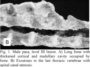
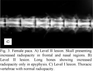
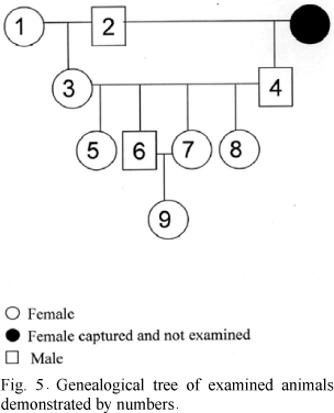
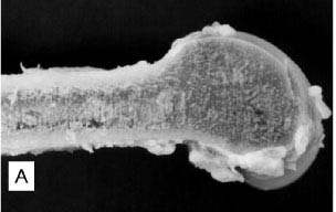
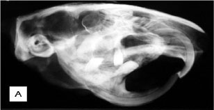
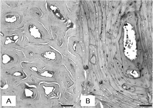
Nine cases of familial osteopetrosis were studied in Agouti paca rodents maintained in captivity. Animals were distributed in three groups depending on the severity of their skeletal lesions. Based upon clinical, radiological, and microscopic findings, it was concluded that one animal had level I lesions, three animals had level II lesions, and five animals had level III osteopetrosis and osteonecrosis. Throughout the entire axial and appendicular skeleton, there was an increased amount of both trabecular and cortical bone tissue. All analyzed bones showed thickened cortex and reduced medullary canals. Bone trabeculae were thick and confluent. Cortex showed a narrowing of Haversian canals. Numerous cementing lines resulted in typical mosaic patterns. Osteocytes were pycnotic. Osteonecrosis was characterized by the disappearance of osteocytes and bone matrix decomposition.
Agouti paca; familial disease; osteonecrosis; osteopetrosis; skeleton

 Familial osteopetrosis in Agouti paca: report of nine cases