Abstract
PURPOSE:
To determine the antioxidant and anti-inflammatory effects of alfa lipoic acid (ALA) on the liver injury induced by methotrexate (MTX) in rats.
METHODS:
Thirty two rats were randomly assigned into four equal groups; control, ALA, MTX and MTX with ALA groups. Liver injury was performed with a single dose of MTX (20 mg/kg) to groups 3 and 4. The ALA was administered intraperitonealy for five days in groups 2 and 4. The other rats received saline injection. At the sixth day the rats decapitated, blood and liver tissue samples were removed for TNF-α, IL-1β, malondialdehyde, glutathione, myeloperoxidase and sodium potassium-adenosine triphosphatase levels measurement and histological examination.
RESULTS:
MTX administration caused a significant decrease in tissue GSH, and tissue Na+, K+ ATPase activity and which was accompanied with significant increases in tissue MDA and MPO activity. Moreover the pro-inflammatory cytokines (TNF-α, IL- β) were significantly increased in the MTX group. On the other hand, ALA treatment reversed all these biochemical indices as well as histopathological alterations induced by MTX.
CONCLUSION:
Alfa lipoic acid ameliorates methotrexate induced oxidative damage of liver in rats with its anti-inflammatory and antioxidant effects.
Thioctic Acid; Methotrexate; Liver; Rats
Introduction
Methotrexate (MTX) is an effective cytotoxic drug and has been widely used in chemotherapeutic based treatments for malignancies primarily in leukaemias11. Bleyer WA. Methotrexate: clinical pharmacology, current status and therapeutic guidelines. Cancer Treat Rev. 1977 Jun;4(2):87-101. PMID: 329989. , 22. Widemann BC, Balis FM, Kempf-Bielack B, Bielack S, Pratt CB, Ferrari S, Bacci G, Craft AW, Adamson PC. High-dose methotrexate-induced nephrotoxicity in patients with osteosarcoma. Cancer. 2004 May;100(10):2222-32. PMID: 15139068. as well as inflammatory diseases including psoriasis and rheumatoid arthritis33. Braun J, Rau R. An update on methotrexate. Curr Opin Rheumatol. 2009 May;21(3):216-23. doi: 10.1097/BOR.0b013e328329c79d.
https://doi.org/10.1097/BOR.0b013e328329...
4. Kose E, Sapmaz HI, Sarihan E, Vardi N, Turkoz Y, Ekinci N. Beneficial effects of montelukast against methotrexate-induced liver toxicity: a biochemical and histological study. ScientificWorldJournal. 2012;2012:987508. doi:10.1100/2012/987508.
https://doi.org/10.1100/2012/987508...
5. Soliman ME. Evaluation of the possible protective role of folic acid on the liver toxicity ınduced experimentally by methotrexate in adult male albino rats. Egypt J Histol. 2009;32:118-28. - 66. Dalaklioglu S, Genc GE, Aksoy NH, Akcit F, Gumuslu S. Resveratrol ameliorates methotrexate-induced hepatotoxicity in rats via inhibition of lipid peroxidation. Hum Exp Toxicol. 2013 Jun;32(6):662-71. doi: 10.1177/0960327112468178.
https://doi.org/10.1177/0960327112468178...
. Long-term methotrexate use, or its use in high doses, may cause hepatic steatosis, cholestasis, fibrosis and cirrhosis77. Chládek J, Martínková J, Sispera L. An in vitro study on methotrexate hydroxylation in rat and human liver. Physiol Res. 1997;46(5):371-9. PMID: 9728483.. Accordingly the dose of methotrexate should be lowered or the drug should be discontinued in case of hepatic toxicity which causes delay in the treatment of the disease. On the other hand much attention is now being paid to factors that may enhance the effectiveness of existing drugs while reducing their unwanted side effects.
Alfa-lipoic acid (ALA) is described as a therapeutic agent in a number of conditions related to liver disease, including alcohol-induced damage, mushroompoisoning, metal intoxification, CCl4 poisoning, and hyperdynamic circulation in biliary cirrhosis88. Bustamante J, Lodge JK, Marcocci L, Tritschler HJ, Packer L, Rihn BH. Alpha-lipoic acid in liver metabolism and disease. Free Radic Biol Med. 1998 Apr;24(6):1023-39. PMID: 9607614.
9. Marley R, Holt S, Fernando B, Harry D, Anand R, Goodier D, Davies S, Moore K. Lipoic acid prevents development of the hyperdynamic circulation in anesthetized rats with biliary cirrhosis. Hepatology. 1999 May;29(5):1358-63. PMID: 10216116. - 1010. Bludovská M, Kotyzová D, Koutenský J, Eybl V. The influence of alpha-lipoic acid on the toxicity of cadmium. Gen Physiol Biophys. 1999 Oct;18 Spec No:28-32. PMID: 10703716.. The effect of ALA against methotrexate toxicity on the liver is not yet clearly.
Furthermore the effect of ALA on MTX induced liver injury has not been studied before. Thus, in this present study we aimed to investigate whether the ALA has any effect on treatment against MTX induced oxidative injury on the liver in rats.
Methods
The experimental protocols were approved by the animal care and use committee of İnönü University Faculty of Medicine.
Thirty two Wistar albino rats of both sexes of 200-250 g were used in this experiment. The rats maintained at a constant temperature (22°C) with a 12-h light-dark cycle and randomly divided into four groups. Group 1 (control group): rats in this group received only physiological saline. Group 2 (α-lipoic acid group): rats in this group received α-lipoic acid (Sigma, St Louis, USA) for five days intraperitoneally (60 mmol/kg). Group 3 (Methotrexate group): rats received a single dose of MTX (Onco-Tain; Faulding Pharmaceutics Plc, Leamington Spa, UK) intraperitoneally (20 mg/ kg). Group 4 (Methotrexate group-α-lipoic acid group): rats received a single dose of MTX and also received α-lipoic acid for five days. Alfa-lipoic acid was dissolved in 0.1% dimethyl sulfoxide (DMSO). At the end of the experiment rats were decapitated and blood samples were obtained for the measurement of tumour necrosis factor-alpha (TNF-α) and interleukin-1-beta (IL-1β). The levels of malondialdehyde (MDA) and glutathione (GSH), as well as myeloperoxidase (MPO) and sodiumpotassium adenosine triphosphatase (Na+/K+-ATPase) activity were alo analysed in the liver tissues. Furthermore the degree of inflammation and histopathologic damage (necrosis, inflammation, vacuolization and vascular congestion) were evaluated via histological examination under a light microscope.
Measurement of malondialdehyde and glutathione levels
To determine the MDA and GSH levels, liver tissue samples were homogenized in ice cold 150mm KCl. The MDA levels (nmol MDA/g tissue) were assayed for the products of lipid peroxidation1111. Buege JA, Aust SD. Microsomal lipid peroxidation. Methods enzymol. 1978;52:302-10.. The GSH levels (mg GSH/g tissue) were measured by spectrophotometric method using Ellman's reagent1212. Beutler E. Glutathione in red blood cell metabolism. A manual of biochemical methods. New York: Grune &Stratton; 1975. p.112-4..
Measurement of myeloperoxidase activity
Tissue-associated MPO (U/g tissue) activity was measured according to the procedure reported by Hillegas et al.1313. Hillegass LM, Griswold DE, Brickson B, Albrightson-Winslow C. Assessment of myeloperoxidase activity in whole rat kidney. J Pharmacol Methods. 1990 Dec;24(4):285-95. PMID: 1963456.Liver tissue samples were homogenized in 50mm potassium phosphate buffer (PB, pH 6.0) and homogenates were centrifuged at 41 400g for 10 min; pellets were suspended in 50mm PB containing 0.5% hexadecyltrimethylammonium bromide. After three cycles of freezing and thawing, with sonication between the cycles, the samples were centrifuged at 41 400g for 10 min. Volumes of 0.3 ml were added to 2.3 ml of reaction mixture containing 50mm PB, o-dianisidine, and 20mm H2O2 solution. One unit of enzyme activity was defined as the amount of MPO that caused a change in the absorbance measured at 460 nm for 3 min.
Measurement of Na+/K+-ATPase activity
The measurement of Na+/K+-ATPase activity was based on the measurement of inorganic phosphate produced from 3mm disodium adenosine triphosphate added to the incubation medium1414. Reading HW, Isbir T. The role of cation-activated ATPases in transmitter release from the rat iris. Q J Exp Physiol Cogn Med Sci. 1980 Apr;65(2):105-16. PMID: 6251506.. The medium (containing in mm: 100 NaCl, 5 KCL, 6 MgCl2, 0.1 EDTAand 30 Tris HCL (pH 7.4)) was incubated at 37°C in water bath for 5 min. Following this preincubation period, Na2ATP, at a final concentration of 3mm, was added into each tube and incubated at 37°C for 30 min. After the incubation, the tubes were placed in an ice bath to stop the reaction. The mixture was then centrifuged at 3500g, and Pi in the supernatant fraction was determined by the method of Fiske and Subarrow1515. Fiske CH, Subbarow Y. The colorimetric determination of phosphorus. J Biol Chem 1925;66:375-400.. The specific activity of the enzyme was expressed as nmol Pi mg-1 protein h-1. The protein concentration of the supernatant was measured by the Lowry method1616. Lowry OH, Rosenbourgh NJ, Farr AL, Randall RJ. Protein measurement with the Folin phenol reagent. J Biol Chem. 1951 Nov;193(1):265-75. PMID: 14907713..
Biochemical analysis
The plasma TNF-α and IL-1β were analysed using the enzyme-linked immunosorbent assay (ELISA) kits (Biosource International, Nivelles, Belgium). These kits were particularly selected because of their high degree of sensitivity, specificity and inter-assay and intra-assay precision, and due to requiring a small amount of plasma sample.
Histological evaluation
Each liver samples were processed for light microscopic examination. The samples were placed in 10% neutral formalin for 48 h and prepared for routine parafin embedding. Tissue samples were cut into 5 µm thick sections, mounted on slides and stained with hematoxylin- eosin (H&E).
The degree of inflammation and histopathologic damage (necrosis, inflammation, vacuolization and vascular congestion) was expressed within each liver section (Table 1), classified on a scale of 0-3 (0, absent; 1, mild; 2, moderate; 3, severe) with a maximum score of 121717. Gul M, Kayhan B, Elbe H, Dogan Z, Otlu A. Histological and biochemical effects of dexmedetomidine on liver during an inflammatory bowel disease. Ultrastruct Pathol. 2013 Oct 17. [Epub ahead of print] PubMed PMID: 24134660..
The degree of inflammation and histopathologic damage (necrosis, inflammation, vacuolization and vascular congestion) was expressed as means within each liver section of groups was shown.
Statistical analysis
Statistical analysis was performed by GraphPad Prism 3.0 (GraphPad Software, San Diego, USA). The data were expressed as mean±standard error of the mean (SEM). Group comparisons were performed with the analysis of variance followed by Tukey's tests. The p<0.05 was considered as statistically significant.
Results
In the MTX group, TNF-α levels were significantly increased (p<0.001) when compared to control group, while this MTX-induced rise in serum TNF-α level was abolished (p<0.001) with α-lipoic acid treatment. Similarly IL-1 β proinflammatory cytokine, was also increased in the MTX group (p<0.001), however when rats were treated with α-lipoic acid following MTX administration, these cytokines were back to control levels (Table 2).
Effect of a-lipoic acid (ALA) treatment on some biochemical parameters in the serum of control, methotrexate (MTX), MTX-ALA (a-lipoic acid) groups. Each group consists of 8 animals. Groups of data were compared with an analysis of variance (ANOVA) followed by Tukey's multiple comparison tests.
In accordance with these findings, levels of the major cellular antioxidant GSH of liver samples in MTX group were significantly lower than those of the group (p<0.001). On the other hand, α-lipoic acid treatment to MTX group restored the GSH levels in all tissues (p<0.01, Figure 1).
Glutathione (GSH) levels in the liver tissues of control, methotrexate (MTX), MTX-ALA (a-lipoic acid) groups. Each group consists of eight animals. Groups of data were compared with an analysis of variance (ANOVA) followed by Tukey's multiple comparison tests. *** p<0.001; compared to control group. ++ p<0.01; compared to MTX group.
The mean level of MDA, which is a major degradation product of lipid peroxidation, was increased in all tissues after MTX administration when compared with the control group (p<0.001), while α-lipoic acid treatment to the MTX group caused a marked decrease in MDA levels (p<0.01, Figure 2).
Malondialdehyde (MDA) levels in the liver tissues of control, methotrexate (MTX), MTX-ALA (a-lipoic acid) groups. Each group consists of eight animals. Groups of data were compared with an analysis of variance (ANOVA) followed by Tukey's multiple comparison tests. ***p<0.001; compared to control group. ++ p<0.01; compared to MTX group
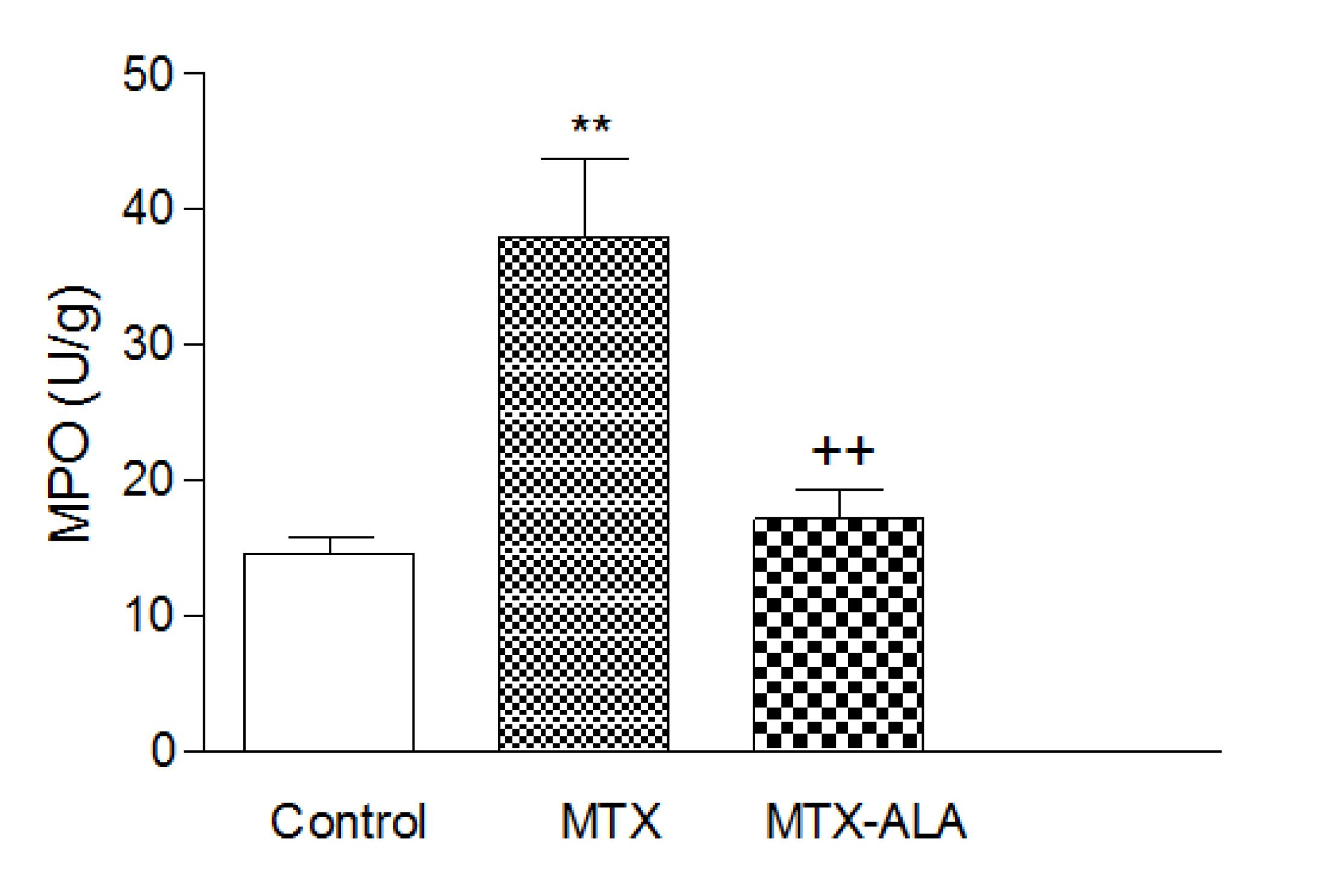
Myeloperoxidase activity, which is accepted as an indicator of neutrophil infiltration, was significantly higher in the liver tissues of the MTX group when compared to control group (p<0.001). On the other hand, α-lipoic acid treatment in MTX group significantly decreased all tissues MPO level (p<0.001, Figure 3), which was found to be not different than that of the control group.
Myeloperoxidase (MPO) activity in the liver tissues of control, methotrexate (MTX), MTX-ALA (a-lipoic acid) groups Each group consists of eight animals. Groups of data were compared with an analysis of variance (ANOVA) followed by Tukey's multiple comparison tests. ***p<0.001; compared to control group. ++ p<0.01, +++ p<0.001; compared to MTX group.
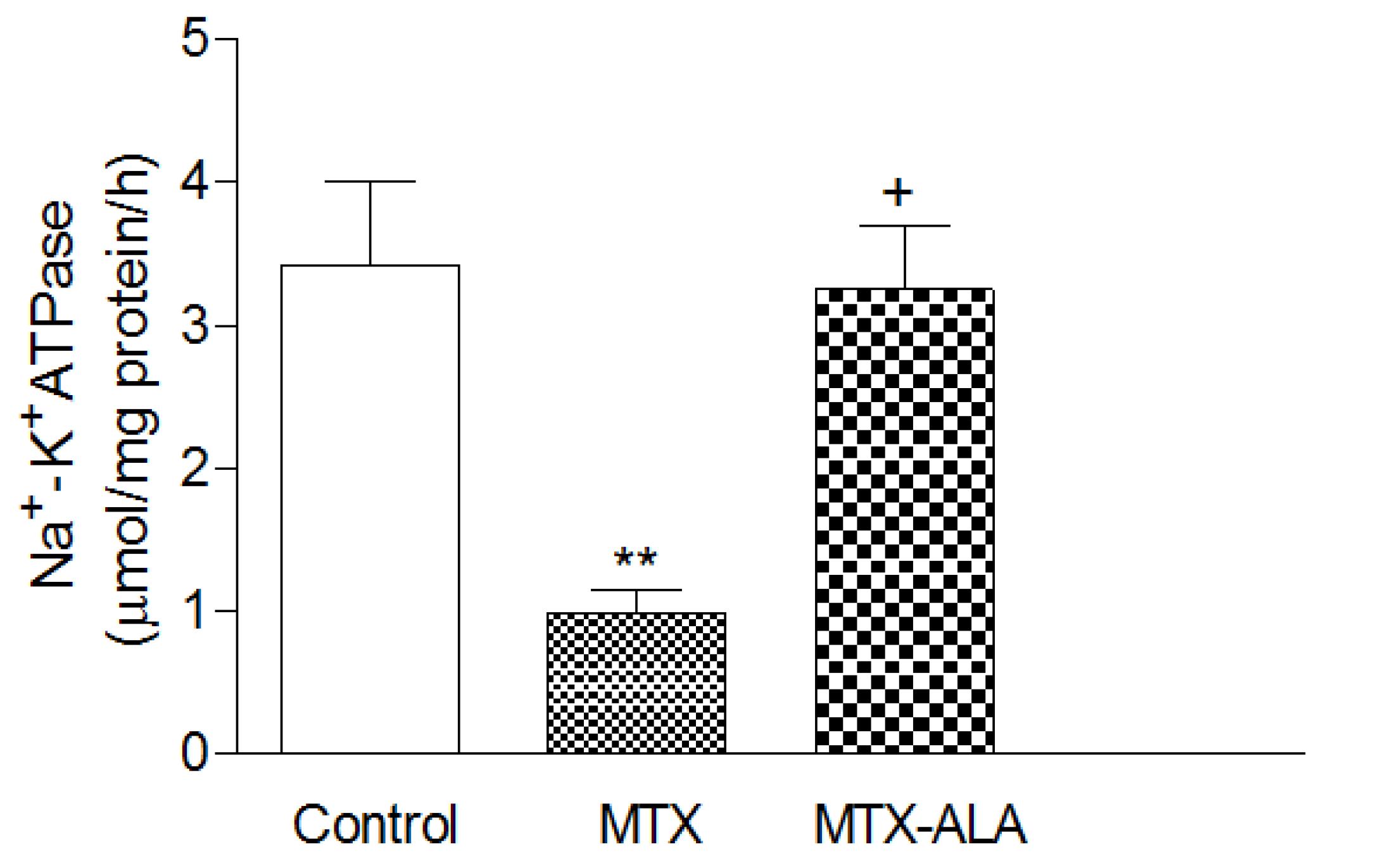
The activity of Na+-K+ ATPase was shown to be significantly decreased in the liver tissue of saline treated MTX group compared with control group; however, α-lipoic acid treatment in MTX group significantly increased all tissues Na+-K+ ATPase activity (p<0.001, Figure 4).
Na+-K+ ATPase activity in the liver tissues of control, methotrexate (MTX), MTX-ALA (a-lipoic acid) groups. Each group consists of eight animals. Groups of data were compared with an analysis of variance (ANOVA) followed by Tukey's multiple comparison tests. **p<0.01, ***p<0.001; compared to control group. ++ p<0.01, +++ p<0.001; compared to MTX group.
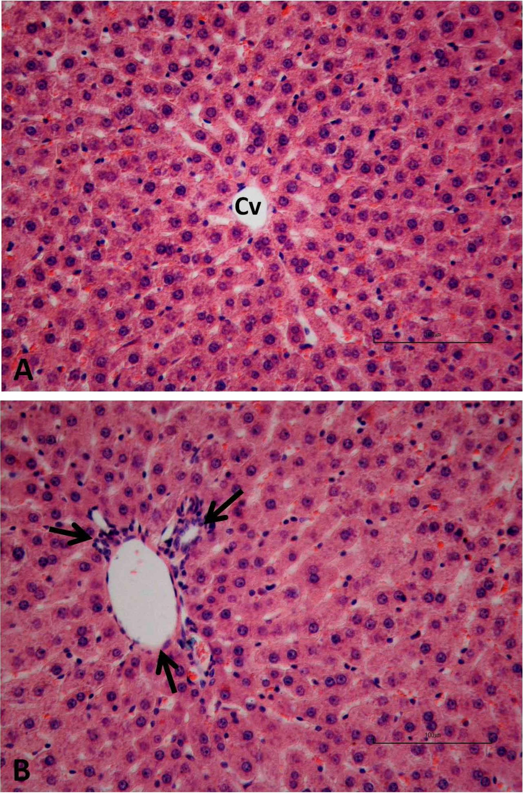
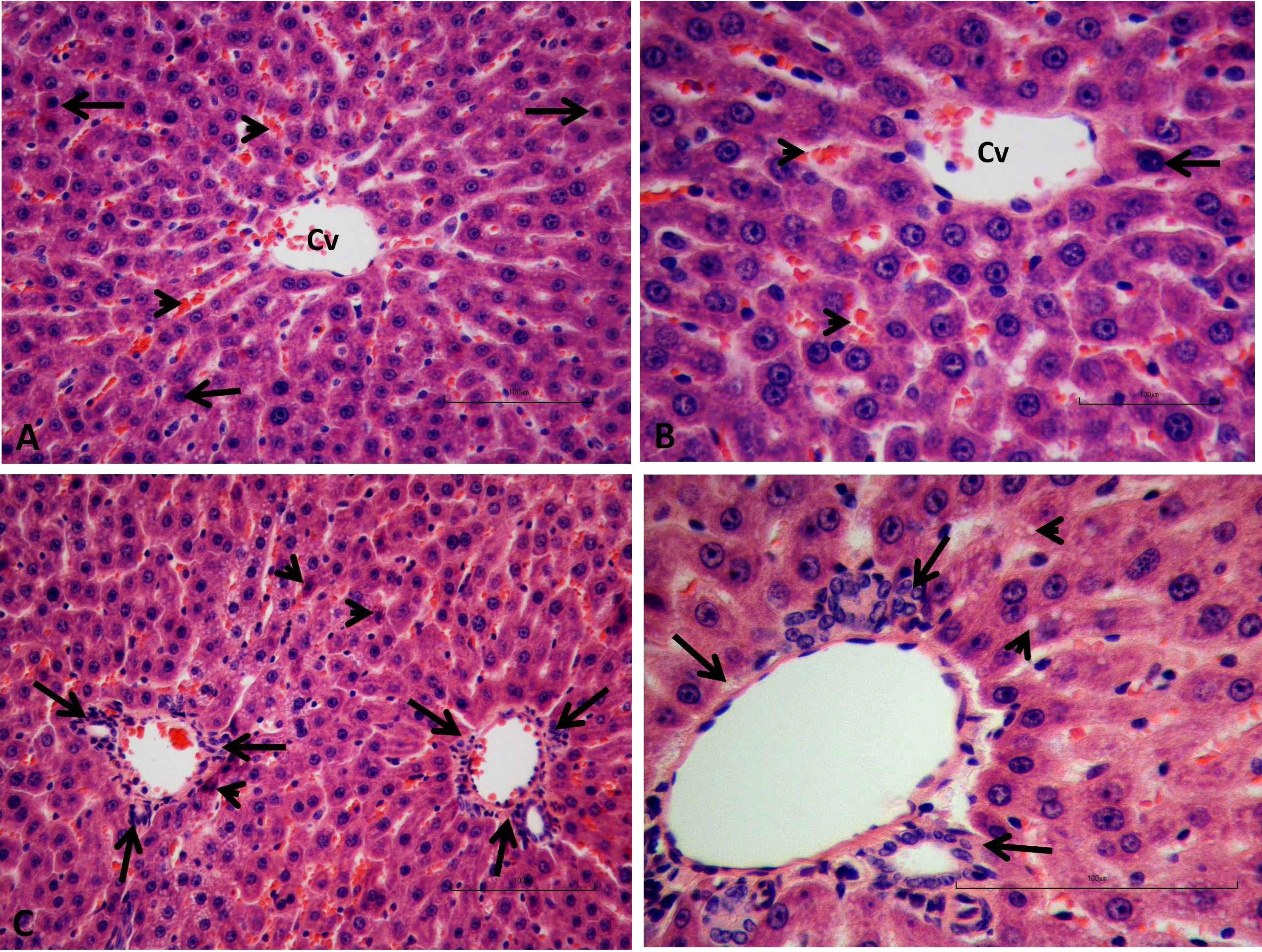
Liver sections from the control (Figure 5A-B) and LA (Figure 6A-B) groups were normal in histological appearance. The liver sections from the MTX treated group showed some histopathological changes such as hepatic necrosis, inflammatory cell infiltration especially in the periportal area and widespread intracellular vacuolization in hepatocytes and dark eosinophilic cytoplasm and heterochromatic, fragmented nuclei in hepatocytes and apoptotic bodies and vascular-sinusoidal congestion (Figure 7A-D).
Control group; Normal histological appearance of liver tissues, (A) central vein (Cv), (B) portal area (arrows). H&E, Scale bar = 100 µm.
ALA group; Normal histological appearance of liver tissues, (A) central vein (Cv), (B) portal area (arrows). H&E, Scale bar = 100 µm.
MTX group; (A) necrosis and inflammatory cell infiltration (aster), sinusoidal congestion (arrows), (B) inflammatory cell infiltration in the periportal area (arrows), (C) widespread intracellular vacuolization in hepatocytes (arrow heads) and vascular congestion (arrow), (D) inflammatory cell infiltration (aster), dark eosinophilic cytoplasm and heterochromatic nuclei in hepatocyte (thick arrow), apoptotic body (thin arrow) and fragmented nuclei in hepatocyte (arrow head). H&E, Scale bar = 100 µm.
However, administration of LA reduced the histopathological damage score significantly in the Mtx+LA group in comparison to the Mtx group. In the Mtx+LA group, histopathological evidence of hepatic damage was markedly reduced. In this group, liver sections showed rare dark eosinophilic cytoplasm and heterochromatic nuclei in hepatocytes, mild intracellular vacuolization in hepatocytes and inflammation was limited in localized areas and mild sinusoidal congestion (Figure 8A-D).
MTX+ALA group; (A, B) central vein (Cv), eosinophilic cytoplasm and heterochromatic nuclei in hepatocyte (arrow), sinusoidal congestion (arrow heads), (C) portal area (arrows) and eosinophilic cytoplasm and heterochromatic nuclei in hepatocyte (arrow heads), (D) portal area (arrows), mild intracellular vacuolization in hepatocytes (arrow heads). H&E, Scale bar = 100 µm.
Discussion
Findings from our study revealed that MTX administration causes oxidative tissue damage, while with its free radical scavenging effect the ALA prevented lipid peroxidation and neutrophil infiltration of the rat liver tissues. Furthermore, ALA treatment decreased the plasma cytokines and improved the liver tissue morphological changes caused by methotrexate.
Methotrexate is an antimetabolite that competitively inhibite the folic acid metabolism thus impairs the DNA synthesis. 7-hydroxymethotrexate (7-OH-MTX) is the major extracellular metabolite of MTX that is metabolized in the liver by an enzymatic system1818. Chládek J, Martínková J, Sispera L. An in vitro study on methotrexate hydroxylation in rat and human liver. Physiol Res. 1997;46(5):371-9. PMID: 9728483.. In the cell MTX store in a polyglutamate form. With the use of MTX intracellular amount of polyglutamate increases on the other hand folic acid levels decreased, that leads to necrosis of hepatocyte1919. Kamen BA, Nylen PA, Camitta BM, Bertino JR. Methotrexate accumulation and folate depletion in cells as a possible mechanism of chronic toxicity to the drug. Br J Haematol. 1981 Nov;49(3):355-60. PMID: 6170307.. Hepatotoxic effect of methotrexate was caused by an increase of its polyglutamate form intracellularly. The hepatotoxic effects of MTX have been reported in many studies11. Bleyer WA. Methotrexate: clinical pharmacology, current status and therapeutic guidelines. Cancer Treat Rev. 1977 Jun;4(2):87-101. PMID: 329989. , 2020. Sener G, Ekşioğlu-Demiralp E, Cetiner M, Ercan F, Yeğen BC. Beta-glucan ameliorates methotrexate-induced oxidative organ injury via its antioxidant and immunomodulatory effects. Eur J Pharmacol. 2006 Aug 7;542(1-3):170-8. PMID: 16793036. , 2121. Uz E, Oktem F, Yilmaz HR, Uzar E, Ozgüner F. The activities of purine-catabolizing enzymes and the level of nitric oxide in rat kidneys subjected to methotrexate: protective effect of caffeic acid phenethyl ester. Mol Cell Biochem. 2005 Sep;277(1-2):165-70. PMID: 16132728..
ALA is found in mitochondria as cofactor of pyruvate dehydrogenase and α-ketoglutarate dehydrogenase2222. Babiak RM, Campello AP, Carnieri EG, Oliveira MB. Methotrexate: pentose cycle and oxidative stress. Cell Biochem Funct. 1998 Dec;16(4):283-93. PMID: 9857491. , 2323. Miyazono Y, Gao F, Horie T. Oxidative stress contributes to methotrexate-induced small intestinal toxicity in rats. Scand J Gastroenterol. 2004 Nov;39(11):1119-27. PMID: 15545171. and is an effective free radical scavenger2424. Ellman GL. Tissue sulfhydryl groups. Arch Biochem Biophys. 1959 May;82(1):70-7. PMID: 13650640..
Lipid peroxidation by free oxygen radicals is an important causes of destruction and oxidative damage to cell membranes these containing unsaturated fatty acids, nucleic acids and proteins2525. Dobrota D, Matejovicova M, Kurella EG, Boldyrev AA. Na/K-ATPase under oxidative stress: molecular mechanisms of injury. Cell Mol Neurobiol. 1999 Feb;19(1):141-9. PMID: 10079973.. It has contribute to develope methotrexate associated tissue damage1010. Bludovská M, Kotyzová D, Koutenský J, Eybl V. The influence of alpha-lipoic acid on the toxicity of cadmium. Gen Physiol Biophys. 1999 Oct;18 Spec No:28-32. PMID: 10703716. , 1111. Buege JA, Aust SD. Microsomal lipid peroxidation. Methods enzymol. 1978;52:302-10. , 1818. Chládek J, Martínková J, Sispera L. An in vitro study on methotrexate hydroxylation in rat and human liver. Physiol Res. 1997;46(5):371-9. PMID: 9728483. , 2121. Uz E, Oktem F, Yilmaz HR, Uzar E, Ozgüner F. The activities of purine-catabolizing enzymes and the level of nitric oxide in rat kidneys subjected to methotrexate: protective effect of caffeic acid phenethyl ester. Mol Cell Biochem. 2005 Sep;277(1-2):165-70. PMID: 16132728..
With the attack of free oxygen radicals lipid peroxidation increase and fail the Na+/K+-ATPase activity2626. Thomas CE, Reed DJ. Radical-induced inactivation of kidney Na+, K(+)-ATPase: sensitivity to membrane lipid peroxidation and the protective effect of vitamin E. Arch Biochem Biophys. 1990 Aug 15;281(1):96-105. PMID: 2166481.. Na+/K+-ATPase is the other target of cellular oxidative tissue damage2525. Dobrota D, Matejovicova M, Kurella EG, Boldyrev AA. Na/K-ATPase under oxidative stress: molecular mechanisms of injury. Cell Mol Neurobiol. 1999 Feb;19(1):141-9. PMID: 10079973.. In this present study, MTX administration caused to a significant liver tissue damage since MDA which was the end product of lipid peroxidation is increased while Na+/K+-ATPase activity is depressed due to damage of cell membrane.
Liver tissue injury was also observed microscopically. On the other hand, following MTX administration, treatment with ALA was significantly reduce the MDA levels and increased the Na+/K+-ATPase enzyme activity, while normal histological appearance was observed in liver tissue.
Glutathione (GSH) plays a particularly important role in the maintenance and regulation of the thiol-redox status of the cell2727. Ballatori N, Krance SM, Notenboom S, Shi S, Tieu K, Hammond CL. Glutathione dysregulation and the etiology and progression of human diseases. Biol Chem. 2009 Mar;390(3):191-214. doi: 10.1515/BC.2009.033.
https://doi.org/10.1515/BC.2009.033...
. Tissue GSH depletion is one of the primary factors permitting liver tissue damage is associated with oxidative stress caused by MTX in our study.
It was expected that free radicals plays an important role in MTX induced liver toxicity2828. Jahovic N, Sener G, Cevik H, Ersoy Y, Arbak S, Yeğen BC. Amelioration of methotrexate-induced enteritis by melatonin in rats. Cell Biochem Funct. 2004 May-Jun;22(3):169-78. PMID: 15124182. , 2929. Jahovic N, Cevik H, Sehirli AO, Yeğen BC, Sener G. Melatonin prevents methotrexate-induced hepatorenal oxidative injury in rats. J Pineal Res. 2003 May;34(4):282-7. PMID: 12662351.. The reactive oxygen metabolites play a role in mediating liver toxicity of some xenobiotics and pathogenesis of organ failure1010. Bludovská M, Kotyzová D, Koutenský J, Eybl V. The influence of alpha-lipoic acid on the toxicity of cadmium. Gen Physiol Biophys. 1999 Oct;18 Spec No:28-32. PMID: 10703716. , 1111. Buege JA, Aust SD. Microsomal lipid peroxidation. Methods enzymol. 1978;52:302-10. , 1818. Chládek J, Martínková J, Sispera L. An in vitro study on methotrexate hydroxylation in rat and human liver. Physiol Res. 1997;46(5):371-9. PMID: 9728483. , 2121. Uz E, Oktem F, Yilmaz HR, Uzar E, Ozgüner F. The activities of purine-catabolizing enzymes and the level of nitric oxide in rat kidneys subjected to methotrexate: protective effect of caffeic acid phenethyl ester. Mol Cell Biochem. 2005 Sep;277(1-2):165-70. PMID: 16132728. , 2727. Ballatori N, Krance SM, Notenboom S, Shi S, Tieu K, Hammond CL. Glutathione dysregulation and the etiology and progression of human diseases. Biol Chem. 2009 Mar;390(3):191-214. doi: 10.1515/BC.2009.033.
https://doi.org/10.1515/BC.2009.033...
. It was reported that ALA protect the nuclear DNA, cell membrane lipids and intracellular proteins from oxidative tissue damage3030. Ozyurt H, Pekmez H, Parlaktas BS, Kus I, Ozyurt B, Sarsilmaz M. Oxidative stress in testicular tissues of rats exposed to cigarette smoke and protective effects of caffeic acid phenethyl ester. Asian J Androl. 2006 Mar;8(2):189-93. PMID: 16491270..
Free oxygen radicals trigger the leukocytes accumulation in tissue and activate the enzyme (including MPO, elastase and protease) secretion of neutrophils thus leads to further tissue damage. Therefore, MPO plays role in oxidant production by neutrophils3131. Winterbourn CC, Vissers MC, Kettle AJ. Myeloperoxidase. Curr Opin Hematol. 2000 Jan;7(1):53-8. PMID: 10608505. , 3232. Donnahoo KK, Meng X, Ayala A, Cain MP, Harken AH, Meldrum DR. Early kidney TNF-alpha expression mediates neutrophil infiltration and injury after renal ischemia-reperfusion. Am J Physiol. 1999 Sep;277(3 Pt 2):R922-9. PMID:10484513.. In our study MPO level which is an index of polimorphonuclear leukocyte infiltration was increased. Systemic inflammatory response indicators; TNF-α and IL-1β levels were also found increased. Increased levels of MPO indicate that neutrophil accumulation contributes to MTX induced oxidative injury in liver tissues. Treatment with ALA decreased the MPO activity and plasma TNF-α and IL-1β.
Conclusion
Alfa lipoic acid can be capable of reducing the methotrexate induced liver oxidative injury through its anti inflammatory and antioxidative effects.
References
-
1Bleyer WA. Methotrexate: clinical pharmacology, current status and therapeutic guidelines. Cancer Treat Rev. 1977 Jun;4(2):87-101. PMID: 329989.
-
2Widemann BC, Balis FM, Kempf-Bielack B, Bielack S, Pratt CB, Ferrari S, Bacci G, Craft AW, Adamson PC. High-dose methotrexate-induced nephrotoxicity in patients with osteosarcoma. Cancer. 2004 May;100(10):2222-32. PMID: 15139068.
-
3Braun J, Rau R. An update on methotrexate. Curr Opin Rheumatol. 2009 May;21(3):216-23. doi: 10.1097/BOR.0b013e328329c79d.
» https://doi.org/10.1097/BOR.0b013e328329c79d -
4Kose E, Sapmaz HI, Sarihan E, Vardi N, Turkoz Y, Ekinci N. Beneficial effects of montelukast against methotrexate-induced liver toxicity: a biochemical and histological study. ScientificWorldJournal. 2012;2012:987508. doi:10.1100/2012/987508.
» https://doi.org/10.1100/2012/987508 -
5Soliman ME. Evaluation of the possible protective role of folic acid on the liver toxicity ınduced experimentally by methotrexate in adult male albino rats. Egypt J Histol. 2009;32:118-28.
-
6Dalaklioglu S, Genc GE, Aksoy NH, Akcit F, Gumuslu S. Resveratrol ameliorates methotrexate-induced hepatotoxicity in rats via inhibition of lipid peroxidation. Hum Exp Toxicol. 2013 Jun;32(6):662-71. doi: 10.1177/0960327112468178.
» https://doi.org/10.1177/0960327112468178 -
7Chládek J, Martínková J, Sispera L. An in vitro study on methotrexate hydroxylation in rat and human liver. Physiol Res. 1997;46(5):371-9. PMID: 9728483.
-
8Bustamante J, Lodge JK, Marcocci L, Tritschler HJ, Packer L, Rihn BH. Alpha-lipoic acid in liver metabolism and disease. Free Radic Biol Med. 1998 Apr;24(6):1023-39. PMID: 9607614.
-
9Marley R, Holt S, Fernando B, Harry D, Anand R, Goodier D, Davies S, Moore K. Lipoic acid prevents development of the hyperdynamic circulation in anesthetized rats with biliary cirrhosis. Hepatology. 1999 May;29(5):1358-63. PMID: 10216116.
-
10Bludovská M, Kotyzová D, Koutenský J, Eybl V. The influence of alpha-lipoic acid on the toxicity of cadmium. Gen Physiol Biophys. 1999 Oct;18 Spec No:28-32. PMID: 10703716.
-
11Buege JA, Aust SD. Microsomal lipid peroxidation. Methods enzymol. 1978;52:302-10.
-
12Beutler E. Glutathione in red blood cell metabolism. A manual of biochemical methods. New York: Grune &Stratton; 1975. p.112-4.
-
13Hillegass LM, Griswold DE, Brickson B, Albrightson-Winslow C. Assessment of myeloperoxidase activity in whole rat kidney. J Pharmacol Methods. 1990 Dec;24(4):285-95. PMID: 1963456.
-
14Reading HW, Isbir T. The role of cation-activated ATPases in transmitter release from the rat iris. Q J Exp Physiol Cogn Med Sci. 1980 Apr;65(2):105-16. PMID: 6251506.
-
15Fiske CH, Subbarow Y. The colorimetric determination of phosphorus. J Biol Chem 1925;66:375-400.
-
16Lowry OH, Rosenbourgh NJ, Farr AL, Randall RJ. Protein measurement with the Folin phenol reagent. J Biol Chem. 1951 Nov;193(1):265-75. PMID: 14907713.
-
17Gul M, Kayhan B, Elbe H, Dogan Z, Otlu A. Histological and biochemical effects of dexmedetomidine on liver during an inflammatory bowel disease. Ultrastruct Pathol. 2013 Oct 17. [Epub ahead of print] PubMed PMID: 24134660.
-
18Chládek J, Martínková J, Sispera L. An in vitro study on methotrexate hydroxylation in rat and human liver. Physiol Res. 1997;46(5):371-9. PMID: 9728483.
-
19Kamen BA, Nylen PA, Camitta BM, Bertino JR. Methotrexate accumulation and folate depletion in cells as a possible mechanism of chronic toxicity to the drug. Br J Haematol. 1981 Nov;49(3):355-60. PMID: 6170307.
-
20Sener G, Ekşioğlu-Demiralp E, Cetiner M, Ercan F, Yeğen BC. Beta-glucan ameliorates methotrexate-induced oxidative organ injury via its antioxidant and immunomodulatory effects. Eur J Pharmacol. 2006 Aug 7;542(1-3):170-8. PMID: 16793036.
-
21Uz E, Oktem F, Yilmaz HR, Uzar E, Ozgüner F. The activities of purine-catabolizing enzymes and the level of nitric oxide in rat kidneys subjected to methotrexate: protective effect of caffeic acid phenethyl ester. Mol Cell Biochem. 2005 Sep;277(1-2):165-70. PMID: 16132728.
-
22Babiak RM, Campello AP, Carnieri EG, Oliveira MB. Methotrexate: pentose cycle and oxidative stress. Cell Biochem Funct. 1998 Dec;16(4):283-93. PMID: 9857491.
-
23Miyazono Y, Gao F, Horie T. Oxidative stress contributes to methotrexate-induced small intestinal toxicity in rats. Scand J Gastroenterol. 2004 Nov;39(11):1119-27. PMID: 15545171.
-
24Ellman GL. Tissue sulfhydryl groups. Arch Biochem Biophys. 1959 May;82(1):70-7. PMID: 13650640.
-
25Dobrota D, Matejovicova M, Kurella EG, Boldyrev AA. Na/K-ATPase under oxidative stress: molecular mechanisms of injury. Cell Mol Neurobiol. 1999 Feb;19(1):141-9. PMID: 10079973.
-
26Thomas CE, Reed DJ. Radical-induced inactivation of kidney Na+, K(+)-ATPase: sensitivity to membrane lipid peroxidation and the protective effect of vitamin E. Arch Biochem Biophys. 1990 Aug 15;281(1):96-105. PMID: 2166481.
-
27Ballatori N, Krance SM, Notenboom S, Shi S, Tieu K, Hammond CL. Glutathione dysregulation and the etiology and progression of human diseases. Biol Chem. 2009 Mar;390(3):191-214. doi: 10.1515/BC.2009.033.
» https://doi.org/10.1515/BC.2009.033 -
28Jahovic N, Sener G, Cevik H, Ersoy Y, Arbak S, Yeğen BC. Amelioration of methotrexate-induced enteritis by melatonin in rats. Cell Biochem Funct. 2004 May-Jun;22(3):169-78. PMID: 15124182.
-
29Jahovic N, Cevik H, Sehirli AO, Yeğen BC, Sener G. Melatonin prevents methotrexate-induced hepatorenal oxidative injury in rats. J Pineal Res. 2003 May;34(4):282-7. PMID: 12662351.
-
30Ozyurt H, Pekmez H, Parlaktas BS, Kus I, Ozyurt B, Sarsilmaz M. Oxidative stress in testicular tissues of rats exposed to cigarette smoke and protective effects of caffeic acid phenethyl ester. Asian J Androl. 2006 Mar;8(2):189-93. PMID: 16491270.
-
31Winterbourn CC, Vissers MC, Kettle AJ. Myeloperoxidase. Curr Opin Hematol. 2000 Jan;7(1):53-8. PMID: 10608505.
-
32Donnahoo KK, Meng X, Ayala A, Cain MP, Harken AH, Meldrum DR. Early kidney TNF-alpha expression mediates neutrophil infiltration and injury after renal ischemia-reperfusion. Am J Physiol. 1999 Sep;277(3 Pt 2):R922-9. PMID:10484513.
-
Financial source: none
-
1
Research performed at Research Laboratory, Inonü University of Medicine, Malatya, Turkey.
Publication Dates
-
Publication in this collection
Apr 2015
History
-
Received
10 Dec 2014 -
Reviewed
11 Feb 2015 -
Accepted
12 Mar 2015


