Abstracts
The sensibility and precision of the Nash and Moe and Raimondi methods were evaluated in this study for the measurement of the rotation of the thoracic and the lumbar vertebra. Three spine surgeons evaluated, independently, the x-rays of a thoracic vertebra (T9) and of a lumbar vertebra (L2) with varying rotational degrees from 0º to 60º and established values in agreement with the Nash and Moe method and the Raimondi method. The agreement among the examiners as to a certain method, the variation of the measures obtained for the thoracic and the lumbar vertebra, starting from a same known actual rotation and the correlation between a known true value of vertebral rotation and its forecast prepared through the methods used in the study were perused. The results showed good agreement among the examiners for the Nash and Moe method, so much for the thoracic vertebra (average k = 0,66), as for the lumbar (average k = 0,80). Using the Raimondi method there was no significant difference among the examiners for the thoracic vertebra. However, there was a low reproducibility of the method for the lumbar vertebra. For a same rotation of the thoracic and lumbar vertebra the results were non-concordant for the method of Nash and Moe, and for the Raimondi method the values observed for the thoracic vertebra were significantly larger than the ones for the lumbar vertebra. The correlation between the true values and the estimated values for the Raimondi method for the thoracic vertebra showed that there was a significant difference produced in function of the rotation up to 20º degrees, however for the lumbar vertebra the obtained values were very close to the actual.
Rotation; Spine; Scoliosis; Evaluation method
Neste estudo foram avaliados a sensibilidade e precisão dos métodos de Nash e Moe e de Raimondi para a medida da rotação da vértebra torácica e lombar.Três cirurgiões de coluna avaliaram, independentemente, as radiografias de uma vértebra torácica (T9) e de uma vértebra lombar (L2) com graus de rotação que variaram de 0º a 60º e estabeleceram valores de acordo com o método de Nash e Moe e o método de Raimondi.Foram estudadas a concordância entre os examinadores para um determinado método, a variação das medidas obtidas na vértebra torácica e lombar a partir de uma mesma rotação real conhecida e a correlação entre um valor real conhecido de rotação vertebral e a sua estimativa pelos métodos utilizados no estudo . Os resultados mostraram boa concordância entre os examinadores para o método de Nash e Moe, tanto para a vértebra torácica (k médio = 0,66), quanto para a lombar (k médio = 0,80). Pelo método de Raimondi não houve diferença significativa entre os examinadores para a vértebra torácica, no entanto, para a vértebra lombar houve baixa reprodutibilidade do método.Para uma mesma rotação na vértebra torácica e lombar os resultados foram não concordantes pelo método de Nash e Moe, e pelo método de Raimondi os valores observados para a vértebra torácica foram significativamente maiores que os da vértebra lombar. A correlação entre os valores reais e as estimados pelo método de Raimondi para a vértebra torácica mostrou que houve diferença significativa produzida em função da rotação até 20º graus, já para a vértebra lombar, os valores obtidos foram muito próximos do real.
Rotação; Coluna vertebral; Escoliose; Método de avaliação
ORIGINAL ARTICLE
Comparative study of the measurements of the vertebral rotation using Nash & Moe and Raimondi methods
Helton L. A. DefinoI; Paulo Henrique Mendes de AraújoII
IAssociate Professor of the Locomotion Apparatus' Biomechanics, Rehabilitation and Medicine Department of the Ribeirao Preto School of Medicine - USP
IIResident Doctor of the Orthopedics Discipline at the Clinicas Hospital in Ribeirao Preto
Correspondence Correspondence to Av. Bandeirantes 3900 - Ribeirão Preto CEP 14049-900 - São Paulo - Sp email: hladefin@fmrp.usp.br
SUMMARY
The sensibility and precision of the Nash and Moe and Raimondi methods were evaluated in this study for the measurement of the rotation of the thoracic and the lumbar vertebra. Three spine surgeons evaluated, independently, the x-rays of a thoracic vertebra (T9) and of a lumbar vertebra (L2) with varying rotational degrees from 0º to 60º and established values in agreement with the Nash and Moe method and the Raimondi method. The agreement among the examiners as to a certain method, the variation of the measures obtained for the thoracic and the lumbar vertebra, starting from a same known actual rotation and the correlation between a known true value of vertebral rotation and its forecast prepared through the methods used in the study were perused. The results showed good agreement among the examiners for the Nash and Moe method, so much for the thoracic vertebra (average k = 0,66), as for the lumbar (average k = 0,80). Using the Raimondi method there was no significant difference among the examiners for the thoracic vertebra. However, there was a low reproducibility of the method for the lumbar vertebra. For a same rotation of the thoracic and lumbar vertebra the results were non-concordant for the method of Nash and Moe, and for the Raimondi method the values observed for the thoracic vertebra were significantly larger than the ones for the lumbar vertebra. The correlation between the true values and the estimated values for the Raimondi method for the thoracic vertebra showed that there was a significant difference produced in function of the rotation up to 20º degrees, however for the lumbar vertebra the obtained values were very close to the actual.
Key words: Rotation; Spine; Scoliosis; Evaluation method.
INTRODUCTION
Scoliosis is a complex three-dimensional deformity of the trunk, the lateral deviation and the rotation of the vertebral bodies among the several pathological components of the deformity are emphasized (11,12). The rotation of the vertebral bodies occurs to the side of the curve convexity and its clinical manifestation with the deformity of the ribs in the thoracic column or of the thorny processes in the lumbar column is called a hump or a gibbus (4). The rotational grade of the vertebrae in the scoliosis is related to the cosmetic and aesthetic alterations of the patients, and measurements were developed, in addition to the clinical methods for its evaluation, using conventional X-rays, highlighting the methods described by Cobb, Nash & Moe, Perdriolle e Raimondi (2,3,8,9,10).
The vertebral rotation has been a very emphasized parameter in the patients' evaluation and of the results of the therapeutic procedures, and the objective of our study was to evaluate the degree of precision and the agreement as to the measure, among different examiners, related to two methods (Nash & Moe and Raimondi methods), that have been used in clinical practice for the evaluation of the vertebral rotation in scoliosis patients.
MATERIAL AND METHODS
In this study we used one thoracic vertebra (T9) and one lumbar vertebra (L2) from an adult individual and without any alterations to its morphology.
A device elaborated, in which the vertebrae were individually fastened, the degree of its rotation could be measured by goniometry, and x-rays were taken in AP using the conventional technique, from the neutral position (0º) up to 60º of rotation, with a 5º interval of between the x-rays. (Figure 1)
The x-rays were appraised independently by 3 spine surgeons, and it was proposed that each one examined the x-rays and determined the measurements of the vertebral rotations in agreement with the Nash & Moe and Raimondi methods.
The method of Nash & Moe is based on the relationship between the vertebral pedicles and the center of the vertebral body in the x-rays, in anteroposterior, and the rotation is classified in five different degrees according to the removal of the pedicles. There is no vertebral rotation in the situations in that the pedicles are halfway of the lateral margins of the vertebral bodies, and it is considered as a degree 0º. As the projection of the pedicle of the apical vertebra moves towards the median line in the x-rays in AP, the rotational degree progresses in the evaluation scale, reaching the largest value (degree IV) when it crosses that line. (Figure 2)
The Raimondi method uses the projection of the vertebral pedicles and the width of the vertebra as a reference for the measuring. The largest axis of the pedicle is demarcated and measured on the side of the curve convexity, and the distance of the longitudinal line from the pedicle to the border of the vertebra on the convex side is measured. Those two values are transported to the ruler, and the value of the rotation is, thus, obtained. (Figure 3)
The measures of the vertebral rotation of the thoracic vertebra and of the lumbar vertebra determined by the selected methods were compared with the values of the actual known rotation, and the values of the measures obtained in the 3 examiners' mensuration were compared.
The statistical study of the values of the measures of the rotation obtained by the 3 examiners was performed through a test "t of Student", "Qui-square" and kappa coefficient, depending on the type of the studied variable.
RESULTS
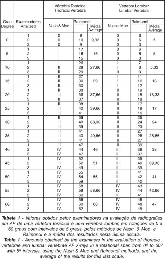
The values of each examiner's individual measures by the Nash & Moe method and by the Raimondi method coefficient are illustrated in Table 1.
The agreement among the individual values of the rotation obtained through Nash & Moe method was compared by analysis among the pairs of examiners, using the coefficient Kappa (K) that varies from 0 to 1. The agreement is considered excellent for k>0,80 values; good for values between 0,60 and 0,80; regular between 0,40 and 0,60; and weak for values under 0,40. Those values are illustrated in Table 2, and a larger agreement degree was observed among the examiners in the measures of the lumbar vertebra (average coefficient of 0,80) in relation to the thoracic vertebra, using the Nash & Moe method.
The comparison of the individual rotation results obtained using the Raimondi method was performed using the "t of Student" test, considering the significance level (p) smaller than 0,05, and are illustrated in Tables 3 and 4. Agreement of the measures of the thoracic vertebra was not observed among the examiners, and in the lumbar spine the agreement of the measures was observed among two pairs of examiners.
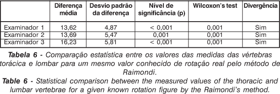
The comparison among the true and known rotation value of the vertebra and the examiners' individual values obtained with the evaluation methods used in the study are illustrated in Tables 5 and 6. For the Nash & Moe method an agreement of the values was observed, according to the coefficient kappa, between the two examiners' measure and the true values of the thoracic and lumbar vertebra rotation. For the Raimondi method, using the "t of Student" test and the Wilcoxon test, it was observed that the values of the examiners' measures diverged from the true value of the thoracic and lumbar vertebra rotations.
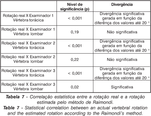
The correlation among the true and known rotation value for vertebra, and the value estimated with the use of the Raimondi method is illustrated in Table 7, and using the Qui-square test for the analysis of the adherence of the results, with no divergence of the measures observed only in the measure of the lumbar vertebra rotation executed by one of the examiners.
DISCUSSION
The vertebral rotation to the side of the convexity of the scoliotic curve is one of the radiography characteristics of scoliosis, and its magnitude has been correlated with the prognostic, cosmetic alterations and the patients' treatment. The method for the mensuration of the vertebral rotation should have low costs, be precise and reproducible(1,5,7,13).
In that study we observed a statistically significant difference among the values measured by the different examiners when analyzing a same x-ray by the same method. For the Nash & Moe method(6) the largest differences occurred in the analysis of the x-rays of the thoracic vertebra, compared with the mensuration of the lumbar vertebra. However, a good agreement was observed among the examiners. For the Raimondi method(13) there was good agreement among the examiners in the mensuration of the thoracic vertebra, and a lot of divergence in the results of the mensuration of the lumbar vertebra, evidencing a low reproducibility of the method in the studied vertebra.
The comparison of the results obtained by the 2 methods, considering the true value and known value of the rotation of the vertebrae, showed that the values were not in agreement. In the Nash & Moe method the obtained values showed a precarious correlation among the vertebrae, and in the Raimondi scale the values observed in the analysis of the known values of the thoracic vertebra were significantly larger than the ones of the lumbar vertebra. Those observations indicate that the existent anatomical differences among the thoracic and lumbar vertebrae can lead to false interpretations in the analysis of the vertebral rotations, taking into consideration the mensuration methods used in the study.
The comparison of the known value of the vertebral rotation and the value observed through the mensuration by the Raimondi method showed a significant difference of the values in the measure of the thoracic vertebra, and in the lumbar vertebra only the measurement of an examiner obtained results very close to the actual values. The results indicate that for the light degrees of rotation of the thoracic vertebra, the mensuration of the rotation would be jeopardized by the use of the Raimondi method. It was not possible to establish this comparison for the Nash method, because that method uses a graduated and not a numeric scale. However, different rotational values received the same graduation for the same examiner using that scale, making evident its imprecision.
It is important to point out that in our study vertebrae were used without morphologic alterations, and that in those conditions the obtained results didn't demonstrate precision in the measurements obtained with the methods used, and it is presumed that an extension of that correlation lack between the actual rotational values and the values supplied by the radiographic mensuration in the presence of morphologic alterations of the scoliosis patients' vertebrae may occur.
CONCLUSIONS
The anatomical differences among the thoracic and the lumbar vertebrae influenced in the precision of the measurement of the vertebral rotation and the measures of the vertebral rotation obtained by the Nash & Moe and Raimondi methods don't present a close correlation nor do they reproduce the actual and known values of the thoracic or lumbar vertebrae rotation .
REFERÊNCIAS BIBLIOGRÁFICAS
Trabalho recebido em 10/04/2004.
Aprovado em 20/06/2004.
Work performed at the Department of Biomechanics, Medicine and Rehabilitation of the Locomotor Apparatus, Faculty of Medicine University of São Paulo, Ribeirão Preto, SP, Brazil
- 1. Adams W. Lectures on the pathology and treatment of lateral and other forms of curvature of the spine. Churchill and Sons, London, 1865.
- 2. Bunnel WP. Vertebral rotation: simple method of measurement on routine radiographs (abstract). Orthop Trans 9: 114, 1985.
- 3. Drerup B. Principles of managment of vertebral rotation from frontal projections of pedicles. J Biomech 17: 923-935, 1984.
- 4. Filho TEPB, Lech, O Exame Físico em Ortopedia. São Paulo, Sarvier, 27-28, 2001.
- 5. Goçen, S Havitçioglu, H. Alici, E A new method to measure rotation from CT scans. Eur Spine J 8: 261-265, 1999.
- 6. Nash CL, Moe JH A study of vertebral rotation. J Bone Joint Surg (Am) 51: 223-229, 1969.
- 7. Omeroglu H, Ozekin O, Biçimoglu A Mesurement of vertebral rotation in idiopathic scoliosis using the Perdriolle torsionmeter: a clinical study on intraobserver and interobserver error. Eur Spine J 5: 167-171, 1996.
- 8. Perdriolle R, Vidal J Thoracic idiopathic scolioses curve evolution and prognosis. Spine 10: 785-791, 1985.
- 9. Richards, SB Measurement error in assessment of vertebral rotation using the Perdriolle torsion meter. Spine 17: 513-517, 1992.
- 10. Russel GG, Raso, VJ A comparison of four computerized methods for measuring vertebral rotation. Spine 15: 24-27, 1990.
- 11. Sevastik B, Xiong B, Sevastik J, Hedlund R, Suliman I Vertebral rotation and pedicle length asymmetry in the normal adult spine. Eur Spine 4: 95-97, 1995.
- 12. Stokes IAF, Three-dimensional terminology of spinal deformity: a report presented to the scoliosis research society by the scoliosis research society working group on 3-D terminology of spinal deformity. Spine 19: 236-248, 1994.
- 13. Weiss HR, Mesurement of vertebral rotation. Perdriolle versus Raimondi. Eur Spine J 4: 34-38, 1995.
Publication Dates
-
Publication in this collection
16 Nov 2004 -
Date of issue
Sept 2004
History
-
Accepted
20 June 2004 -
Received
10 Apr 2004


