
 COMPARATIVE EVALUATION OF FIRST- AND ADAPTED SECOND-GENERATION BONE CEMENTATION IN POST-CURETTAGE BONE DEFECT FILLING IN AN ANIMAL MODEL
Introduction: Bone cementation is fundamental for orthopedic stability, and different techniques have been proposed to optimize the bone–cement interface.
Objective: To evaluate whether adapted second-generation cementation improves osteointegration compared with the first-generation technique.
Methods: Bone defects created in the femora and tibiae of goats were filled with polymethylmethacrylate using either the first-generation technique (Group 1) or an adaptation of the second-generation technique (Group 2). Cementation quality and the bone–cement interface were assessed by radiography, CT, and photographic analysis, evaluated by orthopedic examiners. Statistical analyses employed the chi-square and Mann–Whitney U tests.
Results: No statistical differences were observed between the groups regarding cementation quality (radiography and CT) or osteointegration (photographic analysis) (p > 0.05). On CT, the mean number of bubbles was higher in the second-generation (6.05 vs. 3.95), whereas the proportion of samples with ≥5 bubbles was higher in the first-generation (50% vs. 30%). Photographic analysis showed that the second-generation group had a higher mean number of interdigitations (2.4 vs. 1.6) and a greater frequency of rough interfaces (50% vs. 25%). However, the first-generation group presented greater interdigitation depth (0.40 mm vs. 0.35 mm).
Conclusion: Adapted second-generation cementation showed no clear advantage over the first-generation technique. Level of evidence II; Investigation of treatment outcomes in an animal model.
Keywords:
Bone Cements; Curettage; Polymethyl Methacrylate; Bone Neoplasms
 Thumbnail
 Thumbnail
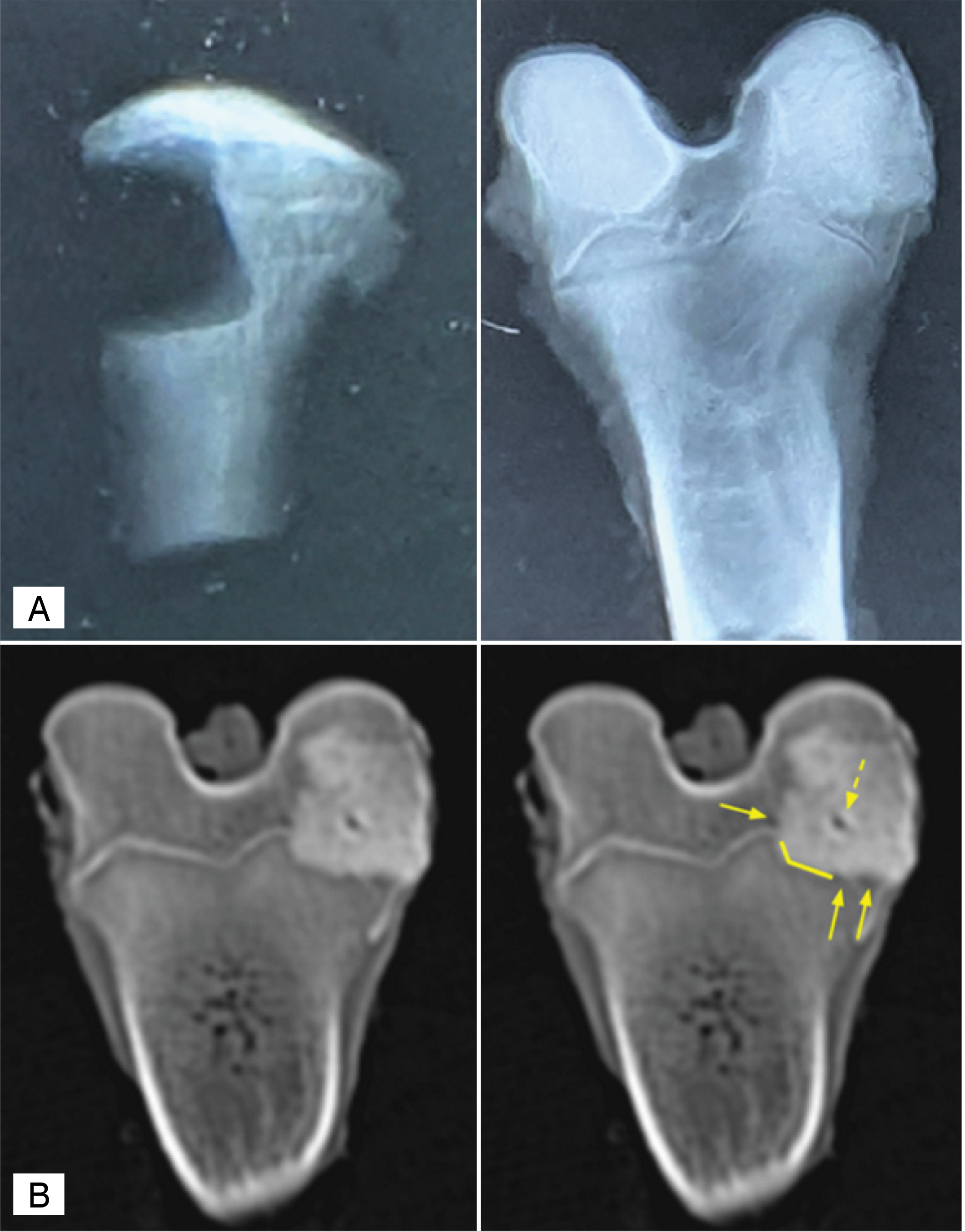 A. Simple X-ray of a bone defect in the metaphyseal region of the tibia, and of a bone defect filled with PMMA in the epiphyseal region of the femur. B. Bone defect in CT: the solid arrow indicates the space between cement and bone (lacuna), the solid line shows the interface between the materials (transition), and the dashed arrow indicates the presence of bubbles.
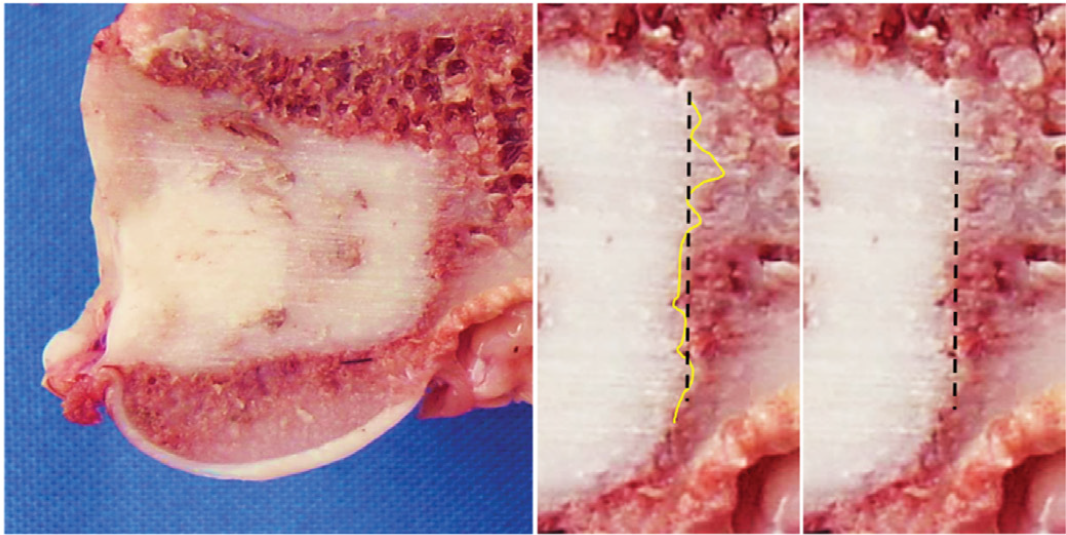 Photographic analysis in the coronal plane of the samples. In the linear dotted line, the central surface of 10 mm of the bone defect; and in the yellow line, the assessment of the interdigitations (protrusions).