Abstract
OBJECTIVE: To investigate the variations in blood glucose levels, hemodynamic effects and patient anxiety scores during tooth extraction in patients with type 2 diabetes mellitus T2DM and coronary disease under local anesthesia with 2% lidocaine with or without epinephrine.
STUDY DESIGN: This is a prospective randomized study of 70 patients with T2DM with coronary disease who underwent oral surgery. The study was double blind with respect to the glycemia measurements. Blood glucose levels were continuously monitored for 24 hours using the MiniMed Continuous Glucose Monitoring System. Patients were randomized into two groups: 35 patients received 5.4 mL of 2% lidocaine, and 35 patients received 5.4 mL of 2% lidocaine with 1:100,000 epinephrine. Hemodynamic parameters (blood pressure and heart rate) and anxiety levels were also evaluated.
RESULTS: There was no difference in blood glucose levels between the groups at each time point evaluated. Surprisingly, both groups demonstrated a significant decrease in blood glucose levels over time. The groups showed no significant differences in hemodynamic and anxiety status parameters.
CONCLUSION: The administration of 5.4 mL of 2% lidocaine with epinephrine neither caused hyperglycemia nor had any significant impact on hemodynamic or anxiety parameters. However, lower blood glucose levels were observed. This is the first report using continuous blood glucose monitoring to show the benefits and lack of side effects of local anesthesia with epinephrine in patients with type 2 diabetes mellitus and coronary disease.
Diabetes Mellitus; Coronary Disease; Dentistry; Local Anesthesia; Epinephrine; Lidocaine
INTRODUCTION
Diabetes mellitus (DM) is recognized as a worldwide epidemic, and the rate of type 2 diabetes mellitus T2DM is increasing. T2DM is a chronic disease that occurs when the pancreas does not produce enough insulin or cannot act effectively. In type 1 diabetes, there is a serious lack of insulin production. Furthermore, insulin resistance occurs because the insulin receptors are not adequately functioning. Persistent hyperglycemia leads to serious damage to target organs, as well as to oral manifestations due to microvascular abnormalities. Thus, the prevalence and severity of periodontal diseases are greater in individuals with diabetes, and these patients must frequently undergo invasive oral procedures. However, there is no established protocol for these procedures in the literature (1-5). In these cases, oral surgery requires special precautions, such as stress control and the use of a safe and effective anesthetic solution. Endogenous catecholamine secreted during psychological stress can lead to an undesirable increase of the heart rate, blood pressure, and blood glucose level (6-8). In addition, there is no consensus regarding the safety of local anesthetic solutions. The inclusion of vasoconstrictors in local anesthetic solutions offers unquestionable advantages. Vasoconstrictors promote longer-lasting anesthesia, reduce the toxic effects by delaying the absorption of anesthetics and reduce local bleeding (9,10). Epinephrine is the most commonly used vasoconstrictor. There is controversy regarding whether epinephrine, as a local anesthetic vasoconstrictor, causes adverse hemodynamic effects in cardiac patients (11-19). Furthermore, this agent has been reported to be problematic with regard to hyperglycemic effects. Therefore, some authors recommend the use of epinephrine-free local anesthetics for T2DM patients (20-22). However, other authors conclude that epinephrine vasoconstriction is not contraindicated in patients with T2DM (23,24). Therefore, the aim of this study was to investigate variations in blood glucose levels, hemodynamic parameters, and anxiety levels during tooth extraction under local anesthesia with plain 2% lidocaine versus 2% lidocaine with 1:100,000 epinephrine in patients with T2DM and coronary disease.
PATIENTS AND METHODS
This prospective randomized study was conducted from September 2009 to November 2012 in patients with T2DM and coronary disease from the Heart Institute (InCor) of Hospital das Clínicas da Faculdade de Medicina da Universidade de São Paulo (HCFMUSP). The study was approved by the local ethics committee under the protocol no 1033/08, and informed consent was obtained from all patients before inclusion in the study. This study was registered at clinicaltrials.gov (NCT02173067).
We enrolled adult patients with pharmacologically controlled T2DM (via the use of insulin and/or hypoglycemic agents) and coronary disease who needed to have at least one posterior maxillary tooth extracted according to an oral examination and panoramic radiography.
To assess the levels of blood glucose, the MiniMed Continuous Glucose Monitoring (CGMS, Medtronic Diabetes) was employed. This device is able to monitor average glucose measurements taken every 5 minutes. The MiniMed monitor was installed in the morning on the day before the surgery. The patient then went home and returned to the hospital for the surgery 24 hours later. The same dentist carried out the surgery in all patients and was not provided any information regarding the blood glucose levels of the patients during the protocol. Therefore, in this sense, the study was double blind. Subjects were randomly divided into 2 groups: the LCA group received 2% lidocaine with 1:100,000 epinephrine, and the LSA group received plain 2% lidocaine. The same volume of anesthetic (5.4 mL) was applied using a standard technique (25). The MiniMed monitor was removed an hour after the surgery, and the data were downloaded into a computer using vMonGluco Client software.
Standardized times and periods evaluated
T0 (first value recorded by the MiniMed), T1 (1 hour before the procedure), T2 (5 minutes before the procedure), T3 (after anesthesia injection), T4 (end of procedure), and T5 (1 hour after the procedure). We considered the basal period (BP) to be from T0 to T1 and the procedure period (PP) to be from T1 to T5.
The systolic blood pressure (SBP), diastolic blood pressure (DBP) and heart rate (HR) were recorded by an automatic sphygmomanometer (Microlife APA-P00001) at T1, T2, T3, and T5.
The anxiety level was measured by the Buchanan and Niven (26) facial scale at T1, T2, and T5.
If the extracted tooth was a potential source of infection, 500 mg of amoxicillin was prescribed to be taken every 8 hours for 7 days; 300 mg of clindamycin was prescribed to amoxicillin-allergic individuals. For cases with a high or moderate risk of infective endocarditis, according to recommendations by the American Heart Association (27), a single dose of 2 g of amoxicillin was administered before the procedure. Patients were instructed to maintain their current medications, including oral anticoagulants and antiplatelet agents.
Statistical Analysis
Data are presented as the mean ± standard deviation for normally distributed data and as the median and range for non-normally distributed data. To compare means between the 2 groups, we used t tests; when the assumption of normality of the data was rejected, we used the nonparametric Mann-Whitney U test. Analysis of variance (ANOVA) for repeated measurements was performed to compare the groups according to the times and periods of study (28). A p value <0.05 was considered significant.
RESULTS
A total of 400 patients were initially evaluated. Among these individuals, 179 were edentulous patients, 74 were partial edentulous patients, 46 did not require tooth extraction, and 28 refused to participate, all of whom were excluded. The remaining 70 patients (50 male, 20 female) were included (mean age, 63.4±8.3 years (44-83), body mass index, 28.0±5.0 kg/m2).
The subjects were randomly divided into 2 groups: an LSA group (n = 35) and an LCA group (n = 35). Table 1 shows the clinical characteristics of the 2 groups. There was no difference in gender between the groups (p = 0.290). Prophylactic antibiotics were administered to 3 patients, with no difference between groups (p = 1.000). Amoxicillin was administered over 7 days to 30 subjects, and clindamycin was administered to 1 individual; there was no significant difference between the LSA and LCA groups for this parameter (p = 0.336). There was also no significant difference between the groups (p = 0.207) according to the duration of the surgery.
Age, body mass index, length of diabetes, glycosylated hemoglobin, time of insulin use in each group.
Blood Glucose Levels
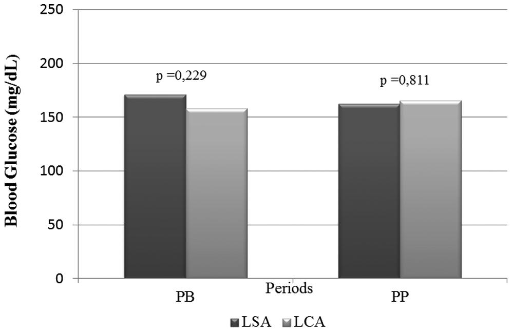
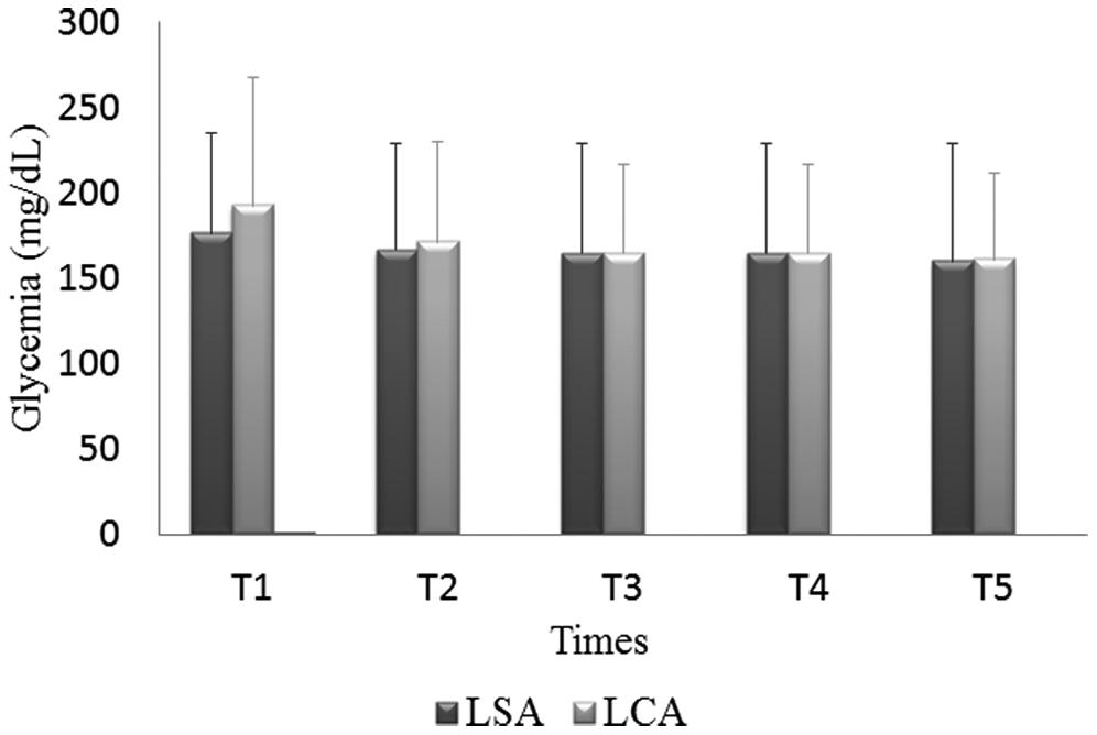
There was no significant difference in the blood glucose levels during either period between the two groups (Fig 1). There was also no significant difference in the mean blood glucose levels in the LSA and LCA groups at any time (p = 0.748). Surprisingly, both groups demonstrated a significant decrease in blood glucose levels (p<0.001) over time (Fig 2).
Means of blood glucose levels in basal period (PB) and procedural period (PP) in LSA and LCA group.
All participants used oral hypoglycemic drugs and/or insulin therapy. There were 20 patients in the LSA group and 17 in the LCA group who were receiving insulin therapy, with no difference between the groups (p = 0.473). Figure 3 illustrates the blood glucose analysis in 4 subgroups with respect to the use of insulin during the trial interval. There was no significant difference between the subgroups (p = 0.737).
Means of blood glucose levels in subgroups with respect to the use the insulin or not in LSA and LCA group.
Blood Pressure, Heart Rate, and Anxiety Levels
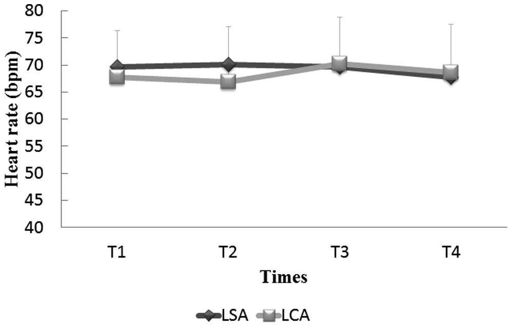
Figures 4 and 5 show that there were no significant differences in the SBP (p = 0.176), DBP (p = 0.913), and HR (p = 0.570) between the groups at T1, T2, T3, or T5.
The anxiety level in the LSA and LCA groups did not significantly change over time (T1, p = 0.953; T2, p = 0.954; and T5, p = 0.823). We observed a significant decrease in the anxiety level (p<0.001) at T1, T2 and T5.
DISCUSSION
This study assessed the variations in blood glucose levels in patients with T2DM and coronary disease who required oral surgery using plain 2% lidocaine or 2% lidocaine with 1:100,000 epinephrine. There was no significant difference in the blood glucose levels between the LSA and LCA groups. Surprisingly, a significant decrease in the blood glucose level was observed over time in both groups. This finding must be further explored.
Bortoluzzi et al (29) did not verify changes in blood glucose levels in healthy patients. However, the anesthetic solution used was different (2% mepivacaine with adrenaline) and was at a lower dosage than that used in the present study (2 × 3 cartridges). Tily and Thomas (30) found the same results in the healthy and diabetic groups regardless of the applied volume. According to Shcaira et al (31), there was a difference (p = 0.00003) in blood glucose levels between the diabetic and healthy groups, with higher values in the diabetic group. Most authors recommend the use of epinephrine-free anesthetics for T2DM patients (20-22). Meechan (32) detected a significant increase in glucose values during oral surgery when using epinephrine. In addition, Meechan and Welbury (33) noted an increase in blood glucose levels in 20 patients undergoing oral surgery with intravenous midazolam. This increase was observed only in the group that was receiving 4.4 mL of lidocaine with 1:80,000 epinephrine. Nakamura et al (34) found relevant elevations in blood glucose levels after local anesthesia with epinephrine. Kalra et al (35) observed a significant increase in blood glucose levels when comparing diabetic and healthy groups (p<0.005) 20 minutes following the injection of lidocaine with epinephrine. Our study shows that the findings of above studies are not definitive; the discrepancy might be attributed to differences in the patient characteristics and the methods used to evaluated the variances. However, this study is more reliable because the blood glucose levels were continuously monitored.
In this study, all patients had pharmacologically controlled T2DM with coronary disease and hypertension. Moreover, the blood glucose measurement methods were different; the above-mentioned studies used a glucose meter or drew blood samples at predetermined times, whereas our study was the first to perform continuous glucose monitoring from 24 hours before surgery to 1 hour after surgery, with the mean glucose levels recorded every 5 minutes. Specifically, an average of 30 glucose readings were obtained for each subject only during the surgery. Therefore, our results seem to be more accurate. Nevertheless, we took care to standardize the anesthetic technique and the volumes applied; this choice provides greater credibility to our results. The volume of 5.4 mL was selected based on the experience of the InCor Dentistry Unit, which routinely uses plain lidocaine in patients with cardiovascular disease. Notably, both of the anesthetic regimens (LSA and LCA) were effective and did not require supplementation in either group. Additionally, our results show a significant decrease in blood glucose levels (p<0.001) over the study period. The levels at T1 were significantly different compared with those at T2 (p = 0.003), T3 (p<0.001), T4 (p<0.001), and T5 (p<0.001). This observation has not been previously described in the literature.
All participants used oral hypoglycemic drugs and/or insulin therapy. Medications were not suspended, and their doses were not changed to accommodate the oral surgery. Oral anticoagulant and antiplatelet agents were maintained, although some authors have suggested suspending them (36).
We also evaluated the hemodynamic effects (BP and HR) and anxiety levels of the patients. The use of epinephrine as a local anesthetic for cardiac patients is controversial. Most studies have shown adverse hemodynamic effects (16-18), whereas other authors recommend a maximum dose of 0.04 mg (23).
In the present study, there were no differences between groups in the SBP (p = 0.176), DBP (p = 0.913) and HR (p = 0.570). These results corroborate those of a study by Neves et al (37) that used 1.8 mL or 3.6 mL of plain lidocaine and lidocaine with 1:100,000 epinephrine prior to procedures on coronary patients. These findings are also in accordance with those of Conrado et al (38), who compared patients undergoing dental extraction with 3% mepivacaine and 2% mepivacaine with 1:100,000 epinephrine. Takahashi et al (39) investigated the influence of varying concentrations of exogenous epinephrine combined with a local anesthetic on endogenous plasma catecholamine levels and did not observe a significant difference in SBP, DBP, and HR among groups. A study by Meral et al (10) used the same anesthetic solution that we used but at a lower volume (2.0 mL of plain lidocaine with 1:100,000 epinephrine). These subjects also underwent extractions, although they were otherwise healthy. There were no significant differences in the BP. With respect to the HR, the authors reported a significant increase in both groups, which differed from the findings of our study. We can attribute our results to the level of anesthetic used due to the time required to administer the local anesthesia.
Anxiety and fear are common during oral procedures. Psychological stress can lead to adverse cardiovascular and metabolic effects. The use of a vasoconstrictor in a local anesthetic solution promotes longer-lasting anesthesia and likely decreases stress (9,10). For this reason, many researchers have their subjects undergo conscious sedation or general anesthesia to compare local anesthetic solutions (31-33). We performed the surgeries in a private dental care office without any sedation. Our results show that the anxiety levels did not significantly change in the LSA and LCA groups during oral surgery.
The results observed in this study allow us to conclude that oral surgery performed in an outpatient clinic on patients with diabetes and coronary disease using local anesthesia at the assessed volume (5.4 mL) is safe, regardless of whether epinephrine (1:100,000) is included. Furthermore, the use of epinephrine does not interfere with blood glucose levels, hemodynamic parameters, or the emotional state. In contrast, we observed lower glucose blood levels when epinephrine was included in the anesthetic solution.
This study contributes to dental practice in unique and severe cases by providing scientific support for surgical procedures in patients with diabetes and coronary disease. Furthermore, it clarifies the theory that the use of epinephrine in combination with anesthetics not increases the risk of hyperglycemia and hemodynamic repercussions.
ACKNOWLEDGEMENTS
This work was funded by the Foundation for Research Support of São Paulo, FAPESP, Process Number 08/57612-0.
REFERENCES
-
1Shlossman M, Knowler WC, Pettitt DJ, Genco RJ. Type 2 diabetes
mellitus and periodontal disease. J Am Dent Assoc. 1990;121(4):532-6,
http://dx.doi.org/10.14219/jada.archive.1990.0211.
» http://dx.doi.org/10.14219/jada.archive.1990.0211 -
2Preshaw PM, Alba AL, Herrera D, Jepsen S, Konstantinidis A,
Makrilakis K, et al. Periodontitis and diabetes: a two-way relationship.
Diabetologia. 2012;55(1):21-31,
http://dx.doi.org/10.1007/s00125-011-2342-y.
» http://dx.doi.org/10.1007/s00125-011-2342-y -
3Papapanou PN. Periodontal diseases: epidemiology. Ann
Periodontol. 1996;1(1):1-36,
http://dx.doi.org/10.1902/annals.1996.1.1.1.
» http://dx.doi.org/10.1902/annals.1996.1.1.1 -
4Lalla RV, D'Ambrosio JA. Dental management considerations
for the patient with diabetes mellitus. J Am Dent
Assoc.2001;132(10):1425-32,
http://dx.doi.org/10.14219/jada.archive.2001.0059.
» http://dx.doi.org/10.14219/jada.archive.2001.0059 -
5Albandar JM. Global risk factors and risk indicators for
periodontal diseases. Periodontol. 2002;9:177-206,
http://dx.doi.org/10.1034/j.1600-0757.2002.290109.x.
» http://dx.doi.org/10.1034/j.1600-0757.2002.290109.x - 6Barcellos IF, Halfon VLC, Oliveira LF, Barcellos Filho I. Conduta odontológica em paciente diabético. Rev Bras Odontol. 2000;57(6):407-10.
- 7Galili D, Findler M, Garfunkel AA. Oral and dental complications associated with diabetes and their treatment. Compendium. 1994;15(4):496-508.
- 8Rhodus NL, Vibeto BM, Hamamoto DT. Glycemic control in patients with diabetes mellitus upon admission to a dental clinic: considerations for dental management. Quintessence Int. 2005;36(6):474-82.
- 9Mariano RC, Santana SI, Coura GS. Análise comparativa do efeito anestésico da lidocaína 2% e da prilocaína 3%. BCI. 2000;7(27):15-9.
-
10Meral G, Tasar F, Sayin F, Saysel M, Kir S, Karabulut E. Effects
of lidocaine with and without epinephrine on plasma epinephrine and lidocaine
concentrations and hemodynamic values during third molar surgery. Oral surgery,
oral medicine, oral pathology, oral radiology, and endodontics.
2005;100(2):e25-30,
http://dx.doi.org/10.1016/j.tripleo.2005.03.031.
» http://dx.doi.org/10.1016/j.tripleo.2005.03.031 -
11Knoll-Kohler E, Fortsch G. Pulpal anesthesia dependent on
epinephrine dose in 2% lidocaine. A randomized controlled double-blind
crossover study. Oral Surg Oral Med Oral Pathol. 1992;73(5):537-40,
http://dx.doi.org/10.1016/0030-4220(92)90091-4.
» http://dx.doi.org/10.1016/0030-4220(92)90091-4 -
12Knoll-Kohler E, Knoller M, Brandt K, Becker J. Cardiohemodynamic
and serum catecholamine response to surgical removal of impacted mandibular
third molars under local anesthesia: a randomized double-blind parallel group
and crossover study. J Oral Maxillofac Surg. 1991;49(9):957-62,
http://dx.doi.org/10.1016/0278-2391(91)90059-U.
» http://dx.doi.org/10.1016/0278-2391(91)90059-U - 13Niwa H, Hirota Y, Sibutani T, Idohji Y, Hori T, Sugiyama K, et al. The effects of epinephrine and norepinephrine administered during local anesthesia on left ventricular diastolic function. Anesth Prog. 1991;38(6):221-6.
-
14Cioffi GA, Chernow B, Glahn RP, Terezhalmy GT, Lake CR. The
hemodynamic and plasma catecholamine responses to routine restorative dental
care. J Am Dent Assoc. 1985;111(1):67-70,
http://dx.doi.org/10.14219/jada.archive.1985.0038.
» http://dx.doi.org/10.14219/jada.archive.1985.0038 - 15Meyer FU. Hemodynamic changes of local dental anesthesia in normotensive and hypertensive subjects. Int J Clin Pharmacol Ther Toxicol. 1986;24(9):477-81.
-
16Goldstein DS, Dionne R, Sweet J, Gracely R, Brewer HB Jr., Gregg
R, et al. Circulatory, plasma catecholamine, cortisol, lipid, and psychological
responses to a real-life stress (third molar extractions): effects of diazepam
sedation and of inclusion of epinephrine with the local anesthetic. Psychosom
Med. 1982;44(3):259-72,
http://dx.doi.org/10.1097/00006842-198207000-00004.
» http://dx.doi.org/10.1097/00006842-198207000-00004 -
17Tolas AG, Pflug AE, Halter JB. Arterial plasma epinephrine
concentrations and hemodynamic responses after dental injection of local
anesthetic with epinephrine. J Am Dent Assoc. 1982;104(1):41-3,
http://dx.doi.org/10.14219/jada.archive.1982.0114.
» http://dx.doi.org/10.14219/jada.archive.1982.0114 - 18Chernow B, Balestrieri F, Ferguson CD, Terezhalmy GT, Fletcher JR, et al. Local dental anesthesia with epinephrine. Minimal effects on the sympathetic nervous system or on hemodynamic variables. Arch Intern Med. 1983;143(11):2141-3.
- 19Dionne RA, Goldstein DS, Wirdzek PR. Effects of diazepam premedication and epinephrine-containing local anesthetic on cardiovascular and plasma catecholamine responses to oral surgery. Anesth Analg. 1984;63(7):640-6.
-
20Vernillo AT. Diabetes mellitus: Relevance to dental treatment.
Oral surgery, oral medicine, oral pathology, oral radiology, and endodontics.
2001;91(3):263-70, http://dx.doi.org/10.1067/moe.2001.114002.
» http://dx.doi.org/10.1067/moe.2001.114002 - 21Orso VA, Pagnoncelli RM. O perfil do paciente diabético e o tratamento odontológico. Revista Odonto Ciência. 2002;17(36):206-13.
- 22Mistro FZ, Kignel S, Cardoso DS, Morais ES. Diabetes mellitus: revisão e considerações no tratamento odontológico. Revista Paulista de Odontologia. 2003;25(6):15-8.
- 23Haas DA. An update on local anesthetics in dentistry. J Can Dent Assoc. 2002;68(9):546-51.
- 24Levin JA, Muzyka BC, Glick M. Dental management of patients with diabetes mellitus. Compend Contin Educ Dent. 1996;17(1):82-90.
- 25Malamed SF. Técnicas de anestesia maxilar. In: Malamed SF. Manual de anestesia local. 4aed. Rio de Janeiro: Guanabara Koogan; 2001.p.144-71.
- 26Buchanan H, Niven N. Validation of a Facial Image Scale to assess child dental anxiety. Int J Paediatr Dent. 2002;12(1):47-52.
- 27Dajani AS, Taubert KA, Wilson W, Bolger AF, Bayer A, Ferrieri P, et al. Prevention of bacterial endocarditis. Recommendations by the American Heart Association. Circulation. 1997;96(1):358-66.
- 28Timm NH. Multivariate analysis with applications in educations and psychology. Monterrey: Brooks/Cole; 1957.
-
29Bortoluzzi MC, Manfro R, Nardi A. Glucose levels and hemodynamic
changes in patients submitted to routine dental treatment with and without local
anesthesia. Clinics. 2010;65(10):975-8,
http://dx.doi.org/10.1590/S1807-59322010001000009.
» http://dx.doi.org/10.1590/S1807-59322010001000009 -
30Tily FE, Thomas S. Glycemic effect of administration of
epinephrine-containing local anaesthesia in patients undergoing dental
extraction, a comparison between healthy and diabetic patients. Int Dent J.
2007;57(2):77-83,
http://dx.doi.org/10.1111/j.1875-595X.2007.tb00442.x.
» http://dx.doi.org/10.1111/j.1875-595X.2007.tb00442.x - 31Schaira VR, Ranali J, Saad MJ, de Oliveira PC, Ambrosano GM, Volpato MC. Influence of diazepam on blood glucose levels in nondiabetic and non-insulin-dependent diabetic subjects under dental treatment with local anesthesia. Anesth Prog. 2004;51(1):14-8.
- 32Meechan JG. Epinephrine, magnesium, and dental local anesthetic solutions. Anesth Prog.1996;43(4):99-102.
- 33Meechan JG, Welbury RR. Metabolic responses to oral surgery under local anesthesia and sedation with intravenous midazolam: the effects of two different local anesthetics. Anesth Prog. 1992;39(1-2):9-12.
-
34Nakamura Y, Matsumura K, Miura K, Kurokawa H, Abe I, Takata Y.
Cardiovascular and sympathetic responses to dental surgery with local
anesthesia. Hypertens Res. 2001;24(3):209-14,
http://dx.doi.org/10.1291/hypres.24.209.
» http://dx.doi.org/10.1291/hypres.24.209 -
35Kalra P, Rana AS, Peravali RK, Gupta D, Jain G. Comparative
evaluation of local anaesthesia with adrenaline and without adrenaline on blood
glucose concentration in patients undergoing tooth extractions.
J Maxillofac Oral Surg. 2011;10(3):230-5,
http://dx.doi.org/10.1007/s12663-011-0239-4.
» http://dx.doi.org/10.1007/s12663-011-0239-4 -
36Vernillo AT. Dental considerations for the treatment of patients
with diabetes mellitus. J Am Dent Assoc. 2003;134 Spec No:24S-33S,
http://dx.doi.org/10.14219/jada.archive.2003.0366.
» http://dx.doi.org/10.14219/jada.archive.2003.0366 -
37Neves RS, Neves IL, Giorgi DM, Grupi CJ, Cesar LA, Hueb W, et
al. Effects of epinephrine in local dental anesthesia in patients with coronary
artery disease. Arq Bras Cardiol. 2007;88(5):545-51,
http://dx.doi.org/10.1590/S0066-782X2007000500008.
» http://dx.doi.org/10.1590/S0066-782X2007000500008 -
38Conrado VC, de Andrade J, de Angelis GA, de Andrade AC, Timerman
L, Andrade MM, et al. Cardiovascular effects of local anesthesia with
vasoconstrictor during dental extraction in coronary patients. Arq Bras Cardiol.
2007;88(5):507-13,
http://dx.doi.org/10.1590/S0066-782X2007000500002.
» http://dx.doi.org/10.1590/S0066-782X2007000500002 -
39Takahashi Y, Nakano M, Sano K, Kanri T. The effects of
epinephrine in local anesthetics on plasma catecholamine and hemodynamic
responses. Odontology. 2005;93(1):72-9,
http://dx.doi.org/10.1007/s10266-005-0044-y.
» http://dx.doi.org/10.1007/s10266-005-0044-y
Publication Dates
-
Publication in this collection
Mar 2015
History
-
Received
26 Aug 2014 -
Reviewed
26 Sept 2014 -
Accepted
5 Jan 2015

 Local anesthesia with epinephrine is safe and effective for oral
surgery in patients with type 2 diabetes mellitus and coronary disease: a
prospective randomized study


