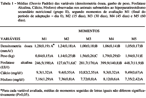
The aim of this study was to evaluate the modification of bone mineral density, as well as the serum biochemistry variation in the nutritional secondary hyperparathyroidism. Ten crossbreed cats, initial aging between 2 and 3 months, and weighing 820 grams were used. After 10 days of adaptation, they were fed with raw beef heart for 60 days. At the end of adaptation time and every 15 days, exams were realized. The method of optical densitometry in radiographic images of the right radius and ulna was used. There was no statistical difference in the bone mineral densitometry between the end of adaptation period and with 15 days of consuming a diet of beef heart. At 30 days the bone density decreased statistically, and it was in the same level at 45 and 60 days. There was no statistical difference in the serum calcium and phosphorus concentrations in all observation time. Serum alkaline phosphatase concentration varied and it was increased above normal variation in the 45th and 60th day of the diet. It was possible to conclude that bone densitometry in radiographic images is an efficient method to evaluate bone demineralization, and calcium, phosphorus and alkaline phosphatase serum biochemistry analysis are limited value.
kittens; hyperparathyroidism; nutritional; densitometry


