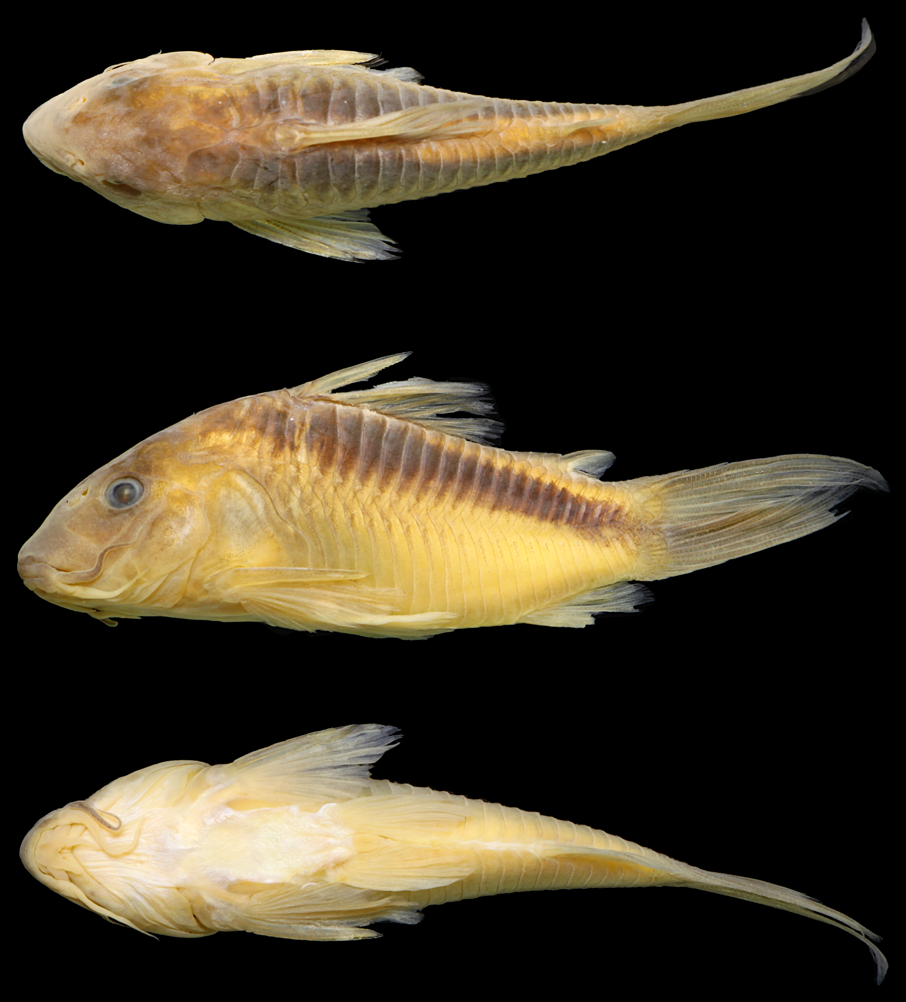
FIGURE 1 |
Osteogaster oharai, holotype, MNRJ 55884, 43.4 mm SL, Aripuanã, Mato Grosso, Brazil, stream tributary of the rio Aripuanã, upstream Salto das Andorinhas and Dardanelos waterfalls, rio Madeira basin.
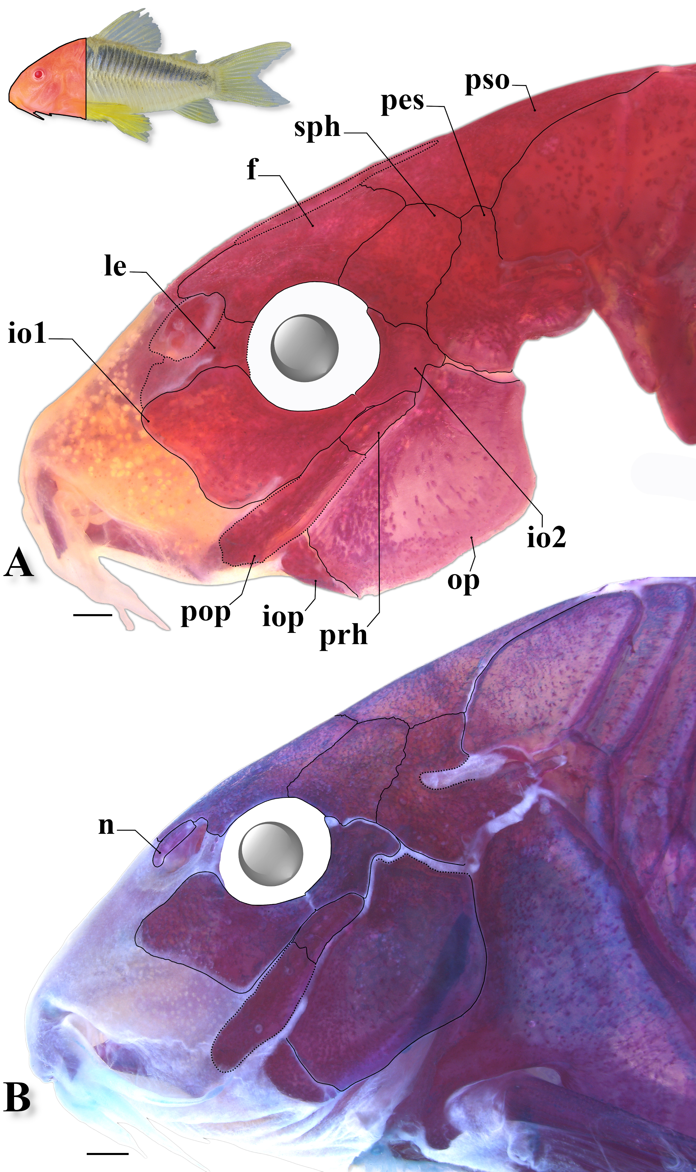
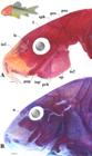
FIGURE 2 |
Head osteological pattern of Osteogaster oharai, showing the general morphology in lateral view of (A) CPUFMT 4889, 44.7 mm SL, and (B) CITL 1504, 41.1 mm SL. Abbreviations: f: frontal, io1–2: infraorbital 1 and 2, iop: interopercle, le: lateral ethmoid, n: nasal, me: mesethmoid, op: opercle, pes: pterotic-extrascapular, pop: preopercle, prh: posterodorsal ridge of hyomandibula, pso: parieto-supraoccipital, sph: sphenotic. Area where the illustrated bones are located in fish’s body marked in red in the miniature photo of an uncatalogued specimen in life (lateral view). Scale bars = 1 mm.
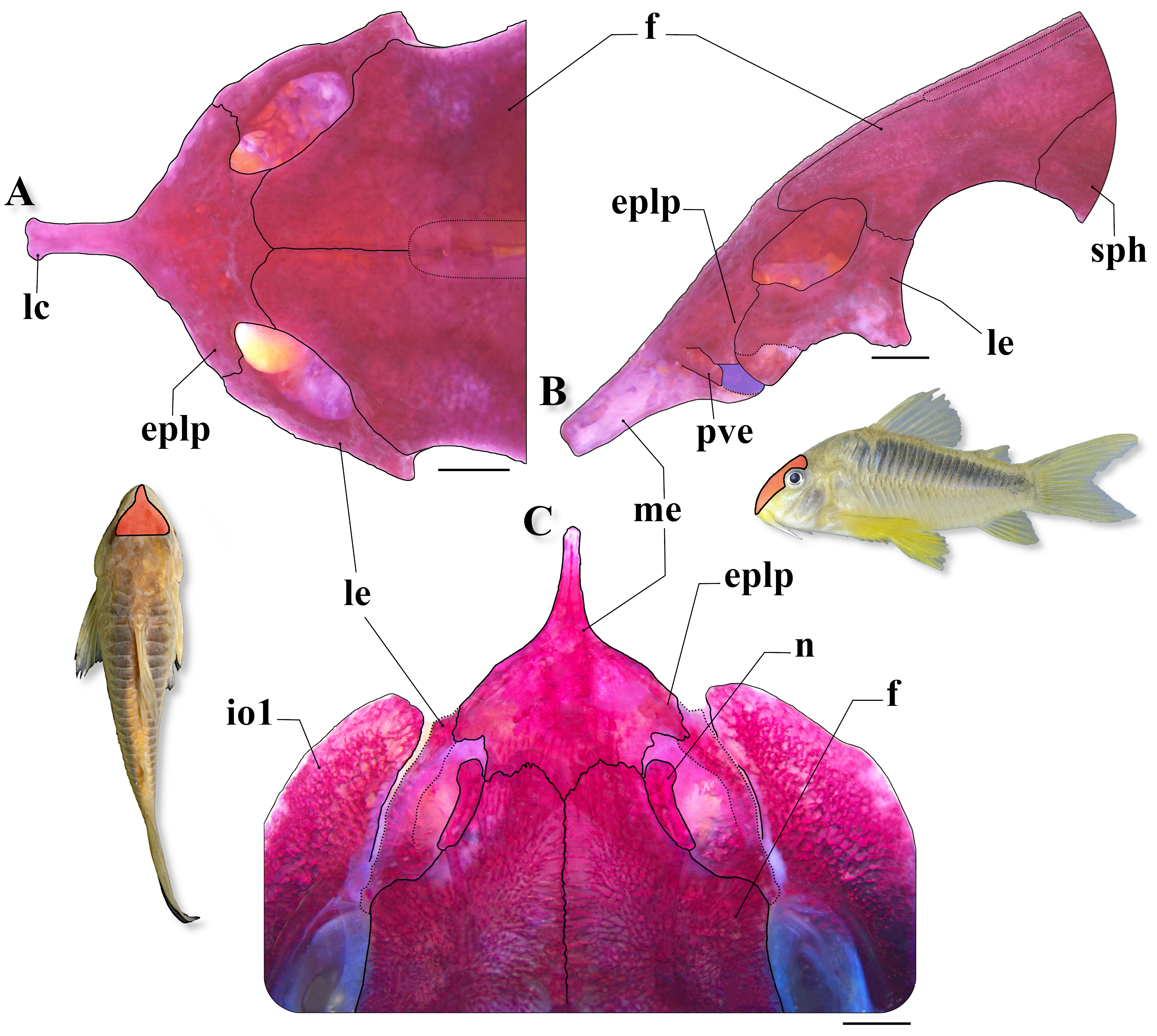
FIGURE 3 |
Cranial osteology of Osteogaster oharai, showing the detail of the mesethmoid in dorsal (A) and lateral (B) views (CPUFMT 4889, 44.7 mm SL; paratype), and (C) the anterior portion of neurocranium in dorsal view (MNRJ 55728, 40.9 mm SL; non-type specimen). Abbreviations: eplp: external projection of lateral posterodorsal expansion of mesethmoid, f: frontal, io1: infraorbital 1, lc: lateral cornua, le: lateral ethmoid, me: mesethmoid, n: nasal, pes: pterotic-extrascapular, pve: ventrolateral expansion of posterior portion of mesethmoid, sph: sphenotic. Area where the illustrated bones in (A) and (B) are located in fish’s body marked in red in the miniature photo of an uncatalogued specimen in life (lateral view), and in (C) in the miniature photo of the preserved holotype (MNRJ 55884, 43.4 mm SL) in dorsal view. Scale bars = 1 mm.
FIGURE 4 |
Cranial osteology in a c&s paratype of Osteogaster oharai (CPUFMT 4889, 44.7 mm SL), showing (A) the upper jaw in frontal view (= dorsal view considering everted mouth), (B) the suspensorium plus operculum in lateral view, and (C) the palatine in dorsal view. Abbreviations: aa: angulo-articular, d: dentary, hym: hyomandibula, iop: interopercle, mp: metapterygoid, mx: maxilla, op: opercle, pmx: premaxilla, pop: preopercle, prh: posterodorsal ridge of hyomandibula, q: quadrate. Red arrow indicates laminar process on posterolateral portion of maxilla; yellow and green arrows indicate anteroventral expansion and posterodorsal process of dentary, respectively; orange and blue arrows indicate dorsal laminar expansion and posteroventral process of anguloarticular, respectively. Dotted line in (C) outlining dorsolateral longitudinal laminar expansion of palatine. Area where the illustrated bones are located in fish’s body marked in red in the miniature photo of an uncatalogued specimen in life (lateral view). Scale bars = 1 mm.
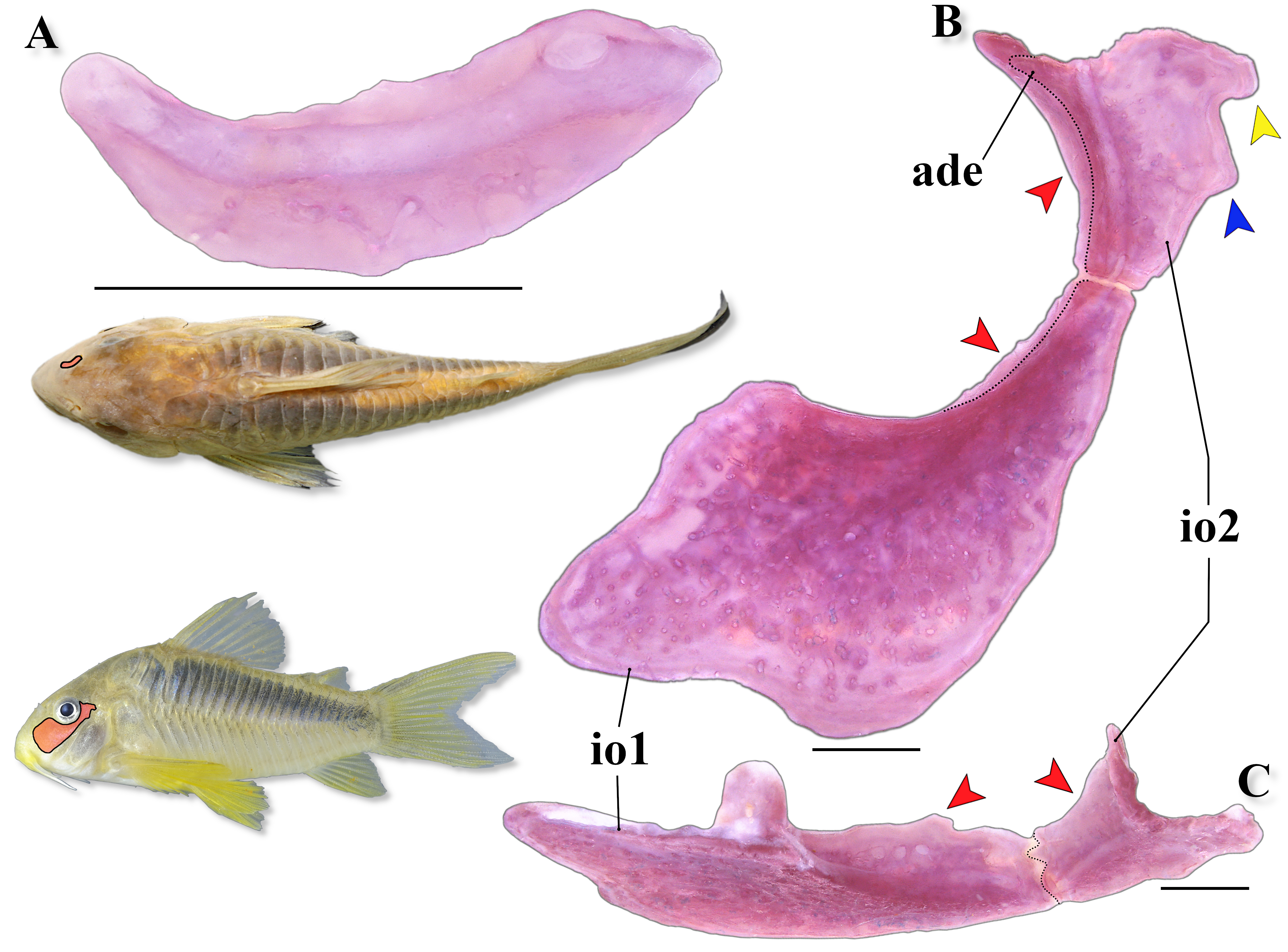
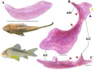
FIGURE 5 |
Cranial osteology in a c&s paratype of Osteogaster oharai (CPUFMT 4889, 44.7 mm SL), showing (A) the nasal bone in dorsal view, and the infraorbital series in lateral (B) and dorsal (C) views. Abbreviations: ade: anterodorsal edge of infraorbirtal 2, io1–2: infraorbital 1 and 2. Red arrows indicate the inner laminar expansion of both infraorbitals; yellow and blue arrows indicate posterodorsal and posterior-middle expansions of infraorbital 2, respectively. Area where the illustrated bones in (A) are located in fish’s body marked in red in the miniature photo of the preserved holotype (MNRJ 55884, 43.4 mm SL) in dorsal view, and in (B) plus (C) in the miniature photo of an uncatalogued specimen in life (lateral view). Scale bars = 1 mm.
FIGURE 6 |
Top of head and predorsal region of trunk of Osteogaster oharai in dorsal view, showing the variations related to the contact between the parieto-supraoccipital and nuchal plate. A. Parieto-supraoccipital close but not in contact with nuchal plate in a c&s paratype (CPUFMT 4889, 35.8 mm SL); B. Posterior process apparently not in contact with nuchal plate externally, but in contact under thick layer of skin in a c&s non-type specimen (CITL 1504, 41.1 mm SL); C. Posterior process in direct contact with nuchal plate externally in a c&s paratype (CPUFMT 4889, 44.7 mm SL); D. Posterior process relatively distant from the nuchal plate externally, first dorsolateral body plate contacting its counterpart between both structures in a c&s non-type specimen (MNRJ 55728, 35.6 mm SL). Abbreviations: f: frontal, n: nasal, np: nuchal plate, me: mesethmoid, pes: pterotic-extrascapular, pso: parieto-supraoccipital, sph: sphenotic. Area where the illustrated bones are located in fish’s body marked in red in the miniature photo of the preserved holotype (MNRJ 55884, 43.4 mm SL) in dorsal view. Scale bars = 1 mm.
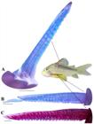
FIGURE 7 |
Lateral view of (A) the dorsal-fin spine and dorsal view of (B) the left pectoral-fin spine of a c&s paratype (CPUFMT 4889, 44.7 mm SL), and of (C) the left pectoral-fin spine of a non-type specimen (MNRJ 55728, 41.1 mm SL) of Osteogaster oharai, showing their serration patterns. Area where the illustrated bones are located in fish’s body marked in red in the miniature photo of an uncatalogued specimen in life (lateral view). Scale bars = 1 mm.
FIGURE 8 |
Pelvic girdle in c&s specimens of Osteogaster oharai, showing its morphological variations in dorsal view, depicting (A) a paratype (CPUFMT 4889, 44.7 mm SL), and three non-type specimens, (B) MNRJ 55728, 35.6 mm SL, (C) MNRJ 55728, 40.9 mm SL, and (D) MNRJ 55728 41.1 mm SL. Abbreviations: bp: basipterygium, pae: anterior external process, pai: anterior internal process, pi: dorsal ischiac process. Pelvic girdle position shown in red in the miniature photo of the preserved holotype (MNRJ 55884, 43.4 mm SL) in ventral view. Scale bars = 1 mm.
FIGURE 9 |
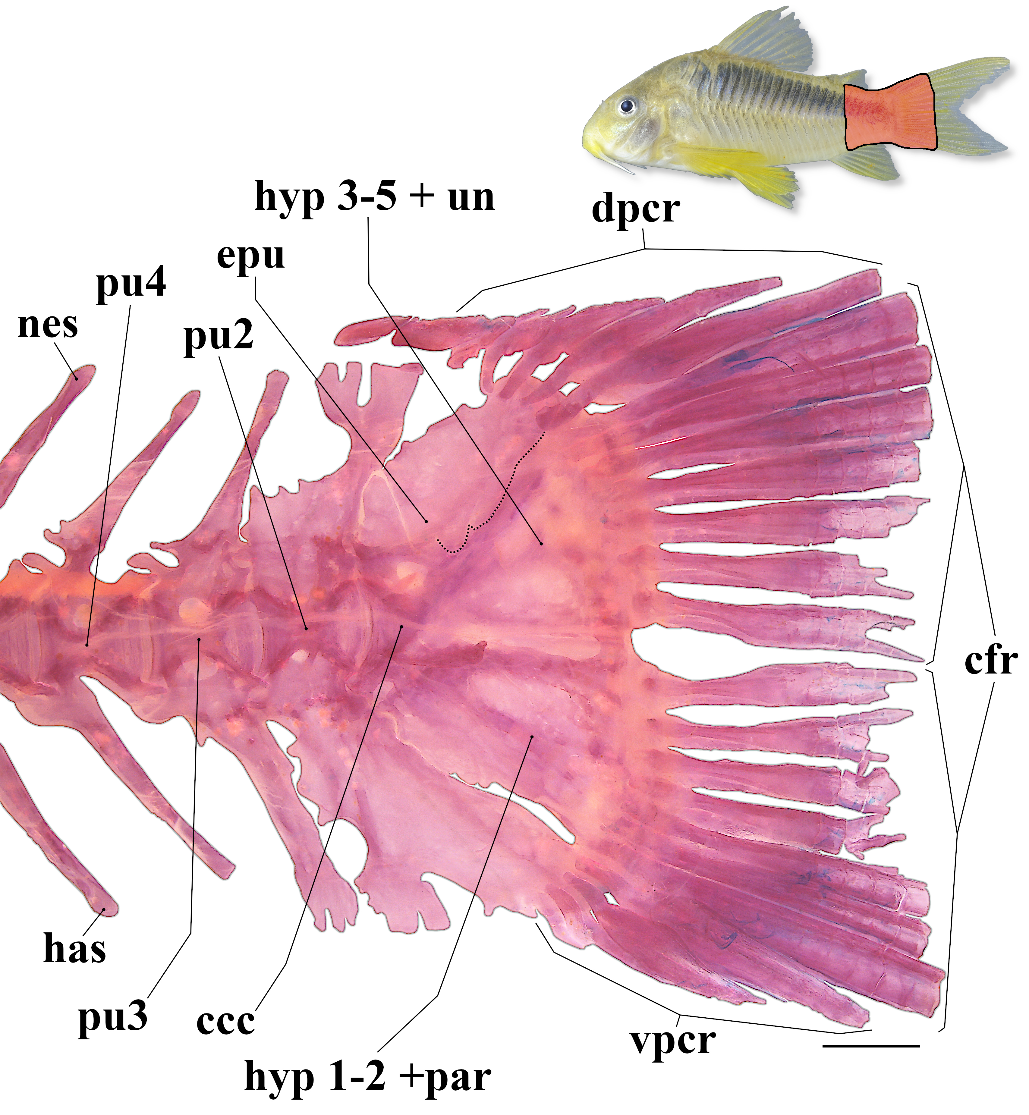
General morphology of caudal skeleton in a c&s paratype of Osteogaster oharai (CPUFMT 4889, 44.7 mm SL). Abbreviations: ccc: compound caudal centrum, cfr: caudal-fin principal rays, dpcr: dorsal procurrent rays, epu: epural, has: haemal spine, hyp 1–5: hypurals 1 to 5, nes: neural spine, par: parhypural, pu 2–4: preural centra 2 to 4, un: uroneural, vpcr: ventral procurrent rays. Area where the illustrated bones are located in fish’s body marked in red in the miniature photo of an uncatalogued specimen in life (lateral view). Scale bar = 1 mm.
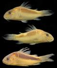
FIGURE 10 |
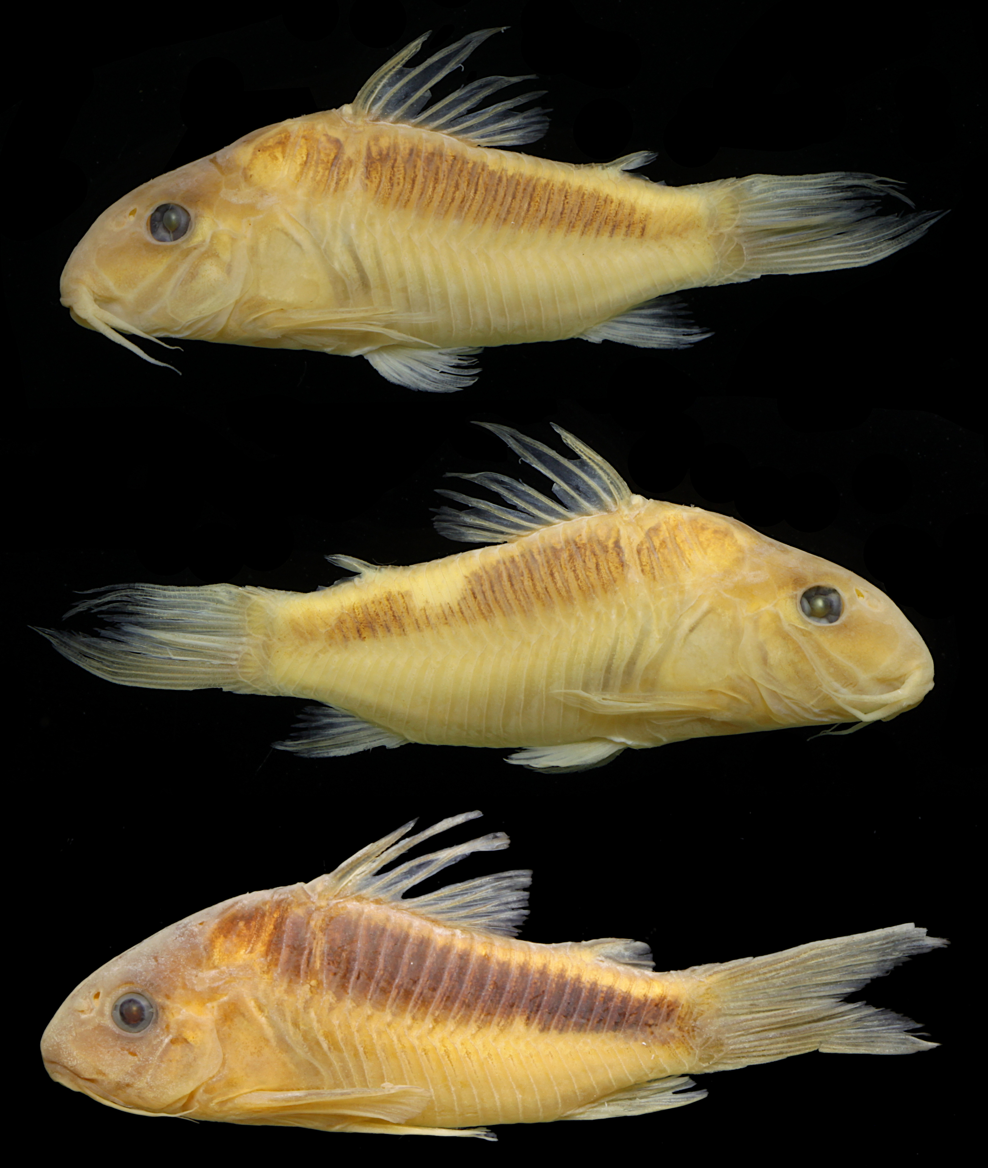
Variations on snout shape and color pattern in paratypes of Osteogaster oharai, showing left and right side of specimen UFRO-ICT 21931, 42.5 mm SL, and left side of specimen CPUFMT 5015, 35.4 mm SL.
FIGURE 11 |
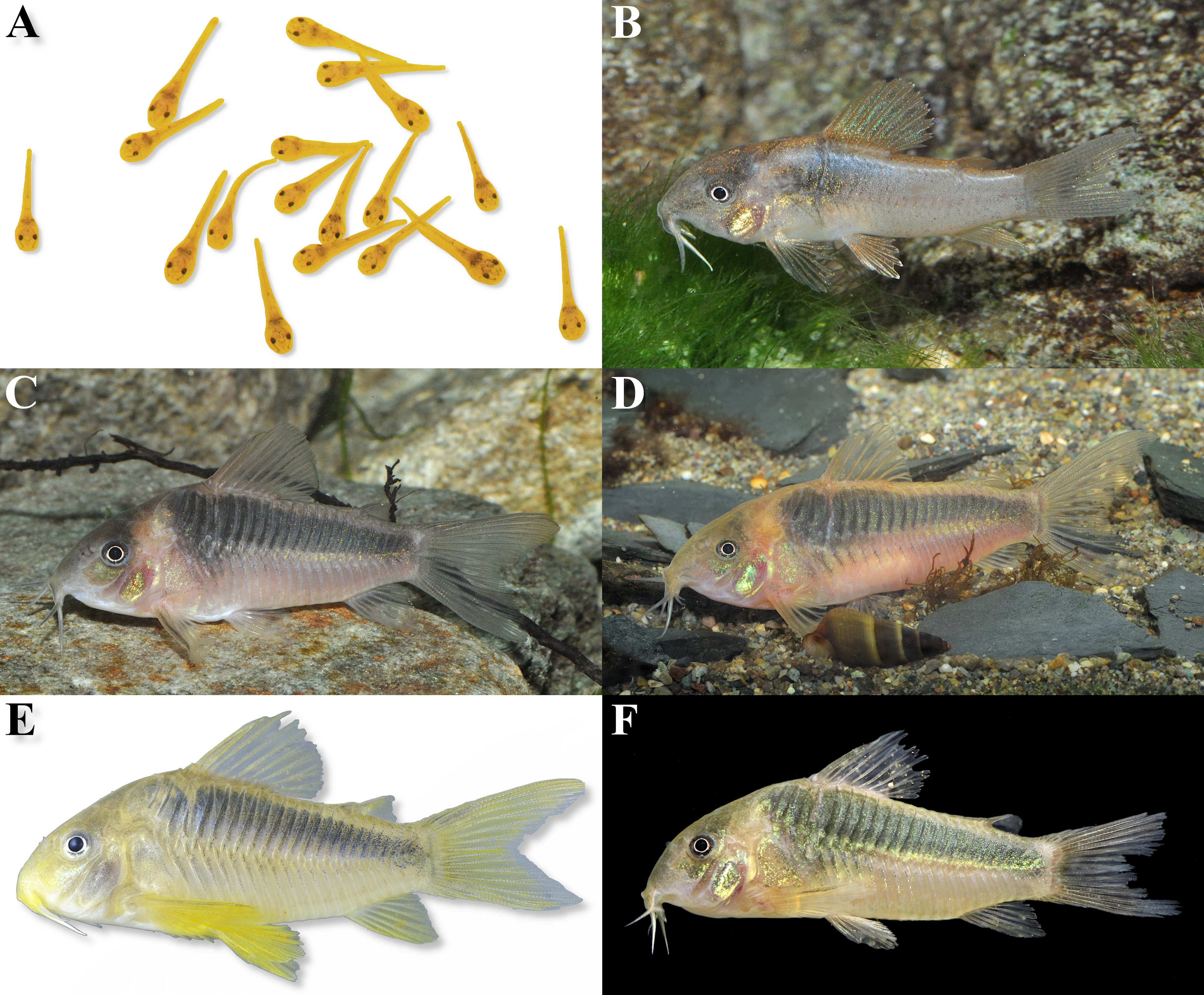
Variation in general body shape and color pattern in life of uncatalogued and unmeasured aquarium specimens of Osteogaster oharai, showing (A) specimens in larval stage, (B) a juvenile specimen, and (C–F) adult specimens. Except for specimen (F), which is from the rio Juruena basin, remaining specimens are from the rio Aripuanã basin. Photos (A–C) by Ingo Seidel, (D) by Hans Evers, (E) by Willian Ohara, and (F) by LFCT.
FIGURE 12 |
Map showing the known geographic records of Osteogaster oharai (yellow star: type-locality, a stream tributary of the rio Aripuanã; purple circles: additional records in the rio Aripuanã basin; red triangles: records in the rio Juruena basin) in Mato Grosso State, Brazil.
FIGURE 13 |
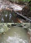
Collecting sites of Osteogaster oharai, showing (A) a small/shallow pool of an intermittent stream, and (B) a larger/deeper stream, the igarapé do Boi Preto, where the specimens UFRO-ICT 22449 were captured, both tributaries of the rio Juruena basin, Mato Grosso, Brazil.
FIGURE 14 |
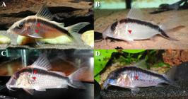
Specimens of Corydoradinae displaying a “pigmentation gap” just ventral to dorsal-fin spine base and/or a bright whitish vertically elongated patch just posterior to cleithrum (indicated by red arrows). (A) Brochis arcuata, (B) B. bethanae, (C) Corydoras narcissus, and (D) Hoplisoma granti. Photos (A and D) by Hans Evers, (B and C) by Steven Grant.
FIGURE 15 |
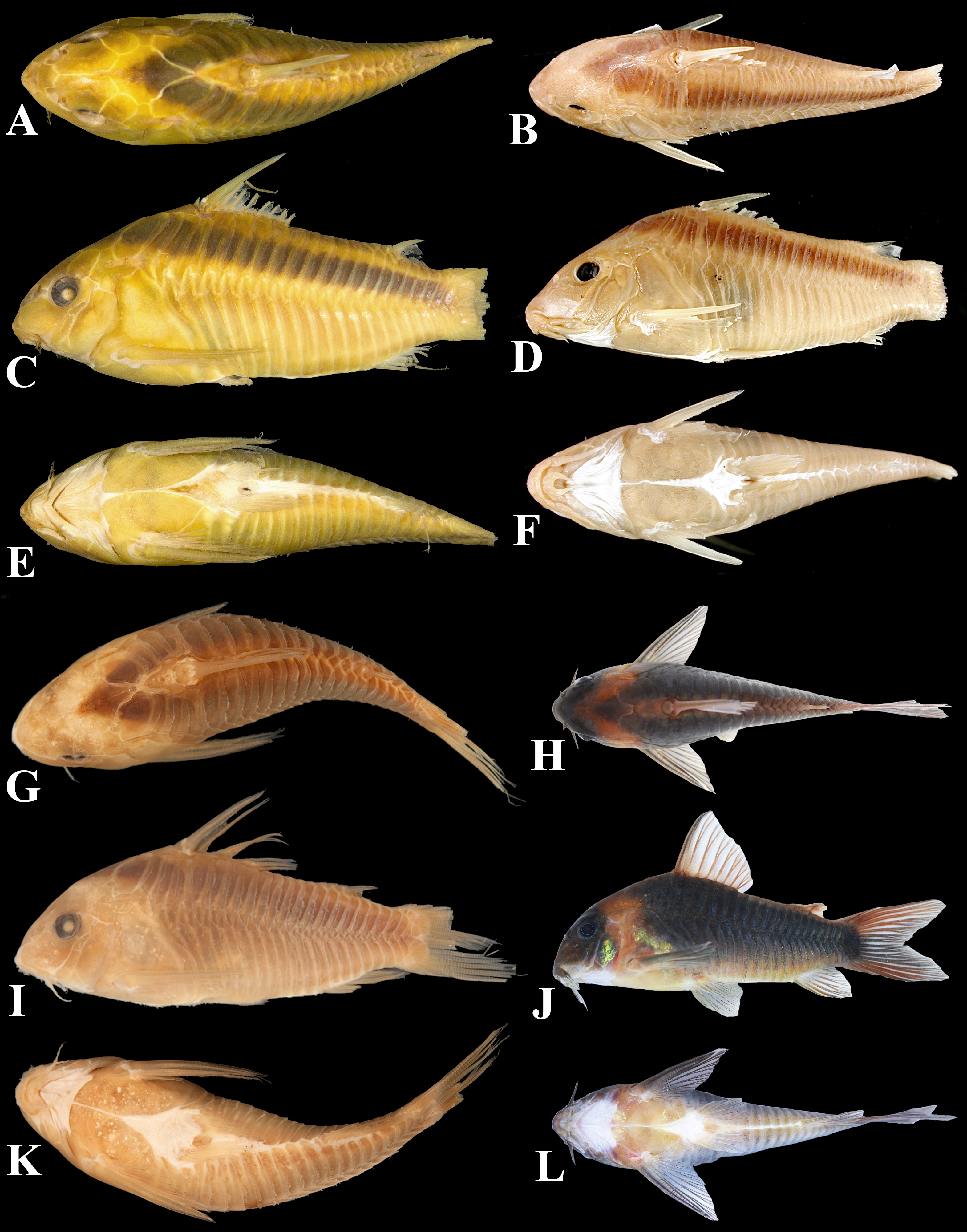
General morphology and color pattern of other striped species of Osteogaster, showing (A, C and E, in dorsal, lateral and ventral views, respectively) O. rabauti (MNRJ 3626, 46.0 mm SL, the holotype of Corydoras myersi), (B, D and F, in dorsal, lateral and ventral views, respectively) O. zygata (CAS 24756, unmeasured, the holotype of Corydoras zygatus), (G, I and K, in dorsal, lateral and ventral views, respectively) O. eques (MCZ 8204, unmeasured, paralectotype of Corydoras eques), and (H, J and L, in dorsal, lateral and ventral views, respectively) an unpreserved aquarium specimen of O. eques photographed alive. Photos (A, C and E) by Victor de Brito, (B, D and F) ©California Academy of Sciences, (G, I and K) ©President and Fellows of Harvard College, and (H, J and L) by Willian Ohara.

 A new striped Osteogaster (Siluriformes: Callichthyidae) from the Madeira and Tapajós river basins, Brazil, with a new generic diagnosis and an identification key to the species of Osteogaster
 Thumbnail
 Thumbnail
 Thumbnail
 Thumbnail
 Thumbnail
 Thumbnail
 Thumbnail
 Thumbnail
 Thumbnail
 Thumbnail
 Thumbnail
 Thumbnail
 Thumbnail
 Thumbnail
 Thumbnail