ABSTRACT:
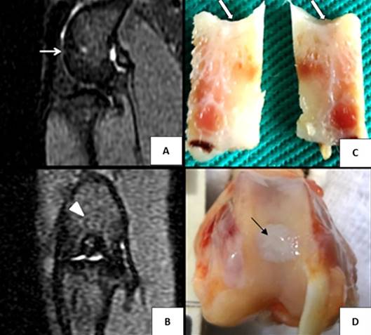
This study aimed to determine the characteristics and applicability of magnetic resonance imaging (MRI) in the evaluation of autogenous osteochondral graft in intact or macerated format, with or without insulin-like growth factor type 1 (IGF-1) used in repair of cartilage lesions induced in rabbits. Nine New Zealand rabbits were used, in which 18 stifle joints underwent grafting procedure in the femoral trochlear groove. These were divided into four groups, referred as intact osteochondral graft + IGF-1 (n=5), intact osteochondral graft + saline solution (n=4), macerated osteochondral graft + IGF-1 (n=5) and macerated osteochondral graft + saline solution (n=4). Animals were euthanized 12 weeks after surgery and the joints were subjected to MRI using a high magnetic field scanner of 1.5 Tesla. In addition, samples of grafting sites were subjected to anatomopathological examination. The MRI was effective as a noninvasive method to evaluate the repair tissue in osteochondral grafts in articular cartilage of the femur of rabbits by providing complementary data to macroscopic and histological examinations. Through these images and anatomopathological examinations satisfactory results were observed in relation to the repair process of autogenous osteochondral grafts in cartilage of rabbits, regardless of its format or the addition of IGF-1.
INDEX TERMS:
Magnetic resonance imaging; autogenous osteochondral graft; articular cartilage; insulin-like growth factor I; rabbits
 Thumbnail
 Thumbnail
 Thumbnail