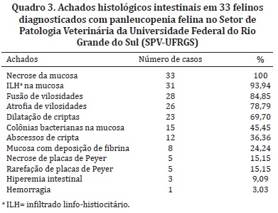
Feline panleukopenia is an important infectocontagious disease of domestic feline, especially in animals under 1 year. This paper describes the clinical-pathological findings and the immunohistochemical diagnosis of 33 cases of feline panleukopenia. The most important clinical signs were vomiting, diarrhea, and anorexia. The main gross findings observed were reddening of intestinal mucosa (16/33), evidentiation of Peyer patches (14/33), and liquefied intestinal content (7/33). The most consistent histological findings were necrosis (33/33) and lymphohistiocytic inflammatory infiltrate in the intestinal mucosa (31/33), villus fusion (27/33) and villus atrophy (26/33). In the hematopoietic tissues, the findings were characterized mainly by necrosis and tissue depletion. Parvovirus positive immunohistochemichal results were obtained in 84.85% of the cases analyzed. The best organ for viral detection was the intestine, with 84.85% of labeling in the immunohistochemichal technique. The spleen showed the best result among lymphoid organs, with 47.37% of the sections positive. This study presents most important lesions in the small intestine and in lymphoid organs and the immunohistochemistry proved good results in the detection of parvovirus.
Feline; panleukopenia; intestine; necrosis; immunohistochemistry; cats

 Pathologic and immunohistochemical findings of domestic cats with feline panleukopenia


