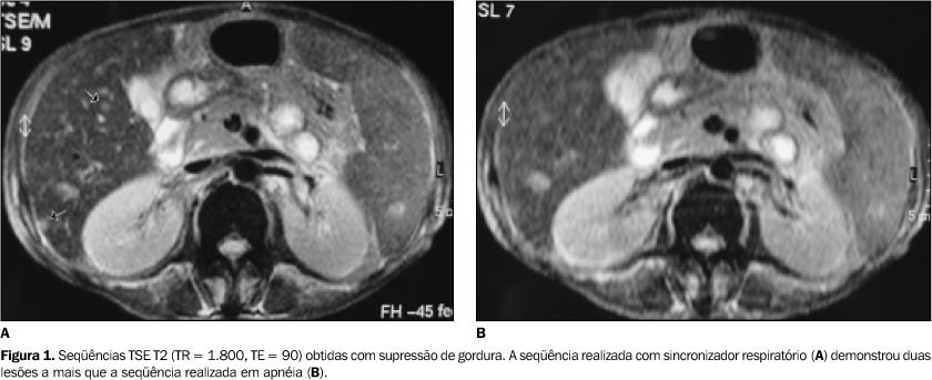
Since the early 1980's several magnetic resonance imaging pulse sequences have been developed in order to determine the optimum imaging technique for the detection and characterization of hepatic lesions. T2-weighted images play an important role in the evaluation of the liver and present equal or greater sensitivity than enhanced computed tomography for the detection of liver lesions. New techniques for obtaining T-2 weighted images have been developed in the attempt to optimize the method. These techniques have improved the image quality by shortening examination time, reducing motion artifacts, and improving contrast-to-noise ratio. The effectiveness of the different techniques (fat suppression, breath-hold, respiratory-triggered and phased-array coils) has been tested in many comparative studies, although the results are controversial. In this article we review the literature and discuss the several T2-weighted image techniques, particularly with regard to sensitivity to detect focal liver lesions.
Magnetic resonance imaging; Pulse sequences; Liver tumors; Liver


