
 Microwave pretreatment of a zinc sulfide ore: mineral exposure, textural reorganization and bioleaching performance
Abstract
Microwave pretreatment has been proposed as an alternative strategy to enhance mineral accessibility in low-grade polymetallic sulfide ores while reducing the energy demand associated with comminution. This study investigates the effects of microwave heating on area-based mineral liberation, textural reorganization, and bioleaching performance of a Zn-Pb ore from Mato Grosso State, Brazil. Samples were treated at 1400 W for 5 min and characterized by X-ray diffraction (XRD), ICP OES, scanning electron microscopy with energy-dispersive spectroscopy (SEM-EDS), specific surface area analysis (gas adsorption), and automated mineralogy (MLA). Bioleaching experiments were conducted using Acidithiobacillus ferrooxidans under controlled conditions. Microwave treatment induced significant microcracking and increased area-based mineral liberation in the coarser fractions (>0.5 mm), without measurable reduction in sulfide grain size, indicating predominantly intergranular fracturing. MLA results showed that microwave irradiation not only increased mineral surface exposure but also selectively modified mineral associations, particularly by reducing sphalerite-galena contacts and increasing associations with pyrrhotite and chalcopyrite in the coarsest fraction. These changes resulted in enhanced zinc extraction during bioleaching in only this size range. In contrast, no improvement was observed in the finer fractions (<0.5 mm), where sphalerite was already largely liberated. The results demonstrate that the effectiveness of microwave pretreatment is strongly particle size-dependent and controlled by both area-based mineral liberation and the reorganization of mineral interfaces, rather than solely by the generation of additional surface area.
Keywords:
galvanic interactions; sulfide liberation; microwave-induced microcracking; automated mineralogy; mineral characterization.
1. Introduction
With the gradual depletion of high-grade ore reserves, bioleaching has gained prominence as a viable alternative for processing complex and low-grade ores with lower operating and capital costs (Garcia Jr.; Urenha, 2001). However, the efficiency of this process strongly depends on the surface exposure of metallic minerals, which, when encapsulated within silicate gangue, exhibit limited bio-oxidation kinetics and therefore require particle size reduction to increase accessible surface area (Chen et al., 2020; Free, 2013) In this context, internal microfractures emerge as a promising alternative, as they facilitate the penetration of oxidizing agents and enable significant metal recovery even in coarse particle size fractions (Ghorbani et al., 2013; von Michaelis, 2019).
Several technologies have been investigated to intensify microfracture generation, including high-pressure grinding rolls (HPGR), electro-fragmentation, and selective fragmentation processes such as SELFRAG (Gomes et al., 2014; Kodali et al., 2011; Tang et al., 2020; Yin et al., 2017; Yin; Chen, 2021; Zhong et al., 2023). Microwave heating represents an additional and promising alternative, both due to its mineralogical selectivity and its potential to reduce comminution energy demand (Batchelor et al., 2016; Olubambi, 2009; Olubambi et al., 2007). Its principle is based on the differential absorption of electromagnetic energy by minerals with distinct dielectric properties (Al-Harahsheh; Kingman, 2004), leading to contrasting thermal expansions that may exceed the mechanical strength of phase boundaries and generate microfractures (Kingman; Rowson, 1998; Lu et al., 2020). Although Haque (1999) initially classified minerals according to their heating rates, subsequent studies demonstrated that factors, such as thermal conductivity, mineral texture, particle size distribution, and equipment geometry play a decisive role in the process (Trigos et al., 2022).
Studies applied to polymetallic ores have shown that microwave heating can enhance metallurgical recovery (Bai et al., 2022; Charikinya; Bradshaw, 2017; Farahat; Elmahdy; Hirajima, 2017). Olubambi et al., (2007) demonstrated that microfractures tend to form when highly microwave-absorbing sulfide minerals, such as pyrite, galena, and sphalerite, are embedded in a weakly absorbing silicate matrix, generating thermal contrasts sufficient to induce stress and fracturing. However, despite the recognition of this mechanism, most studies have documented its effects only indirectly through improvements in metallurgical recovery, without quantitatively evaluating the mineralogical and textural changes induced by microwave treatment or how these changes modify mineral interfaces.
This gap is relevant because microwave heating may not only generate microfractures but also modify mineral textures and the distribution of sulfide contacts, potentially influencing electrochemical interactions during bioleaching. However, the extent to which these textural changes affect galvanic interactions among sulfide minerals remains poorly understood. Such rearrangements may influence mineral liberation and the subsequent bioleaching behavior of sulfide ores.
The advent of automated mineralogy has enabled high-resolution quantification of fracture patterns, mineral exposure, mineral associations, and mineral interfaces, allowing an integrated interpretation of the relationship between microfracturing and metallurgical performance (Andrews, 1988; Fandrich et al., 2007; Johnson; Hallberg, 2016; Lee, 2011). Recent studies demonstrate that sulfide exposure generated by microfractures is a key factor controlling bioleaching kinetics (Ghorbani et al., 2012; Zhong et al., 2023) and that beneficial effects are strongly particle-size dependent (Tavares, 2005; Yin; Chen, 2021).
In this context, the present study advances along two main fronts: (i) to quantify the mineralogical consequences of microwave-induced micro-fracturing, including changes in mineral exposure and mineral interfaces, using automated mineralogy (MLA) and BET across four particle-size fractions (2.38-1.41; 1.41-0.500; 0.50-0.149; 0.149 mm) after microwave heating; and (ii) to evaluate how these textural effects, including the redistribution of sulfide contacts, which may influence electrochemical interactions during bioleaching.
2. Materials and methods
The experimental methodology was structured into four main stages: ore preparation, microwave thermal treatment, chemical-mineralogical and morphological characterization, and bioleaching tests. The overall flowchart of the experimental procedure is presented in Figure 1.

General flowchart of the experimental procedure adopted in this study, comprising the stages of ore preparation, microwave treatment, chemical-mineralogical and morphological characterization, and bioleaching tests
The ROM sample (30 kg; top size = 40 mm) was subjected to the following steps: (1) jaw crusher for primary crushing; (2) roll crusher for secondary crushing; (3) Jones riffle splitter for homogenization and sample mass reduction; (4) set of sieves for particle size classification into the fractions 2.38-1.41, 1.41-0.50, 0.50-0.149, and 0.149 mm; (5) microwave heating system used for thermal treatment of the samples; (6) magnetic separation using a Frantz isodynamic separator; (7) X-ray diffraction (XRD); (8) scanning electron microscopy (SEM-EDS) coupled with MLA; (9) specific surface area analyzer; (10) ICP OES spectrometer for chemical analysis of solutions; and (11) orbital shaker incubator used in the bioleaching experiments. The untreated (TQ) and treated (MW/1400 W) particle size fractions were subsequently evaluated in terms of their biohydrometallurgical behavior.
A 30 kg run-of-mine (ROM) sample of zinc sulfide ore (top size of 40 mm) from a mine in northern Mato Grosso State, Brazil, was crushed in successive stages using jaw and roll crushers to <5 mm, homogenized and reduced by riffle splitting. Aliquots of 60 g were subsequently obtained for four particle size fractions: 2.38-1.41, 1.41-0.50, 0.50-0.149 and 0.149 mm.
Mineralogical characterization included magnetic separation of the 1.41-0.50 mm fraction using a Frantz LB-1 magnetic barrier separator operated at successive currents of 0.0, 0.2, 0.4, and 0.6 A. Magnetic products obtained at each separation stage and the final non-magnetic product were collected separately, pulverized in a dry planetary mill equipped with a chrome-steel grinding vessel, and prepared for X-ray diffraction (XRD) analysis using the back-loading technique. X-ray diffraction measurements were performed using a Malvern Panalytical Empyrean diffractometer equipped with Cu-Kα radiation and an X’Celerator PSD detector, operating over a 2θ range of 2.5-70° with a step size of 0.02°. Phase identification was carried out using HighScore Plus software (Malvern Panalytical) with reference patterns from the Crystallography Open Database (COD). Chemical analyses were conducted after digestion of 0.25 g of each particle size fraction in aqua regia combined with HF, with quantification by ICP OES (Agilent 725).
Microwave heating experiments were carried out using 60 g of each size fraction in a MARS 6™ system (two 1 kW magnetrons operating at 2450 MHz), at an applied power of 1400 W for 5 min. This operating condition was selected based on the operational characteristics of the MARS 6™ system, providing intense microwave heating while remaining within the safe operating limits of the equipment and reaction vessels, in accordance with previous studies employing high-power microwave pretreatment of sulfide ores (Olubambi et al., 2007). Sample temperature was not monitored because the experimental setup was not equipped with microwave-compatible temperature sensors.
Bioleaching experiments employed Acidithiobacillus ferrooxidans (SISGEN registration code AA84667) cultivated in a selective Fe2+-rich medium (pH 1.8-2.0, 34 °C). Tests were performed in triplicate in 250 mL Erlenmeyer flasks at 180 rpm, with 10% (v/v) inoculum, a pulp density of 2% (w/v), an initial Fe2+ concentration of 5 g.L-1, daily pH adjustment, and continuous monitoring of redox potential (Eh). Dissolved Zn, Pb and Fe concentrations in the leach liquor were determined by ICP OES.
Morphological characterization was performed by scanning electron microscopy coupled with energy-dispersive spectroscopy (SEM-EDS) on polished sections prepared before and after bioleaching. The analyses were used for qualitative assessment of particle morphology, microfracture development, and the occurrence of secondary phases on mineral surfaces. Specific surface area was determined by N2 adsorption using the Brunauer-Emmett-Teller (BET) method in a NOVA 1200E analyzer (Quantachrome Instruments). Prior to analysis, samples were degassed under vacuum at 195 °C for 5 h to remove adsorbed moisture and volatile species. Nitrogen adsorption measurements were performed at 77.3 K using N2 as the adsorbate.
Automated mineralogical analyses were performed using a Mineral Liberation Analyzer (MLA; JKTech, FEI-Thermo Fisher Scientific) coupled to a Quanta 650 FEG scanning electron microscope (FEI-Thermo Fisher Scientific) equipped with a Bruker Esprit energy-dispersive X-ray spectroscopy (EDS) system. Aliquots of each particle-size fraction were obtained by rotary sample splitting. Fractions finer than 0.50 mm were prepared as epoxy-mounted monolayers to minimize particle overlap, whereas fractions coarser than 0.50 mm were embedded in epoxy resin, sectioned, and re-embedded to obtain representative particle cross-sections. Polished sections with a diameter of 30 mm were prepared and carbon coated prior to MLA analysis. The SEM was operated at a working distance of 14 mm, an accelerating voltage of 20 kV, and a spot size of 6. Automated image acquisition was performed in GXMAP mode, performed on phases with a grayscale value above 255, using a pixel size of 1.4 μm. This acquisition mode combines backscattered electron (BSE) imaging with pixel-based X-ray mapping, enabling improved discrimination of mineral phases exhibiting similar BSE contrast and average atomic number (Fandrich et al., 2007). The resulting mineral maps were processed using MLA DataView software for quantitative determination of modal mineralogy, grain-size distribution, mineral associations, particle morphology, and area-based mineral liberation. Depending on particle size fraction and treatment condition, approximately 900 to 3,600 sulfide particles were identified and quantified per sample, totaling more than 16,000 sulfide particles analyzed throughout the study. The total mapped area was 156.3 and 197.0 mm2 for the 2.38-1.41 fraction, 105.4 and 137.6 mm2 for the 1.41-0.50 fraction, 5.58 and 5.09 mm2 for the 0.50-0.149 fraction, and 0.38 and 0.53 mm2 for the 0.149 fraction, for the untreated (TQ) and microwave-treated (1400 W) samples, respectively. Liberation was quantified on an area basis and expressed as the proportion of exposed mineral area relative to the total mineral area identified in polished sections.
3. Results
3.1 Chemical and mineralogical characterization
Chemical characterization by ICP OES (Table 1) revealed a progressive enrichment of Zn, Fe, Pb, and S toward the finer size fractions, reaching up to 5.18 wt.% Zn and 1.30 wt.% Pb in the 0.50-0.149 mm fraction and 29.90 wt.% Fe and 8.89 wt.% S in the 0.149 mm fraction. This behavior consists of an increasing proportion of sulfide minerals, particularly sphalerite, pyrrhotite, and galena. The X-ray diffraction patterns of the magnetic and non-magnetic products obtained from the 1.41-0.50 mm fraction after Frantz magnetic separation (Figure 2) confirmed the mineral assemblage identified by MLA. The magnetic fractions were characterized by dominant pyrrhotite peaks, while sphalerite was consistently identified as the sole Zn-bearing mineral. Galena was detected at lower intensities, reflecting its lower modal abundance. Quartz, chlorite, and phlogopite were identified as the main gangue minerals, consistent with the chemical composition of the ore. Automated mineralogical analyses further revealed the occurrence of finely disseminated chalcopyrite associated with pyrrhotite and sphalerite, despite its low overall abundance (<0.33 wt.% Cu).

X-ray diffraction patterns of ore samples after magnetic separation in a Frantz magnetic barrier separator for the 1.41-0.50 mm fraction at currents of 0, 0.2, 0.4, and 0.6 A. Symbols: ▲ quartz; ★ pyrrhotite; Υ chlorite; ● phlogopite; ♦ galena; ▼ sphalerite.
3.2 Effect of microwave treatment on particle size and mineral exposure
The cumulative particle size distributions (Figure 3) show that microwave heating affected exclusively the >0.5 mm fractions, in which a shift of the curves toward smaller equivalent diameters was observed, indicating partial breakage of composite particles. In the <0.50 mm fractions, the curves for microwave-treated (1400 W/5 min) and untreated (TQ) samples remained nearly superimposed. Despite the reduction in composite particle size observed in the coarse fractions, the size distributions of sphalerite grains and of the bulk sulfide minerals did not exhibit any measurable decrease, indicating the absence of transgranular fracture.

Cumulative size distributions of bulk particles (a-d), bulk sulfides (e-h), and sphalerite grains (i-l). Untreated samples are represented by the solid black line, whereas microwave-treated samples (1400 W/5 min) are represented by the dashed red line. The same logarithmic scales are used in all panels.
MLA-based area liberation analyses indicate that microwave pretreatment increased the area proportion of sphalerite only in the coarser fractions, being largely ineffective in the finer fractions. This behavior is consistent with a fracture regime dominated by chipping and partial intergranular opening, characterized by selective removal of edges without grain fragmentation (Semsari Parapari; Parian; Rosenkranz, 2020). The increase in area-based liberation may also reflect a reduction in the thickness of adjacent layers and the development of internal porosity, which enhances solution accessibility to the interior of the particles (Ram et al., 2020). The liberation curves (Figure 4) and MLA images (Figure 5) show that, in the finer fractions, sphalerite is already largely liberated, occurring predominantly as free grains. The proportion of free sphalerite reached 74% in the 0.149 mm fraction and approximately 23% in the 0.50-0.149 mm fraction, indicating a high degree of mineral exposure regardless of microwave treatment. In contrast, in the coarser fractions, sphalerite remained predominantly locked within mixed particles, with only 0.4% free grains in the 2.38-1.41 mm fraction and 2.3% in the 1.41-0.50 mm fraction. Under these textural conditions, where sphalerite remains predominantly locked within composite particles, microwave treatment becomes more effective in promoting the opening of mineral-gangue interfaces and increasing mineral exposure.

Area-based liberation curves of sphalerite (a-d) and total sulfides (e-h) for untreated (TQ- solid black line) and microwave-treated samples (1400 W/5 minsolid red line) across different particle size fractions. The same axes and scales are used in all panels.

MLA images of particles with the highest sphalerite area in the following size fractions: (a) 2.38-1.41, (b) 1.41-0.50, (c) 0.50-0.149 and (d) 0.149 mm. Colors represent the different mineral phases. A higher proportion of area-liberated sphalerite is observed in the <0.5 mm fractions.
3.3 Textural reorganization and mineral associations
Mineralogical association analyses (Table 2) showed that microwave pretreatment modified both sphalerite liberation and the complexity of mineral associations, although the magnitude of these changes depended on particle size. The greatest effect was observed in the 1.41-0.50 fraction, where free sphalerite increased from 2.3 to 8.6%, binary associations increased from 20 to 29%, and ternary associations decreased from 78 to 63%. In the 0.50-0.149 fraction, binary associations also increased from 31 to 48% at the expense of ternary associations, which decreased from 46 to 30%, whereas free sphalerite remained nearly unchanged (23 vs. 22%). In contrast, the 2.38-1.41 fraction remained dominated by ternary composite particles (83%), despite the increase in free sphalerite from 0.4 to 2.4%. Mineral contact distributions further revealed granulometry-dependent changes after microwave treatment. In the 2.38-1.41 fraction, binary sphalerite-galena contacts decreased from 1.7 to 0.3%, whereas sphalerite-pyrrhotite and sphalerite-chalcopyrite associations increased from 0.05 to 0.8% and from 0.01 to 0.03%, respectively. In the 1.41-0.50 fraction, binary sphalerite-galena associations increased from 4.8 to 14.5%, whereas sphalerite-pyrrhotite associations decreased from 6.6 to 1.4%. In the 0.50-0.149 fraction, binary sphalerite-galena and sphalerite-pyrrhotite associations increased from 8.9 to 23.0% and from 12.4 to 17.5%, respectively, while sphalerite-quartz associations decreased from 2.9 to 0.3%.
MLA-derived sphalerite liberation and mineral association characteristics for each particle-size fraction before (TQ) and after micr wave pretreatment (1400 W for 5 min). Free = free sphalerite particles; PB = particles containing sphalerite associated with one additional mineral phase (binary composite particles).; PT = particles containing sphalerite associated with two or more additional mineral phases (ternary or more complex composite particles).
The granulometry-dependent response observed in the surface metrics obtained by MLA reflects fundamental differences in the fracture mechanisms induced by microwave pretreatment (Figure 6). In the coarser fractions (>0.50 mm), the simultaneous increase in ∆PSSA (Particle Surface Specific Area), ∆PSLS (Particle Surface Liberation Surface) and ∆PSFS (Particle Surface Free Surface), particularly for pyrrhotite and chalcopyrite, indicates effective generation of new external surfaces and opening of mineral-gangue interfaces. This behavior is consistent with the pronounced increases in specific surface area measured by gas adsorption in these fractions (Table 3), suggesting that microwave-induced fracturing occurs predominantly through intergranular cracking connected to the external particle surface, thereby increasing both the N2-accessible surface area and the mineral exposure quantified by MLA.

Percentage variation of surface metrics by mineral phase after microwave pretreatment (1400 W/5 min) relative to the untreated condition (TQ) for four particle size fractions.
In contrast, in the finer fractions (<0.50 mm), the increments recorded by the MLA metrics were more heterogeneous and, in many cases, limited, despite a measurable increase in gas-based surface area. This decoupling between MLA and gas adsorption results indicates that, at these size ranges, microwave treatment promotes predominantly intragranular micro-fracturing and internal porosity development, which contribute to the gas-accessible surface area but do not translate into a significant opening of mineral-gangue interfaces or an increase in free external surface detectable by MLA. SEM-EDS images (Figure 7) support this interpretation by showing wide, connected fractures crossing multiple mineral phases in the coarse fractions, whereas in the fine fractions discrete microcracks confined to the interior of individual grains predominate.

SEM/BSE images (with overlaid EDS maps) of untreated (TQ) samples (a) and microwave-treated samples (1400 W/5 min) (b). Columns correspond to particle size classes (in mm): (i) 2.38-1.41, (ii) 1.41-0.50, (iii) 0.50-0.149, and (iv) 0.149. Arrows indicate inter and transgranular microfractures. Symbols: ▲ quartz, ★ pyrrhotite, ♦ galena, ▼ sphalerite. A higher density of microfractures is observed in the 1400 W/5 min samples, particularly along sphalerite-galena and sphalerite-pyrrhotite interfaces.
3.4 Bioleaching performance
Biotic tests exhibited Zn extractions significantly higher than those obtained under abiotic conditions, which remained below 7% in all cases (Figure 8). The highest Zn extraction was observed in the 0.149 mm fraction, reaching approximately 30%. Among the microwave-treated samples, only the 2.38-1.41 mm fraction showed a clear increase in Zn extraction relative to the untreated sample. In the 1.41-0.50 mm fraction, Zn extractions were similar for the untreated (TQ) and microwave-treated (1400 W/5 min) samples; however, the treated sample exhibited higher residual Pb concentrations in solution after the tenth day (Figure 10).

Evolution of Zn extraction (a-d) and redox potential, Eh (e-h), during bioleaching tests and abiotic controls for untreated (TQ) and microwave-treated samples (MW/1400 W/5 min) across different particle size fractions: (a,e) 2.38-1.41; (b,f) 1.41-0.50; (c,g) 0.50-0.149; and (d,h) 0.149 mm.
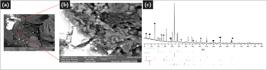
(a-b) SEM/BSE images of the 2.38-1.41 mm fraction residue after bioleaching, showing sphalerite particles coated by oxidation products; the enlarged detail highlights a rough surface morphology with fine adherent precipitates. (c) XRD pattern confirming plumbojarosite (★) and gangue phases: quartz (▲), chlorite (Υ), and phlogopite (●), consistent with Pb and Fe precipitation under acidic oxidizing.

Scanning electron microscopy images in backscattered electron mode (SEM/BSE) with energy-dispersive X-ray spectroscopy (EDS) analyses of sulfide particles under different experimental conditions: (a) Abiotic system, showing sphalerite associated with gangue minerals, with EDS analyses indicating sphalerite (Zn-S) and quartz (Si-O), and no evident secondary coatings; (b) Biotic system, displaying sphalerite with a Znand S-rich core and an S-enriched rim; EDS peaks confirm the formation of an elemental sulfur (S0) surface layer, consistent with passivation by bio-oxidation; and (c) Biotic system after microwave pretreatment, in which S0 occurs as a more continuous and homogeneous film, associated with a pyrrhotite grain.
Redox potential (Eh) profiles indicated that, in the biotic tests, Eh increased rapidly during the first three days and stabilized at approximately 650 mV, regardless of particle size or microwave pretreatment. In the abiotic tests, Eh remained between 380 and 400 mV throughout the experiment, with no differences between treated and untreated samples. Microwave pretreatment did not alter the redox behavior of the biotic systems, and the Eh curves remained stable over the 24-day test period.
4. Discussion
The characterization of bioleaching residues integrates the effects of microwave pretreatment on particle morphology, free surface area, redistribution of mineral interfaces, and the resulting bioleaching behavior. A limitation of this study is that the sample temperature was not directly monitored during microwave irradiation because microwave-compatible temperature sensors were not available in the experimental setup. Consequently, the effects of microwave pretreatment are interpreted based on the applied power (1400 W), exposure time (5 min), and the resulting mineralogical, textural, and bioleaching responses rather than on the absolute temperature reached by the samples.
In all biotic tests, the residues exhibited surfaces covered by oxidation products, predominantly plumbojarosite. Under conditions of pH 1.8, 34 °C, and high Fe3+ concentration - favorable to jarosite precipitation (Daoud; Karamanev, 2006) - the formation inof this phase was consistently observed and readily identified by its characteristic yellow coloration (Cruells; Roca, 2022). The progressive decrease in Fe and Pb concentrations in solution (Figure 11) corroborates their removal from the leach liquor and incorporation into stable solid phases. This behavior is exemplified in Figure 9a-c for the 2.38-1.41 mm fraction treated by microwave irradiation (1400 W/5 min), in which SEM/BSE images reveal a microfractured sphalerite grain partially covered by thin, discontinuous plumbojarosite layers, confirmed by X-ray diffraction.

Pb and Fe concentrations in the leach liquor during bioleaching for the 2.38-1.41, 1.41-0.50, 0.50-0.149 and 0.149 mm size fractions under untreated (TQ) and microwave-treated conditions (MW/1400 W/5 min).
Comparison among the residues further highlights the influence of mineralogical and textural conditions together with the oxidative environment established during bioleaching. While Figure 10a shows an intact sphalerite grain in the abiotic experiment, Figures 10b and 10c reveal the persistence of elemental sulfur (S0) in the biotic residues (Fowler; Crundwell, 1999), more pronounced in the microwave-treated samples. The initial addition of 5 g·L-1 Fe2+ directed Acidithiobacillus ferrooxidans toward preferential iron oxidation (Ponce et al., 2012), an effect reinforced by prior adaptation of the inoculum to Fe2+-rich media. Combined with Eh values above 620 mV and the limited duration of the experiments-insufficient for complete S0 oxidation-this redox regime favored the stabilization of elemental sulfur as a surface film.
The 2.38-1.41 mm fraction was the only granulometric class in which a significant difference in Zn extraction was observed between microwave-treated and untreated samples. This behavior is consistent with the relative increase in specific surface area measured by BET, the enhanced free surface area, and the reduction of binary sphalerite-galena contacts, accompanied by increased associations with pyrrhotite and chalcopyrite observed prior to bioleaching. These observations suggest that microwave pretreatment intensified intergranular disaggregation, promoting more reactive sphalerite interfaces. From a mineralogical perspective, the changes observed in the particle association classes and mineral contact distributions (Table 2) are consistent with the improved metallurgical performance observed in this size fraction. The reduction in sphalerite-galena contacts, commonly associated with early sphalerite passivation, together with the redistribution of sulfide associations, is consistent with modifications of mineral interfaces that could influence electrochemical interactions during bioleaching, as proposed in previous studies (Ghorbani et al., 2013b; Olubambi et al., 2007). However, no direct electrochemical measurements were performed in the present study, and therefore the contribution of galvanic interactions could not be directly evaluated.
In the 1.41-0.50 mm fraction, although microwave treatment increased the exposed surface area of sphalerite, the resulting mineral associations led to an unfavorable scenario. The treatment intensified sphalerite-galena contacts, which modified mineral interfaces in a manner consistent with changes that may influence electrochemical interactions between sulfide minerals (Kocabağ, 1985), consistent with the higher residual Pb concentrations in solution after the tenth day of bioleaching. The increased availability of Pb2+ likely favored the formation of sparingly soluble Pb-bearing secondary phases, including plumbojarosite and possibly anglesite (Da Silva; Lastra; Budden, 2003), acting as passivating barriers. Consequently, the initial increase in liberated surface area did not result in higher reactivity, leading to similar Zn extractions in treated and untreated samples.
In the finer fractions (<0.50 mm), Zn extraction was virtually identical between treated and untreated samples, corroborating studies reporting negligible benefits of microwave pretreatment when sphalerite is already largely liberated (Ghorbani et al., 2011). In these size ranges, microwave treatment did not substantially modify the accessibility of reactive mineral surfaces, as the mineral texture already provides high natural exposure. Overall, the results suggest that the benefits of microwave pretreatment are concentrated in the coarser particle-size fractions, where microwave-induced microcracking and reorganization of mineral interfaces increase mineral exposure before the development of passivating secondary phases. These observations are consistent with previous studies on microwave-assisted bioleaching of sulfide ores, although the specific contribution of galvanic interactions could not be directly evaluated in the present work (Charikinya; Bradshaw; Becker, 2015; Charikinya; Bradshaw, 2017).
5. Conclusions
Microwave pretreatment increased the area-based exposure of sphalerite in the 2.38-1.41 and 1.41-0.50 fractions, although with contrasting bioleaching responses. In the 2.38-1.41 fraction, the reorganization of mineral associations, characterized by a reduction in sphalerite-galena contacts and increased associations with pyrrhotite and chalcopyrite, was accompanied by higher Zn extraction. In contrast, the 1.41-0.50 fraction exhibited increased sphalerite-galena contacts and similar Zn extraction to the untreated sample, despite the higher exposed surface area. During bioleaching, the mobilization of Pb and Fe was followed by the precipitation of plumbojarosite, while other Pb-bearing secondary phases may also have formed, contributing to surface coverage.
In the finer fractions (<0.50 mm), where sphalerite was already highly liberated, microwave pretreatment produced little additional increase in mineral exposure and did not improve Zn extraction. Overall, the results suggest that the effectiveness of microwave pretreatment is strongly dependent on particle size and the initial textural characteristics of the ore. Microwave-induced microcracking and mineral interfaces reorganization appear to enhance mineral exposure primarily in the coarser fractions, whereas their contribution becomes limited once a high degree of mineral liberation has already been achieved by comminution. The observed mineralogical changes are consistent with previous studies on microwave-assisted bioleaching and indicate that reorganization of sulfide contacts accompanied the different metallurgical responses observed among particle-size fractions. However, because no direct electrochemical measurements were performed, the possible contribution of galvanic interactions remains interpretative and could not be directly evaluated in the present study.
Funding information
Data availability
The data that support the findings of this study are available from the corresponding author upon reasonable request.
References
- AL-HARAHSHEH, M.; KINGMAN, S. W. Microwave-assisted leaching - a review. Hydrometallurgy, v. 73, no 3-4, p. 189-203, 2004.
- ANDREWS, G. F. The selective adsorption of Thiobacilli to dislocation sites on pyrite surfaces. Biotechnology and Bioengineering, v. 31, n. 4, p. 378-381, 1988.
- BAI, Y. et al. Effect of microwave pretreatment on chalcopyrite dissolution in acid solution. Journal of Materials Research and Technology, v. 16, p. 471-481, 2022.
- BATCHELOR, A. R. et al. Increasing the grind size for effective liberation and flotation of a porphyry copper ore by microwave treatment. Minerals Engineering, v. 94, p. 61-75, 2016.
- CHARIKINYA, E.; BRADSHAW, S. M. An experimental study of the effect of microwave treatment on long term bioleaching of coarse, massive zinc sulphide ore particles. Hydrometallurgy, v. 173, p. 106-114, 2017.
- CHARIKINYA, E.; BRADSHAW, S.; BECKER, M. Characterising and quantifying microwave-induced damage in coarse sphalerite ore particles. Minerals Engineering, v. 82, p. 14-24, 2015.
- CHEN, J. et al. The influence of micro-cracks on copper extraction by bioleaching. Hydrometallurgy, v. 191, p. 105243, 2020.
- CRUELLS, M.; ROCA, A. Jarosites: formation, structure, reactivity and environmental. Metals, v. 12, no 5, p. 802, 2022.
- DA SILVA, G.; LASTRA, M. R.; BUDDEN, J. R. Electrochemical passivation of sphalerite during bacterial oxidation in the presence of galena. Minerals Engineering, v. 16, no 3, p. 199-203, 2003.
- DAOUD, J.; KARAMANEV, D. Formation of jarosite during Fe2+ oxidation by Acidithiobacillus ferrooxidans. Minerals Engineering, v. 19, no 9, p. 960-967, 2006.
- FANDRICH, Rolf et al. Modern SEM-based mineral liberation analysis. International Journal of Mineral Processing, v. 84, n. 14, p. 310-320, 2007.
- FARAHAT, M.; ELMAHDY, A. M.; HIRAJIMA, T. Influence of microwave radiation on the magnetic properties of molybdenite and arsenopyrite. Powder Technology, v. 315, p. 276-281, 2017.
- FOWLER, T. A.; CRUNDWELL, F. K. Leaching of zinc sulfide by Thiobacillus ferrooxidans: bacterial oxidation of the sulfur product layer increases the rate of zinc sulfide dissolution at high concentrations of ferrous ions. Applied and Environmental Microbiology, v. 65, no 12, p. 5285-5292, 1999.
- FREE, M. L. Hydrometallurgy: fundamentals and applications. Hoboken, New Jersey: Wiley-Tms, 2013.
- GARCIA JR., O.; URENHA, L. C. Lixiviação bacteriana de minérios. In: LIMA, Urgel de Almeida et al. (ed.). Biotecnologia industrial - processos fermentativos e enzimáticos. São Paulo: Editora Blucher, 2001. v. 3, p. 485-512.
- GHORBANI, Y. et al. An experimental study of the long-term bioleaching of large sphalerite ore particles in a circulating fluid fixed-bed reactor. Hydrometallurgy, v. 129-130, p. 161-171, 2012.
- GHORBANI, Y. et al. Investigation and modelling of the progression of zinc leaching from large sphalerite ore particles. Hydrometallurgy, v. 131-132, p. 8-23, 2013a.
- GHORBANI, Y. et al. Investigation of the effect of mineralogy as rate-limiting factors in large particle leaching. Minerals Engineering, v. 52, p. 38-51, 2013b.
- GHORBANI, Y. et al. Large particle effects in chemical/biochemical heap leach processes - a review. Minerals Engineering, v. 24, n. 11, p. 1172-1184, 2011.
- GOMES, O. et al. Characterization of particle damage and surface exposure of a copper ore processed by Jaw Crusher, HPGR and electro-dynamic fragmentation. In: CARPENTER, John S. et al. (ed.). Characterization of minerals, metals, and materials: [s. l.], 2014. p. 247-252.
- HAQUE, K. E. Microwave energy for mineral treatment processes - a brief review. International Journal of Mineral Processing, v. 57, n. 1, p. 1-24, 1999.
- JOHNSON, D. B.; HALLBERG, K. B. Techniques for detecting and identifying acidophilic mineral-oxidizing microorganisms. In: RAWLINGS, D. E.; JOHNSON, D. Barrie (ed.). Biomining. Berlin: Springer-Verlag, 2016. p. 237-261.
- KINGMAN, S. W.; ROWSON, N. A. Microwave treatment of minerals - a review. Minerals Engineering, v. 11, n. 11, p. 1081-1087, 1998.
- KOCABAĞ, Duran. The effect of grinding media and galvanic interactions upon the flotation of sulfide minerals. Complex Sulfides, Process Symposium. [s. L.] 1985. v. 5.
- KODALI, P. et al. Particle damage and exposure analysis in HPGR crushing of selected copper ores for column leaching. Minerals Engineering, v. 24, n. 13, p. 1478-1487, 2011.
- LEE, J. Biooxidation of gold ores. In: SOBRAL, Luis Gonzaga Santos.; MONTEIRO DE OLIVEIRA, Débora.; GOMES DE SOUZA, Carlos Eduardo. (ed.). Biohydrometallurgical processes: a practical approach. Centro de Tecnologia Mineral - CETEM, 2011. p. 179-194.
- LU, G. et al. The influence of minerals on the mechanism of microwave-induced fracturing of rocks. Journal of Applied Geophysics, v. 180, p. 104123, 2020.
- OLUBAMBI, P. A. et al. Influence of microwave heating on the processing and dissolution behaviour of low-grade complex sulphide ores. Hydrometallurgy, v. 89, n. 1-2, p. 127-135, 2007.
- OLUBAMBI, P. A. Influence of microwave pretreatment on the bioleaching behaviour of low-grade complex sulphide ores. Hydrometallurgy, v. 95, n. 1-2, p. 159-165, 2009.
- PONCE, J. S. et al. Acidithiobacillus ferrooxidans oxidizes ferrous iron before sulfur likely through transcriptional regulation by the global redox responding RegBA signal transducing system. Hydrometallurgy, v. 127-128, p. 187-194, 2012.
- RAM, Rahul et al. Study of the leaching and pore evolution in large particles of a sulfide ore. Hydrometallurgy, [s. l.], v. 192, p. 105261, 2020.
- SEMSARI PARAPARI, Parisa; PARIAN, Mehdi; ROSENKRANZ, Jan. Breakage process of mineral processing comminution machines - an approach to liberation. Advanced Powder Technology, [s. l.], v. 31, no 9, p. 3669-3685, 2020.
- TANG, Y. et al. Enhancement of gold agitation leaching by HPGR comminution via microstructural modification of gold ore particles. Minerals Engineering, v. 159, p. 106639, 2020.
- TAVARES, L. M. Particle weakening in high-pressure roll grinding. Minerals Engineering, v. 18, no 7, p. 651-657, 2005.
- TRIGOS, L. et al. Natural factors related to the differential heating of aggregates exposed to microwaves. Construction and Building Materials, v. 314, p. 125654, 2022.
- VON MICHAELIS, H. How efficient is HPGR? In: WORLD GOLD CONFERENCE, 2009. The southern African Institute of Mining and Metallurgy.
- YIN, W. et al. Comparison of sample properties and leaching characteristics of gold ore from jaw crusher and HPGR. Minerals Engineering, v. 111, p. 140-147, 2017.
- YIN, W.; CHEN, K. Effect of the particle size and microstructure characteristics of the sample from HPGR on column bioleaching of agglomerated copper ore. Hydrometallurgy, v. 200, p. 105563, 2021.
- ZHONG, W. et al. Understanding the mechanism of microcrack-enhanced bioleaching of copper. Hydrometallurgy, v. 218, p. 106045, 2023.


