
 Mandibular fracture in an elderly patient with late diagnosis – a 3-year follow-up: a case report
ABSTRACT
Mandibular fractures represent a significant clinical challenge in dentistry and oral and maxillofacial surgery, particularly in elderly patients, in whom reduced bone density and tooth loss may contribute to the severity of trauma. These injuries are often associated with accidental falls and impact trauma, leading to pain and functional impairments. Delayed diagnosis of mandibular fractures can result in complications such as infections, inadequate bone healing, and facial asymmetry. Early imaging and thorough clinical examination are therefore crucial to achieving satisfactory treatment decisions, as they enable a detailed assessment of the fracture and the establishment of an effective therapeutic strategy. In addition to imaging evaluation, the categorization of mandibular fractures based on fracture pattern, anatomical location, and trauma mechanism is fundamental for defining the appropriate treatment. In this context, this study aimed to analyze a clinical case of mandibular fracture in an elderly patient, in which the diagnosis was made late.
Indexing terms
Aged; Delayed diagnosis; Mandibular fractures
RESUMO
As fraturas mandibulares representam um obstáculo clínico considerável na odontologia e na cirurgia bucomaxilofacial, sobretudo em pacientes idosos, pois a redução da densidade óssea e a perda dentária podem contribuir para a gravidade do trauma. Essas lesões são comumente ligadas a traumas de impacto, podendo resultar em dores e dificuldades funcionais. O diagnóstico tardio dessas fraturas pode resultar em complicações como infecções, consolidação óssea inadequada e assimetrias faciais. A importância da realização de exames de imagem associados ao exame clínico é crucial para a decisão do tratamento, uma vez que viabiliza uma avaliação detalhada da fratura e o estabelecimento de uma estratégia terapêutica eficaz. Além dos exames de imagem, a categorização das fraturas mandibulares é fundamental para a definição do tratamento. Elas podem ser classificadas de acordo com o traço da fratura, a sua localização anatômica e o mecanismo do trauma. Diante desse contexto, este estudo teve como objetivo analisar um caso clínico de fratura mandibular em um paciente idoso, no qual o diagnóstico foi realizado tardiamente.
Termos de indexação
Idoso; Diagnóstico tardio; Fraturas mandibulares
INTRODUCTION
Population aging represents a significant challenge for public health and the family unit, as it involves functional and motor changes that increase vulnerability to falls and fractures [1]. According to the Ministry of Health [2], the reduction in functional capacity in active elderly individuals results in motor and reflex alterations, which increases the risk of accidents.
In edentulous elderly patients, tooth loss contributes to bone resorption and consequent mandibular atrophy, making the mandible more susceptible to fractures even in low-impact trauma, such as falls from standing height [3,4]. Aging is associated with biological changes that reduce osteogenic capacity, such as a decrease in osteochondral stem cells, as well as reduced osteoblastic activity and changes in the initial inflammatory response, factors that result in slower bone healing and a higher risk of infection or non-union of fractures [5].
Mandibular fractures are among the most frequent injuries of the maxillofacial complex, with the mandibular body being the most commonly affected region [6]. In these situations, bone atrophy may result in complications, especially when diagnosis is delayed [4].
In addition, there is a significant increase in the prevalence of chronic systemic diseases in this age group, such as hypertension, Diabetes Mellitus (DM), stroke, and Chronic Obstructive Pulmonary Disease (COPD). DM is recognized as an independent risk factor for infections, delayed wound healing, and postoperative complications due to microvascular dysfunction, tissue hypoxia, and reduced immune response [7].
Patients with a history of stroke (cerebrovascular accident) present a higher risk of blood pressure instability and neurological events during surgical procedures [8], while emphysema and other chronic pulmonary diseases are associated with respiratory complications and longer hospital stays [9]. These factors highlight the need for careful preoperative evaluation and individualized treatment planning for elderly patients undergoing oral and maxillofacial surgery.
Diagnosis depends on a proper clinical examination associated with imaging exams, with computed tomography considered the gold standard for evaluating fracture lines, adjacent structures, and surgical planning [10]. However, in small hospitals, diagnostic failures often occur due to the lack of oral and maxillofacial surgeons and imaging technology, which may compromise functional recovery [11].
Regarding the treatment of atrophic mandibular fractures, the literature points to fixation with 2.4 mm RECON (reconstruction) plates as the standard, as they provide greater biomechanical stability compared to 2.0 mm plates, which, due to their thickness, are unable to withstand the mechanical loads applied to the fixation material [12,13].
Adequate stability of bone fragments is essential to prevent chronic inflammation and failure of bone healing [14]. Furthermore, rigid fixation (ORIF) with load-bearing locking plates is considered a reliable protocol for the treatment of edentulous atrophic fractures, and autogenous grafts are required when there is significant bone loss [15,16].
Thus, the aim of this study was to discuss and evaluate the postoperative outcomes of oral and maxillofacial surgeries in mandibular fractures in elderly patients, in delayed interventions, based on a clinical case report performed in a public hospital in the southern region of Santa Catarina, Brazil.
CASE REPORT
A male patient, 80 years old, completely edentulous, using complete upper and lower dentures, with residual mandibular bone height below 20 mm, was admitted due to facial trauma. The patient had a history of epilepsy controlled with continuous use of phenobarbital (Gardenal), with no seizures for several years, and no other reported comorbidities.
The accident occurred after he tripped over a stone on his rural property, resulting in a fall from standing height. The patient presented a brief loss of consciousness, but without amnesia or episodes of vomiting. The following day, he sought medical care at a small local hospital in his city, where he was evaluated by a general practitioner. At that time, only analgesics were prescribed, with no imaging studies performed.
After 20 days of persistent pain, facial edema, weight loss due to difficulty in eating, and pain during mastication, the patient returned to the same hospital, where a radiographic examination was finally requested, revealing a fracture of the left mandibular body (figure 1).

The patient was referred to the regional hospital, where a new radiographic examination was performed (figure 2), and he remained hospitalized. After three days, he underwent surgery via a Risdon approach with internal fixation. The time elapsed between the trauma and the surgical intervention was 23 days. The patient was discharged from the hospital on 04/20/2022, without signs of neurological deficits, paresthesia, or difficulty in mouth opening.

The Risdon approach is widely indicated for fractures of the mandibular body and angle, as it allows broad visualization of the surgical field and adequate manipulation of bone fragments, minimizing damage to adjacent neural structures and reducing infection risk by avoiding direct contact with the intraoral microbiota.
In the immediate postoperative period, dipyrone 500 mg, ketoprofen 100 mg, tramadol 50 mg, and cephalexin 500 mg were prescribed for seven days. During hospital follow-up, no postoperative imaging examinations were performed, only hematological tests. The patient did not present infectious or functional complications.
Outpatient follow-up was conducted at the regional hospital, without imaging exams for control. After three years, the patient was reassessed at the clinics of the Universidade do Extremo Sul Catarinense (UNESC), where a computed tomography scan was performed. The examination revealed the presence of a 2.0 mm plate (figure 3).

Computed tomography showing panoramic reconstruction, axial, transverse, and three-dimensional (3D) views.
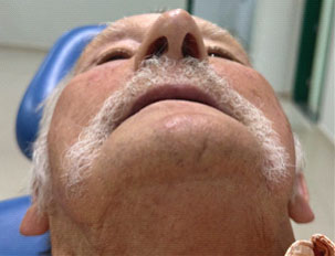
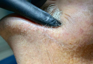
On clinical evaluation, atrophy of the lower alveolar ridge was observed (figure 4), a submental scar resulting from the trauma caused by the fall (figure 5), the presence of a residual submandibular cutaneous scar (figure 6), adequate mouth opening, and absence of functional limitations (figure 7).


On frontal extraoral evaluation, the patient presented preserved facial symmetry and satisfactory aesthetic outcome.
The long-term follow-up of three years revealed adequate bone healing and absence of functional complications. This period is considered significant for evaluation of stability, as fractures in atrophic mandibles present a risk of pseudoarthrosis and progressive bone resorption when there is fixation failure or functional overload.
Currently, the patient is in good general condition, with preserved masticatory function, swallowing, and speech, without complaints or functional limitations, maintaining a good quality of life.
DISCUSSION
According to Batista et al. [3], tooth loss in elderly patients is directly related to mandibular bone atrophy, a factor that increases susceptibility to fractures resulting from simple falls. The reported case confirms this statement, as a completely edentulous patient presented a mandibular fracture after a fall. These findings are also consistent with those reported by Dantas Filho [17], who highlights falls as one of the main causes of fractures in elderly individuals.
According to Almeida Neto et al. [18], the appropriate diagnosis of mandibular fractures depends on high-quality imaging exams, such as computed tomography, which allows detailed analysis of the fracture line. In the present case, however, no imaging examinations were performed during either the initial care or the postoperative follow-up. A similar situation is reported by Soares et al. [10], who emphasize the indispensability of computed tomography for surgical planning.
Another important aspect is the choice of fixation system. According to Cavalcante Neto et al. [12], the 2.4 mm RECON system is considered the gold standard for atrophic mandibles due to its superior mechanical stability. However, in this case, a 2.0 mm plate was used. Despite this, the patient showed satisfactory evolution, with no functional or infectious complications.
The literature supports these findings, demonstrating that 2.4 mm plates present a low rate of major complications (around 3%) and a high clinical success rate, especially in comminuted, displaced fractures or atrophic mandibles [15,19]. This low complication rate confirms one of the fundamental principles of the AO Foundation (Arbeitsgemeinschaft für Osteosynthesefragen), according to which susceptibility to infection is directly related to fragment mobility; inadequate fixation may lead to chronic inflammation, delayed union, non-union, or infection [20].
Considering that the patient presented an edentulous atrophic mandible, the use of a 2.0 mm plate system is less indicated. This type of system is more appropriate for edentulous mandibles that are not severely atrophic, i.e., with bone height greater than 20 mm. In cases of severe atrophy, the remaining bone volume and quality are insufficient to ensure adequate stability and support for screws [12,14]. Thus, load-bearing osteosynthesis using 2.4 mm locking reconstruction plates is the most indicated alternative, as it provides greater mechanical resistance and reduces the risk of fixation failure or displacement [15,21].
Furthermore, Stein et al. [22] reinforce that advanced age is associated with increased postoperative complications and prolonged recovery time. In the present case, despite delayed treatment and absence of complementary imaging, no relevant complications were observed, which contrasts with the findings of these authors.
Recent studies explain that aging directly influences cellular and molecular bone healing processes, affecting osteoblastic differentiation, inflammatory control, and bone callus remodeling [5].
In addition to anatomical changes, systemic comorbidities are determining factors in prognosis. Diabetes mellitus (DM), for example, is recognized as an independent risk factor for postoperative infections and poor wound healing due to microvascular alterations and immune dysfunction that impair blood flow and inflammatory response [7].
In addition to the absence of systemic conditions commonly associated with postoperative complications, such as a history of stroke, pulmonary emphysema, or diabetes mellitus, the patient evolved without significant complications. This outcome may be attributed to good general health status, considering that the only comorbidity was well-controlled epilepsy, and to the patient’s cooperative behavior, which contributed to postoperative care.
Thus, this report confirms part of the evidence already established in the literature, especially regarding the risk of mandibular fractures in edentulous elderly patients, reinforcing the importance of diagnostic and therapeutic protocols that prioritize adequate imaging exams and safer fixation methods for atrophic mandibles.
CONCLUSION
This case study demonstrated the challenges and complexity of treating mandibular fractures in elderly patients with delayed diagnosis. Proper clinical and surgical management, based on well-established protocols adapted to the anatomical conditions of elderly patients, is essential to minimize complications and provide effective functional rehabilitation. The importance of early diagnosis, individualized treatment planning, and the use of techniques that promote bone stability and reduced systemic impact is emphasized.
-
Article aligned with the Good Health and well-being goal of the Sustainable Development Goals (SDGs).
-
How to cite this article
Balcevicz EJM, Savi AVD, Santos FDB. Mandibular fracture in an elderly patient with late diagnosis – a 3-year follow-up: a case report. RGO, Rev Gaúch Odontol. 2026;74:e20260028. http://dx.doi.org/10.1590/1981-86372026002820260016
Data Availability
The research data are available in the body of the document.
REFERENCES
- 1 Veras RP. Envelhecimento populacional contemporâneo: demandas, desafios e inovações. Rev Saúde Pública. 2009;43(3):548-54.
- 2 Ministério da Saúde (Brasil). Envelhecimento e saúde da pessoa idosa. Brasília: Ministério da Saúde; 2023.
-
3 Batista ALA, Silva DA, Souza FT, Andrade WRB, Silva WWM, Sousa YAB, et al. Fatores de risco associados à perda dentária em idosos: uma revisão integrativa. Res Soc Dev. 2021;10(11):e47101119799. doi: http://dx.doi.org/10.33448/rsd-v10i11.19799
» https://doi.org/10.33448/rsd-v10i11.19799 -
4 Castro-Núñez J, Cunningham LL, Van Sickels JE. A historical perspective with current opinion on the management of atrophic mandibular fractures. Oral Surg Oral Med Oral Pathol Oral Radiol. 2017;124(6):e1-e8. doi: https://doi.org/10.1016/j.oooo.2017.09.007
» https://doi.org/10.1016/j.oooo.2017.09.007 -
5 Clark D, Nakamura M, Miclau T, Marcucio R. Effects of aging on fracture healing. Curr Osteoporos Rep. 2017;15(6):601-8. doi: https://doi.org/10.1007/s11914-017-0413-9
» https://doi.org/10.1007/s11914-017-0413-9 -
6 Zanata-Pinheiro LH, Silva BBD, Kulminare AY, Silva FBC, Basso RDCF, Kharmandayan P. Fratura de mandíbula: análise de 50 casos cirúrgicos em um hospital-escola. Rev Bras Cir Plast. 2023;38(4):e0783. doi: https://doi.org/10.5935/2177-1235.2023RBCP0783-PT
» https://doi.org/10.5935/2177-1235.2023RBCP0783-PT -
7 Luo M, Cao Q, Wang D, Tan R, Shi Y, Chen J, et al. The impact of diabetes on postoperative outcomes following spine surgery: a meta-analysis of 40 cohort studies with 2.9 million participants. Int J Surg. 2022;104:106789. doi: https://doi.org/10.1016/j.ijsu.2022.106789
» https://doi.org/10.1016/j.ijsu.2022.106789 -
8 Kendall J, Dhanesh DB, Maxwell BB, Adam H, Ben BA, Mason M, et al. Anesthetic considerations for oral maxillofacial surgery in neurologically injured patients. J Oral Maxillofac Anesth. 2021;1(1):1-15. doi: https://doi.org/ 10.21037/joma-23-31
» https://doi.org/10.21037/joma-23-31 -
9 Loeffelbein DJ, Julinek A, Wolff KD, Kochs E, Haller B, Haseneder R. Perioperative risk factors for postoperative pulmonary complications after major oral and maxillofacial surgery with microvascular reconstruction. J Craniomaxillofac Surg. 2016;44(8):952-7. doi: https://doi.org/10.1016/j.jcms.2016.05.007
» https://doi.org/10.1016/j.jcms.2016.05.007 -
10 Mendonça JCG, Gaetti Jardim EC, Manrique GR, Lopes HB, Freitas GP. Acesso cirúrgico para tratamento de fraturas mandibulares: revisão de literatura. Arch Health Invest. 2013;2(2):19-23. doi: https://doi.org/10.21270/archi.v2i2.134
» https://doi.org/10.21270/archi.v2i2.134 -
11 Sundeepkumar V, Saravanan R, Krishnan B. Infectious complications in compound mandibular fractures undergoing a delayed surgical intervention: a prospective observational study. Br J Oral Maxillofac Surg. 2023;61(4):302-8. doi: https://doi.org/10.1016/j.bjoms.2023.03.008
» https://doi.org/10.1016/j.bjoms.2023.03.008 -
12 Cavalcante Neto AA, Silva FM, Oliveira R, Silva AP. Fratura de mandíbula atrófica: revisão de literatura. Braz J Health Rev. 2024;7(9): e75050, 2024. doi: https://doi.org/10.34119/bjhrv7n9-224
» https://doi.org/10.34119/bjhrv7n9-224 - 13 Ellis E, Price C. Management of atrophic edentulous mandibular fractures. Oral Surg Oral Med Oral Pathol Oral Radiol Endod. 2002;94(6):668-74.
-
14 Vajgel A, Camargo IB, Willmersdorf RB, de Melo TM, Laureano Filho JR, de Holanda Vasconcellos RJ. Comparative finite element analysis of the biomechanical stability of 2.0 fixation plates in atrophic mandibular fractures. J Oral Maxillofac Surg. 2013;71(2):335-42. doi: https://doi.org/10.1016/j.joms.2012.09.019
» https://doi.org/10.1016/j.joms.2012.09.019 -
15 Gerbino G, Cocis S, Roccia F, Novelli G, Canzi G, Sozzi D. Management of atrophic mandibular fractures: An Italian multicentric retrospective study. J Craniomaxillofac Surg. 2018;46(12):2176-81. doi: https://doi.org/10.1016/j.jcms.2018.09.020
» https://doi.org/10.1016/j.jcms.2018.09.020 -
16 Araujo JL, Fares RD. Osteossíntese na mandíbula de baixa qualidade: relato de caso. Cad Odontol UNIFESO. 2025;7(1):94-103. doi: https://doi.org/10.29327/2715939.7.1-9
» https://doi.org/10.29327/2715939.7.1-9 - 17 Dantas Filho MO. Fratura mandibular na pessoa idosa: um relato de experiência. Anais do X Congresso Internacional de Envelhecimento Humano; 2023. Campina Grande: Editora Realize; 2023.
-
18 Almeida Neto LF, Marcelino KP, Souza JAN, Silva JLR, Souza BB, Moreira Junior LC, et al. Surgical treatment of severely displaced atrophic jaw fracture: a case report. Res Soc Dev. 2020;9(7):e7391976458. doi: https://doi.org/10.33448/rsd-v9i9.7391
» https://doi.org/10.33448/rsd-v9i9.7391 - 19 Ellis E, Price C. Treatment of mandibular fractures using 2.4 mm reconstruction plates. J Oral Maxillofac Surg. 2002;60(8):860-65.
- 20 Ehrenfeld M, Manson PN, Prein J. Principles of internal fixation of the craniomaxillofacial skeleton: trauma and orthognathic surgery. 2nd ed. New York: Thieme; 2012.
-
21 Ellis E, Price C. Treatment protocol for fractures of the atrophic mandible. J Oral Maxillofac Surg. 2008;66(3):421-35. doi: https://doi.org/10.1016/j.joms.2007.08.042
» https://doi.org/10.1016/j.joms.2007.08.042 -
22 Lee CC, Caruso DP, Wang TT, Hajibandeh JT, Peacock ZS. Mandibular fracture repair in older adults: is age associated with adverse outcomes? J Oral Maxillofac Surg. 2022;80(6):1040-52. https://doi.org/10.1016/j.joms.2022.01.015
» https://doi.org/10.1016/j.joms.2022.01.015
Edited by
-
Assistant editor
Luciana Butini Oliveira

 Note: Date: April 16, 2022, at the referring hospital. Source: Patient’s medical record.
 Note: April 16, 2022. Source: Patient’s medical record.
 Note: Date: March 26, 2025. Source: Patient’s medical record.
 Source: Patient’s medical record.
 Source: Patient’s medical record.
 Source: Patient’s medical record.
 Source: Patient’s medical record.