 Microscopic investigation of leaf trichome condition in some Namaceae species (Boraginales)
Abstract
The Namaceae family exhibits considerable diversity in leaf trichomes. This study meticulously analyzed the leaves of five species, including two Eriodictyon species, two Nama species, and one Wigandia species using scanning electron microscopy (SEM) to reveal the type and structure of their trichomes. Only non-glandular trichomes were observed, with distinct variations in shape and type among the three genera. Additionally, species distribution maps were prepared using data from GBIF, and an identification key based on trichome characteristics was developed. These findings enhance our understanding of the taxonomic relationships within the Namaceae family. Additionally, the study underscores the potential of trichomes as valuable taxonomic tools for this family and the broader Boraginales order. Furthermore, this study underscores the necessity of using electron microscopy to examine the trichomes of more species within this family to draw more general conclusions.
Key words:
geographic distribution; leaf micromorphology; Namaceae; non-glandular trichomes; scanning electron microscopy (SEM); taxonomy
Resumo
A família Namaceae apresenta uma diversidade considerável nos tricomas foliares. Este estudo analisou minuciosamente as folhas de cinco espécies, incluindo duas espécies de Eriodictyon, duas de Nama e uma de Wigandia, utilizando microscopia eletrônica de varredura (MEV) para revelar o tipo e a estrutura de seus tricomas. Apenas tricomas não glandulares foram observados, com variações distintas na forma e no tipo entre os três gêneros. Além disso, mapas de distribuição das espécies foram elaborados utilizando dados do GBIF, e uma chave de identificação baseada nas características dos tricomas foi desenvolvida. Esses achados ampliam nossa compreensão das relações taxonômicas dentro da família Namaceae. Ademais, o estudo destaca o potencial dos tricomas como ferramentas taxonômicas valiosas para essa família e para a ordem Boraginales como um todo. Além disso, este estudo destaca a necessidade de utilizar a microscopia eletrônica para examinar os tricomas de mais espécies dentro desta família, a fim de se chegar a conclusões mais gerais.
Palavras-chave:
distribuição geográfica; micromorfologia foliar; Namaceae; tricomas não glandulares; microscopia eletrônica de varredura (MEV); taxonomia
Introduction
In the present study, the leaf trichomes of five species of the Namaceae have been examined using an electron microscope to show the necessity of examining this diagnostic character in the family.
In addition, based on the reports recorded in GBIF, a distribution map was drawn for each species and the type of trichome was compared with it to be a model for a more general investigation of the relationship between trichome and geographical area.
The Namaceae family (Boraginales) includes three to four genera: Eriodictyon Benth., Nama L., Turricula J.F.Macbr., and Wigandia Kunth, with a total of 70 accepted species (according to POWO 2024). (Note that the monotypic Turricula parryi (A. Gray) Greene has been recently treated as a species of Eriodictyon; see Vasile et al. 2020.) This family includes annual or perennial herbaceous plants, subshrubs, shrubs or small trees. The largest genus of the family is Nama, with 54 species primarily found in the Hawaiian Islands, western and central United States, extending southwards to Honduras, the Caribbean, western and southern South America. Eriodictyon, another notable genus within this family, comprises nine species distributed from the western United States to northwestern Mexico. Wigandia, with its six species, thrives in regions stretching from Mexico to Venezuela and Peru, including the Caribbean. Turricula, the smallest genus with only one species, is native to southern California and northwestern Mexico (POWO 2024). Namaceae belongs to the Boraginales II group, which also includes Coldeniaceae, Cordiaceae, Ehretiaceae, Heliotropiaceae, Hoplestigmataceae, and Lennoaceae based on plastid and nuclear DNA data as reported in various studies (Luebert et al. 2016; Stull et al. 2015; Weigend et al. 2014). Phylogenetic studies over the past two decades have clarified the relationships among species within this family to some extent. This family is divided into two clades: the first clade comprises Nama with ca. 50 species, while second clade includes Eriodictyon, Turricula, six woody species of Wigandia, and two species of Nama (Hofmann et al. 2016; Luebert et al. 2016). An expanded sampling of Namaceae provided the first detailed insights into its historical biogeography, suggesting it originated in North America with predominant directionality being from North to South America (Vasile et al. 2020).
Despite advances in phylogenetic studies, examining and comparing the morphology of species using existing characters remains one of the most common and often the most suitable methods for classifying and identifying plants (Noroozi et al. 2024). In many cases, trichome characteristics as apomorphic traits have helped botanists to solve complex taxonomic problems (e.g. El Beyrouthy et al. 2008; Ghahremaninejad 2004; Steyn & Vyk 2021).
Due to their bioactive compounds, plants undergo various morphological changes in response to stressful factors caused by climate change (Altındal & Altındal 2021). These changes can include alterations in leaf size, shape, and structure, as well as modifications in root architecture (Pandey et al., 2017). Global warming and associated climatic shifts, such as increased temperatures and altered precipitation patterns, pose significant threats to the survival of many plant species (Janni et al. 2024; Bellard et al. 2012). This is particularly concerning for species with limited geographical ranges and specific habitat requirements. One of the notable impacts of climate change is the effect of water deficit on plant morphology. Studies have shown that reduced water availability leads to a decrease in root water potential, which in turn triggers an increase in trichome density. Trichomes play a crucial role in reducing water loss and providing protection against herbivores and UV radiation. Changes in trichome density and morphology are adaptive responses that help plants conserve water and enhance their survival under drought conditions (Chen et al. 2020; Skalák et al. 2016; Yan et al. 2012). Moreover, climate change can induce a range of other morphological changes in plants, such as alterations in leaf thickness, stomatal density, and root-to-shoot ratio. These changes are often mediated by complex hormonal signaling pathways and involve the activation of various stress-responsive genes (Zhu et al. 2020). Understanding these morphological adaptations is critical for developing strategies to improve plant resilience in the face of changing climatic conditions.
The scanning electron microscope (SEM) is a highly effective magnification instrument with significant scientific and industrial applications. The images produced by SEMs can address a wide range of scientific challenges. In botanical research, electron microscopy has frequently resolved taxonomic issues and revealed new features and characteristics (e.g., Noroozi et al. 2021). In this study, SEM images vividly display the diversity of trichomes in the leaves of the examined Namaceae species.
By combining geographical and molecular data with detailed morphological investigations using electron microscopy, a comprehensive range of information can be obtained to understand the evolutionary trajectories and adaptive significance of various morphological characters, including trichomes. Such integrative approaches are crucial for elucidating the relationships and positions of different species within the phylogenetic tree, assisting botanists in resolving taxonomic ambiguities and predicting the future trajectories of species in response to climate change (Soltis & Soltis 2009). The ability to correlate specific morphological traits with environmental factors and genetic data enhances our understanding of plants’ adaptation to changing conditions, providing valuable insights for conservation strategies and biodiversity management.
Material and Methods
Preparation of specimens and microscopic imaging
Herbarium specimens of two species of Eriodictyon, two species of Nama, and one species of Wigandia were selected for analysis. The specimens were obtained from the Naturhistorisches Museum Wien (W). Trichome morphology was analyzed using scanning electron microscopy (SEM) at the Natural History Museum (NHM), Vienna. For each species, leaf samples were taken from one well-preserved herbarium specimens. From each specimen, three mature leaves were selected. To ensure a representative analysis, observations were made on multiple areas of the leaf blade, including the central region (midrib and lamina) and the margin.
Separated leaves from herbarium samples were rehydrated by placing them in distilled water at approximately 40 °C for 10 minutes to improve turgidity and facilitate observation. It is acknowledged that herbarium preparation may cause collapse or loss of delicate structures, particularly glandular trichomes. However, the consistent observation of robust, three-dimensional non-glandular trichomes across all samples, and the absence of any collapsed structures resembling glandular heads, supports the conclusion that glandular trichomes are likely to be absent in the studied leaf samples of these species.
Leaf samples were attached to SEM stubs using conductive adhesive tabs. To enhance the conductivity of the specimens and prevent charging under the electron beam, the mounted leaves were coated with a thin layer of gold which is a conductive material. This coating is typically applied using a sputter coater, which ensures a uniform and thin conductive layer. The preparation of the specimens involved gold sputtering using a HUMMER V apparatus and Imaging was conducted using a JEOL JXA 6610LV scanning electron microscope (SEM) at the Museum of Natural History in Vienna. Multiple digital images were taken from different regions of each sample to ensure comprehensive coverage. Trichome characteristics, including type (glandular or non-glandular), shape, size, density, and distribution patterns, were documented for each species. Trichome density was assessed qualitatively based on SEM images and categorized as: rare (scattered, easily countable), sparse (present but not covering surface), dense (covering a considerable portion of the surface), and very dense (covering most of the surface, trichomes often overlapping). Additionally, a voucher table listing the information and codes of the specimens used was prepared (Table 1).
Distribution map
To create distribution maps for each species, data points from GBIF (accessed on June 20, 2024: <https://www.gbif.org/>) were extracted and stored in Excel files. GBIF records were filtered to include only those with valid geographic coordinates. Records flagged as having geospatial issues or lacking coordinate precision were excluded. R version 4.4.0 packages including readxl (to read Excel files into R), dplyr (for data manipulation and transformation), leaflet (for creating interactive maps in R), and htmlwidgets (to create interactive web visualizations that can be embedded in R Markdown documents) were used to create distribution maps for each species. The resulting maps are intended as descriptive visualizations of species occurrence and do not support detailed environmental inferences.
Results
Morphology
Examination of five species from three genera of Namaceae showed that only non-glandular trichomes are present in the leaves of these species. However, marked differences in size, shape, and density were observed in the studied specimens. Additionally, some trichomes exhibited surface decoration (grainy), while others did not, providing a potential distinguishing feature. The information regarding the type, distribution, and trichome surface decorations of the species is provided in Table 2.
Eriodictyon californicum (Hook. & Arn.) Decne.
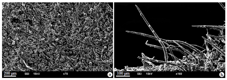
Microscopic examination of the leaf of this species showed that the adaxial surface is without trichomes. On the abaxial surface, there are special recesses. Very short multicellular, uniseriate non-glandular trichomes are visible outside the recesses of the leaves (Fig. 1a). A unique type of trichome is present in the recesses. These trichomes are simple, dense and have bends and twists, with an unadorned surface (Fig. 1b).

Microscopic images of Eriodictyon californicum leaf trichomes - a. short and smooth multicellular trichomes on adaxial surface of the leaves; b. dense twisted trichomes on abaxial surface of the leaves.
Eriodictyon sessilifolium Greene
Microscopic examination of the leaf of this species (Fig. 2a-b) shows that both surfaces of the leaf have long non-glandular simple trichomes that are placed at a short distance from each other. No special decorations can be seen on them. Also, shorter trichomes with curved ends are rarely seen.

Microscopic images of Eriodictyon sessilifolium leaf trichomes - a. adaxial surface of the leaves; b. long erect trichomes on abaxial surface of the leaves.
Nama aretioides (Hook. & Arn.) Brand
Microscopic examination of the leaf of this species (Fig. 3a-d) showed that the adaxial surface has simple horn-shaped trichomes that are conical and multicellular at the base. Their density is low and they can be counted. Their sizes are different, including small and large types, but their shape is almost the same. On the abaxial surface of the leaf, the number of trichomes is many fewer, but on the middle vein and also the margin of the leaf, their number is high. Trichomes can be seen both upright and bent. Their surface ornamentation consists of high-density oval projections that cover the entire surface of the trichome.

Microscopic images of Nama aretioides leaf trichomes - a. abaxial surface on the left side and adaxial surface on the right side; b. trichomes with different thickness are denser in margin of the leaves; c. multicellular base of trichomes; d. oval decorations are sparsely distributed on the trichome surface.
Nama demissa A.Gray
Microscopic analysis of the leaf of this species (Fig. 4a-d) revealed that both the adaxial and abaxial surfaces are adorned with homocytic non-glandular simple trichomes, exhibiting a high density. These trichomes display bends and occasional twists on both surfaces. While some are slightly longer, particularly at the leaf's periphery, the majority maintain a consistent size. Another noteworthy point is that the multicellular base protruding from the leaf surface was not seen in the trichomes of this species. Their surfaces feature densely packed protrusions that lack smoothness, with additional secondary protrusions visible.

Microscopic images of Nama demissa leaf trichomes - a. adaxial surface of the leaves; b. abaxial surface of the leaves; c. cylindrical base without cells protruding from the leaf surface; d. trichome showing surfaces with secondary protrusions.
Wigandia urens (Ruiz & Pav.) Kunth
During the microscopic examination, it was discovered that both surfaces of the leaves of this species are coated with heterocytic multicellular non-glandular trichomes, which closely resemble certain glandular trichomes characterized by a multicellular base and non-uniform base cells (Fig. 5a-d). While there is diversity in the cell shapes of these trichomes, they largely belong to the same category. The trichomes exhibit a high density, with some being longer in specific areas such as the leaf margin. These trichomes lack ornamentation on their surfaces and are predominantly curved or contorted.

Microscopic images of Wigandia urens leaf trichomes - a. high density of trichomes; b. differences in the shape of trichome cells; c. multicellular base protruding from the surface of the leaf; d. absence of surface decorations.
Trichome type and geographical distribution
The distribution map of the studied species, based on GBIF reports (2025), reveals that apart from Wigandia urens which is found from North to South America, and also present in parts of Europe, Southwest Asia, Africa, and Australia, other species are confined to the western and southwestern United States (Fig. 6). According to the map, Eriodictyon californicum and Eriodictyon sessilifolium, which have simple trichomes without a cylindrical base and surface decorations, are distributed in western regions near the ocean coast. In contrast, Nama aretioides and Nama demissa, which have trichomes with a cylindrical base, sometimes multi-cell bases, and surface decorations are found farther from the west coast. Furthermore, Wigandia urens, characterized by its diverse multicellular trichomes, is distributed across multiple continents, predominantly along the margins of continents.

The distribution map of the studied species along with the image of the type of trichomes for each species.
Identification key
Based on microscopic observations and using the observed trichome characters, an identification key can be written for the studied species using only the leaf trichome features, which is as follows.
-
1 Plants with heterocytic multicellular leaf trichome …….…….…….…….…….……. Wigandia urens
-
1' Plants with homocytic multicellular leaf trichome …….…….…….…….…….…….…….…….……. 2
-
2 Trichomes with a cylindrical base and surface decorations (grainy) …….…….…….…….….. 3
-
2' Trichomes without cylindrical base and without surface decorations …….…….…….…….… 4
-
3 Trichomes often with raised multicellular base, low density, oval surface features …….…….…….…….…….…….…….…….…….…….…….…….…….……. Nama aretioides
-
3' Trichomes without raised multicellular base, very dense, without surface features …….…….…….…….…….…….…….…….…….…….…….…….…….……. Nama demissa
-
4 Trichomes short, only on the abaxial surface of the leaf, dense …….…….…….…….…….…….…….…….…….…….…….……. Eriodictyon sessilifolium
-
4' Trichomes long, present on both leaf surfaces, sparse …….……. Eriodictyon californicum
Discussion
This study emphasizes the importance of scanning electron microscopy (SEM) in examining leaf trichomes of the Namaceae family, revealing the diversity of non-glandular trichomes in Eriodictyon, Nama, and Wigandia. SEM provides detailed insights into trichome morphology, such as size, shape, and density, which are crucial for taxonomic clarity in this family and the Boraginales order. These features have also been significant for the taxonomy of families like Asteraceae (Adedeji & Jewoola 2008) and Fabaceae (Zhao et al. 2022). The research highlights SEM's necessity for morphological studies, enabling better species identification and understanding of environmental adaptations, as seen in Lamiaceae (Raymond 2022; Semerdjieva et al. 2023) and Balsaminaceae (He et al. 2023).
The variation in trichome characteristics observed in this study illustrate the potential of these structures as reliable taxonomic tools, aiding in the differentiation and classification of closely related species. Checking trichome structures in detail is crucial as they often serve as key diagnostic features in plant taxonomy, helping to clarify species boundaries and evolutionary relationships. Furthermore, the study highlights the need for a more extensive investigation of Namaceae species to establish a clearer relationship between geographic distribution and trichome types. While only five plant species from this family have been examined using an electron microscope, thereby limiting certainty in determining trichome type ratios across geographical areas and coastal distances, further exploration with electron microscopy could establish patterns for other species within this family and potentially extend to Boraginales species for more comprehensive insights. The initial correlation observed between the specific characteristics of trichomes and their geographical locations can indicate their evolutionary adaptation to environmental conditions. However, the limited sample size in this study necessitates further research to confirm these patterns and extend the findings to other species within the family and the Boraginales order.
Considering the rapid increase in global warming, the evolutionary trajectory of speciation in both advanced and newly emerging species is likely to face significant challenges. Global warming imposes various stress factors, necessitating adaptations for survival. One key adaptation in plants is the development of trichomes, which play a crucial role in reducing transpiration and water loss (Skelton et al. 2017; Wagner 1991). As biotopes and regions worldwide experience increased dryness and higher temperatures, the evolution of trichomes may accelerate to enhance these protective functions. These findings suggest that natural selection favors plants with more efficient trichome configurations under arid conditions. The inability to adapt to these climatic changes may lead to plant extinction (IPCC 2014).
The observed variation in trichome type and density among the studied species, when viewed alongside their geographic distributions, presents a correlative pattern that is consistent with adaptive responses to environmental gradients, such as aridity. For instance, species distributed in less humid regions (Nama aretioides, N. demissa) possess trichomes with surface protrusions, which may enhance their protective function. While these patterns may reflect deep phylogenetic divergence rather than recent adaptation to contemporary climate change, they highlight trichome morphology as a trait potentially influenced by environmental factors. Further intraspecific studies across environmental gradients would be necessary to test adaptive hypotheses more directly.
Specifically, species in less humid regions are likely to develop trichomes with more thickness and surface protrusions as a survival strategy. Conversely, in more humid areas, where water vapor and air humidity are higher, plants tend to exhibit simpler trichomes with less intricate surface decorations. For example, a study was conducted on the response of trichomes in tomato plants under drought conditions and heat stress (Dutta & Phani 2023), which is expected to increase due to climate change. The researchers in this study found that trichome density and morphology changed significantly with drought, with increased trichome density and changes in their shape, possibly as an adaptive mechanism to reduce water loss and protect against overheating.
The trichome traits observed in this study align with findings in other Namaceae and Boraginales genera, highlighting their significance in taxonomic and evolutionary studies. For instance, the use of trichome traits was used as an important factor to name a new species of Nama from Arizona, USA (Crawford & Rink 2024). Also, trichome traits can be very useful for introducing new species and helping to classify them more correctly in plants of the Boraginales order, as these traits have been used in the introduction of three new species of Cryptantha (Rebman & Simpson 2022). Crawford & Rink (2024) have done a comparative work to introduce a new species of Nama from Arizona, U.S.A. In a part of this work, SEM images of the leaf surface are shown. The trichome is considered as a diagnostic character.
In line with the current findings, several recent studies have emphasized the systematic and ecological significance of trichome morphology in the Boraginales order. Similar scanning electron microscopy (SEM)-based investigations conducted on various lineages within Boraginales have yielded comparable results, reinforcing the value of trichomes in plant taxonomy and functional anatomy. For instance, detailed micromorphological analyses of leaf trichomes in Heliotropiaceae (Nazari & Ghahremaninejad 2024), Australian Boraginales (Nazari & Ghahremaninejad 2025a), Cordiaceae (Nazari & Ghahremaninejad 2025b), and Ehretiaceae (Nazari & Ghahremaninejad 2025c) demonstrated a wide diversity of trichome types and surface features, which proved useful in distinguishing closely related taxa. These studies collectively underscore that trichome traits-such as type, size, density, and surface decoration-hold significant systematic potential and may reflect adaptive responses. Together with the present investigation, this growing body of work further establishes SEM-based trichome analysis as a powerful tool for exploring both taxonomic boundaries and morphological adaptations in Boraginales. Overall, this research provides a foundational framework for future studies on the Namaceae family, advocating for the integration of SEM in taxonomic and phylogenetic analyses. Such comprehensive studies are crucial for advancing our understanding of the evolutionary pathways and biogeographic distributions of trichomes, ultimately contributing to a more refined classification system for these and related plant species.
Acknowledgments
We thank Dr. Michael G. Simpson and Dr. John Edmondson for their comments to improve this work. We extend our sincere gratitude to the invaluable contributors that have facilitated this work. Special thanks to Mag. Heimo Rainer, head of the Botany Department of the Natural History Museum of Vienna (W), for his extensive assistance during this project. We also thank Meg. Astrid Hille, Dr. Andreas Berger, Dr. Tanja Schuster, and Dr. Johannes Walter, members of the Department of Botany at the Natural History Museum of Vienna, for their help. Special thanks to Dr. Wencke Wegner, a SEM specialist at the Natural History Museum of Vienna, for supervising the microscopic imaging of the fragments. Our appreciation goes to the Herbarium of Natural History Museum of Vienna (W) for sharing their extensive botanical resources. This work is related to part of Hamid Nazari's Ph.D. thesis at Bu-Ali Sina University, Hamadan, Iran.
References
-
Adedeji O & Jewoola OA (2008) Importance of leaf epidermal characters in the asteraceae family. Notulae Botanicae Horti Agrobotanici Cluj-Napoca 36: 7-18. DOI: 10.15835/nbha362243.
» https://doi.org/10.15835/nbha362243. - Altındal N & Altındal D (2021) The effects of climate change on the alteration of plant traits. In: Choudhary DK, Mishra A & Varma A (eds.) Climate change and the microbiome. Soil Biology. Vol. 63. Springer, Cham. Pp. 299-307. DOI: 10.1007/978-3-030-76863-8_15
-
Bellard C, Bertelsmeier C, Leadley P, Thuiller W & Courchamp F (2012) Impacts of climate change on the future of biodiversity. Ecology Letters 15: 365-377. DOI: 10.1111/j.1461-0248.2011.01736.x.
» https://doi.org/10.1111/j.1461-0248.2011.01736.x. -
Chen JJ, Sun Y, Kopp K, Oki L, Jones SB & Hipps L (2022) Effects of water availability on leaf trichome density and plant growth and development of Shepherdia xutahensis Frontiers of Plant Science 13: 855858. DOI: 10.3389/fpls.2022.855858
» https://doi.org/10.3389/fpls.2022.855858 -
Crawford RA & Rink G (2024) A novel species of Nama (Namaceae) from northern Arizona, U.S.A. Journal of the Botanical Research Institute of Texas 18: 1-5. DOI: 10.17348/jbrit.v18.i1.1335
» https://doi.org/10.17348/jbrit.v18.i1.1335 -
Dutta TK & Phani V (2023) The pervasive impact of global climate change on plant-nematode interaction continuum. Frontiers in Plant Science 14: 1143889. DOI: 10.3389/fpls.2023.1143889.
» https://doi.org/10.3389/fpls.2023.1143889. -
El Beyrouthy M, Arnold N, Delelis-Dusollier A, Dupont F & de Foucault B (2008) Morphologie du trichome des Teucrium du Liban, Acta Botanica Gallica 155: 563-576. DOI: 10.1080/12538078.2008.1051613
» https://doi.org/10.1080/12538078.2008.1051613 -
GBIF: The Global Biodiversity Information Facility (2025) Free and open access to biodiversity data. Available at <https://www.gbif.org/>. Access in September 2025.
» https://www.gbif.org - Ghahremaninejad F (2004) Value of trichome characteristics for separating of bifurcating Astragalus (Fabaceae) on sectional level. Turkish Journal of Botany 28: 241-245.
-
He H, Ma M, Chen X, Li X, Li F, Zhao Q, He X, Tan Y, Qu S, Huang H & Huang M (2023) Studies of the pollen characteristics and the taxonomic significance of Impatiens from the Yunnan-Guizhou Plateau. Folia Horticulturae 35: 347-368. DOI: 10.2478/fhort-2023-0025
» https://doi.org/10.2478/fhort-2023-0025 - Hofmann M, Walden GK, Hilger HH & Weigend M (2016) Hydrophyllaceae. In: Kadereit JW & Bittrich V (eds.) Families and genera of vascular plants XIV. Springer, Berlin, Heidelberg, New York. Pp. 221-238. DOI: 10.1007/978-3-319-28534-4_20
- IPCC (2014) Climate Change 2014: impacts, adaptation, and vulnerability. Part A: global and sectoral aspects. Contribution of Working Group II to the Fifth Assessment Report of the Intergovernmental Panel on Climate Change. Cambridge University Press, Cambridge. Pp. 1-944.
-
Janni M, Maestri E, Gullì M, Marmiroli M & Marmiroli N (2024) Plant responses to climate change, how global warming may impact on food security: a critical review. Frontiers of Plant Science 14: 1297569. DOI: 10.3389/fpls.2023.1297569
» https://doi.org/10.3389/fpls.2023.1297569 -
Luebert F, Cecchi L, Frohlich MW, Gottschling M, Guilliams CM, Hasenstab-Lehman KE, Hilger HH, Miller JS, Mittelbach M, Nazaire M, Nepi M, Nocentini D, Ober D, Olmstead RG, Selvi F, Simpson MG, Sutory K, Valdes B, Walden GK & Weigend M (2016) Familial classification of the Boraginales. Taxon 65: 502-522. DOI: 10.12705/653.5
» https://doi.org/10.12705/653.5 -
Nazari H & Ghahremaninejad F (2024) A SEM study of leaf trichomes in Heliotropiaceae. Iranian Journal of Botany 30: 173-183. DOI: 10.22092/ijb.2024.366196.1475.
» https://doi.org/10.22092/ijb.2024.366196.1475. -
Nazari H & Ghahremaninejad F (2025a) Micromorphological analysis of leaf trichomes in Australian species of Boraginales, Turczaninowia 28: 101-111. DOI: 10.14258/turczaninowia.28.2.10
» https://doi.org/10.14258/turczaninowia.28.2.10 -
Nazari H & Ghahremaninejad F (2025b) Contribution to the systematic value of the leaf trichomes in the Cordiaceae (Boraginales). Revista Mexicana de Biodiversidad 96:e965577. DOI: 10.22201/ib.20078706e.2025.96.5577
» https://doi.org/10.22201/ib.20078706e.2025.96.5577 -
Nazari H & Ghahremaninejad F (2025c) Leaf trichome diversity in Ehretiaceae. Rostaniha 26: 112-123. DOI: 10.22092/bot.j.iran.2025.368150.1410
» https://doi.org/10.22092/bot.j.iran.2025.368150.1410 -
Noroozi M, Ghahremaninejad F, Boglerb D, Witherspoonc MJ, Ryand LG, Millerb S J, Riahia M, Cohen IJ (2021) Parsing a plethora of pollen: the role of pollen size and shape in the evolution of Boraginaceae. Cladistics 38: 204-226. DOI: 10.1111/cla.12488
» https://doi.org/10.1111/cla.12488 -
Noroozi M, Ghahremaninejad F, Riahi M & Cohen JI (2024) Phylogenomics and plastome evolution of Lithospermeae (Boraginaceae). BMC Plant Biology 24: 957. DOI: 10.1186/s12870-024-05665-6.
» https://doi.org/10.1186/s12870-024-05665-6. -
Pandey P, Irulappan V, Bagavathiannan MV & Senthil-Kumar M (2017) Impact of combined abiotic and biotic stresses on plant growth and avenues for crop improvement by exploiting physio-morphological traits. Frontiers of Plant Science 8: 537. DOI: 10.3389/fpls.2017.00537.
» https://doi.org/10.3389/fpls.2017.00537. -
POWO: Plants of the World Online (2024) Plants of the World Online. Available at <https://powo.science.kew.org>. Access in September 2025.
» https://powo.science.kew.org -
Raymond W (2022) An overview of cryo-scanning electron microscopy techniques for plant imaging. Plants 11: 1113. DOI: 10.3390/plants11091113
» https://doi.org/10.3390/plants11091113 -
Rebman JP & Simpson MG (2022) Three new species of Cryptantha (Boraginaceae) from the southern channel islands of California. Madroño 69: 111-134. DOI: 10.3120/0024-9637-69.1.111
» https://doi.org/10.3120/0024-9637-69.1.111 -
Semerdjieva I, Yankova-Tsvetkova E, Zheljazkov VD, Koleva-Valkova LH & Nikolova R (2023) Reproductive capacity and Scanning Electron Microscopy (SEM) analyses of the micromorphological surfaces of three endemic satureja species from Bulgaria. Plants 12: 2436. DOI: 10.3390/plants12132436
» https://doi.org/10.3390/plants12132436 -
Skalák J, Vercruyssen L, Claeys H, Hradilová J, Černý M, Novák O, Plačková L, Saiz-Fernández I, Skaláková P, Coppens F, Dhondt S, Koukalová Š, Zouhar J, Inzé D & Brzobohatý B (2019) Multifaceted activity of cytokinin in leaf development shapes its size and structure in Arabidopsis. The Plant Journal 97: 805-824. DOI: 10.1111/tpj.14285.
» https://doi.org/10.1111/tpj.14285. -
Skelton RP, Brodribb TJ & Choat B (2017) Casting light on xylem vulnerability in an herbaceous species reveals a lack of segmentation. New Phytol 214: 561-569. DOI: 10.1111/nph.14450.
» https://doi.org/10.1111/nph.14450. -
Soltis PS & Soltis DE (2009) The role of hybridization in plant speciation. Annual Review of Plant Biology 60: 561-588. DOI: 10.1146/annurev.arplant.043008.092039
» https://doi.org/10.1146/annurev.arplant.043008.092039 -
Steyn HM & Van Wyk AE (2021) Taxonomic significance of trichomes in the genus Acanthopsis Harv. (Acanthaceae, tribe Acantheae). Adansonia 43: 163-176. DOI: 10.5252/adansonia2021v43a14
» https://doi.org/10.5252/adansonia2021v43a14 -
Stull GW, Stefano RD, Soltis DE & Soltis PS (2015) Resolving basal lamiid phylogeny and the circumscription of Icacinaceae with a plastome-scale data phylogeny and historical biogeography of Hydrophyllaceae and Namaceae 769 set. American Journal of Botany 102: 1794-1813. DOI: 10.3732/ajb.1500298
» https://doi.org/10.3732/ajb.1500298 -
Vasile MA, Jeiter J, Weigend M & Luebert F (2020) Phylogeny and historical biogeography of Hydrophyllaceae and Namaceae, with a special reference to Phacelia and Wigandia. Systematics and Biodiversity 18: 757-770. DOI: 10.1080/14772000.2020.1771471
» https://doi.org/10.1080/14772000.2020.1771471 -
Wagner GJ (1991) Secreting glandular trichomes: more than just hairs. Plant Physiology 96: 675-679. DOI: 10.1104/pp.96.3.675
» https://doi.org/10.1104/pp.96.3.675 -
Weigend M, Luebert F, Gottschling M, Couvreur TLP, Hilger HH & Miller JS (2014) From capsules to nutlets-phylogenetic relationships in the Boraginales. Cladistics 30: 508-518. DOI: 10.1111/cla.12061
» https://doi.org/10.1111/cla.12061 -
Yan W, Zhong Y & Shangguan Z (2016) A meta-analysis of leaf gas exchange and water status responses to drought. Scientific Reports 6: 20917. DOI 10.1038/srep20917
» https://doi.org/10.1038/srep20917 -
Zhao X, Hou Q, Du M, Zhang H, Jia L, Zhang Z, Ma Z & Sun K (2022) Micromorphological leaf epidermal traits as potential taxonomic markers for infrageneric classification of Oxytropis (Fabaceae). PhytoKeys 201: 51-76. DOI: 10.3897/phytokeys.201.85154.
» https://doi.org/10.3897/phytokeys.201.85154. -
Zhu JK (2016) Abiotic stress signaling and responses in plants. Cell 167: 313-324. DOI: 10.1016/j.cell.2016.08.029.
» https://doi.org/10.1016/j.cell.2016.08.029.