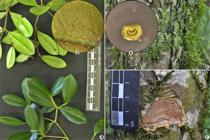
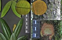
Figure 1
a-d. Blepharocalyx salicifolius – a. branch and leaves; b. external bark; c. internall bark; d. detail of the translucid punctuations on the leaves (abaxial face, magnification of 0.7x, without light).
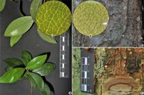
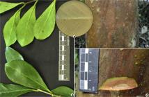
Figure 2
a-e. Myrcia glomerata – a. branch and leaves; b. external bark; c. internal bark; d. detail of the translucid punctuations and leaf indumentum (abaxial face, magnification of 4.5x, without light); e. detail of the petiole (profile, magnification 4,5x, without light).
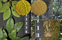
Figure 3
a-e. Campomanesia guaviroba – a. branch and leaves; b. external bark; c. internal bark; d. detail of the indumentum on the axillary tufts (abaxial face, magnification 0.7x, without light); e. detail of the translucid punctuations and indumentum on the leaves (abaxial face, magnification 4.5x, without light).
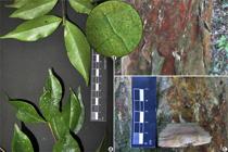
Figure 4
a-d. Campomanesia guazumifolia – a. branch and leaves; b. external bark; c. internal bark; d. detail of translucid punctuations on the leaves (abaxial face, magnification 4.5x, with light).
Figure 5
a-e. Campomanesia xanthocarpa – a. branch and leaves; b. external bark; c. internal bark; d. detail of the translucid punctuations on the leaves (abaxial face, magnification 4.5x, without light); e. detail of the indumentum on the axillary tufts (abaxial face, magnification 0.7x, without light).
Figure 6
a-e. Eugenia chlorophylla – a. branch and leaves; b. external bark; c. internal bark; d. detail of the translucid punctuations on the leaves (adaxial face, magnification 4.5x, without light); e. detail of the waxy on the leaves (adaxial face, magnification 4.5x, without light).
Figure 7
a-d. Eugenia involucrata – a. branch and leaves; b. external bark; c. internal bark; d. detail of the translucid punctuations on the leaves (abaxial face, magnification 4.5x, without light).
Figure 8
a-e. Eugenia platysema – a. branch and leaves; b. external bark; c. internal bark; d. detail of the translucid punctuations on the leaves (adaxial face, magnification 0.7x, with light); e. detail of the double intramarginal vein (adaxial face, magnification 4.5x, with light).
Figure 9
a-e. Eugenia pluriflora – a. branch and leaves; b. external bark; c. internal bark; d. detail of the translucid punctuations on the leaves (abaxial face, magnification 4.5x, without light); e. detail of the petiole (profile, magnification 4,5x, without light).
Figure 10
a-e. Eugenia uniflora – a. branch and leaves; b. external bark; c. internal bark; d. detail of the translucid punctuations on the leaves (abaxial face, magnification 4.5x, without light); e. detail of the petiole (profile, magnification 4.5x, without light).
Figure 11
a-e. Myrceugenia acutiflora – a. branch and leaves; b. external bark; c. internal bark; d. detail of the translucid punctuations on the leaves (adaxial face, magnification 4.5x, with light); e. detail of the sericeous indumentum (abaxial face, magnification 4.5x, without light).
Figure 12
a-d. Myrceugenia euosma – a. branch and leaves; b. external bark; c. internal bark; d. detail of the translucid punctuations on the leaves (adaxial face, magnification 0.7x, with light).
Figure 13
a-d. Myrceugenia glaucescens – a. branch and leaves; b. external bark; c. internal bark; d. detail of the translucid punctuations on the leaves (abaxial face, magnification 4.5x, without light).
Figure 14
a-e. Myrceugenia miersiana – a. branch and leaves; b. external bark; c. internal bark; d. detail of the translucid punctuations on the leaves (8); e. detail of the velvety indumentum (abaxial indumentum, magnification 4.5x, without light).
Figure 15
a-d. Myrceugenia regnelliana – a. branch and leaves; b. external bark; c. internal bark; d. detail of the translucid punctuations on the leaves (abaxial face, magnification 4.5x, without light).
Figure 16
a-d. Myrcia amazonica – a. branch and leaves; b. external bark; c. internal bark; d. detail of the translucid punctuations on the leaves (abaxial face, magnification 4.5x, without light).
Figure 17
a-d. Myrcia guianensis – a. branch and leaves; b. external bark; c. internal bark; d. detail of the translucid punctuations on the leaves (abaxial face, magnification 0.7x, without light).
Figure 18
a-e. Myrcia hatschbachii – a. branch and leaves; b. external bark; c. internal bark; d. detail of the translucid punctuations on the leaves (adaxial face, magnification 0.7x, with light); e. detail of the pilose indumentum (abaxial face, magnification 4.5x, without light).
Figure 19
a-d. Myrcia selloi – a. branch and leaves; b. external bark; c. internal bark; d. detail of the translucid punctuations on the leaves (abaxial face, magnification 4.5x, without light).
Figure 20
a-d. Myrcia palustris – a. branch and leaves; b. external bark; c. internal bark; d. detail of the translucid punctuations on the leaves (adaxial face, magnification 0.7x, with light).
Figure 21
a-d. Myrcia splendens – a. branch and leaves; b. external bark; c. internal bark; d. detail of the translucid punctuations on the leaves (adaxial face, magnification 4.5x, with light).
Figure 22
a-e. Myrcia venulosa – a. branch and leaves; b. external bark; c. internal bark; d. detail of the translucid punctuations on the leaves (adaxial face, magnification 0.7x, with light); e. detail of the lanate indumentum (abaxial face, magnification 4,5x, without light).
Figure 23
a-d. Myrcianthes gigantea – a. branch and leaves; b. external bark; c. internal bark; d. detail of the translucid punctuations on the leaves (adaxial face, magnification 0.7x, with light).
Figure 24
a-d. Myrciaria tenella – a. branch and leaves; b. external bark; c. internal bark; d. detail of the translucid punctuations (abaxial face, magnification 4.5x, without light).
Figure 25
a-d. Myrrhinium atropurpureum – a. branch and leaves; b. external bark; c. internal bark; d. detail of the translucid punctuations on the leaves (abaxial face, magnification 0.7x, without light).
Figure 26
a-e. Pimenta pseudocaryophyllus – a. branch and leaves; b. external bark; c. internal bark; d. detail of the translucid punctuations on the leaves (adaxial face, magnification 0.7x, with light); e. detail of the canescent indumentum (abaxial face, magnification 4.5x, without light).
Figure 27
a-e. Plinia peruviana – a. branch and leaves; b. external bark; c. internal bark; d. detail of the translucid punctuations on the leaves (adaxial face, magnification 4.5x, with light); e. detail of the double intramarginal vein (adaxial face, magnification 4.5x, with light).
Figure 28
a-d. Psidium cattleyanum – a. branch and leaves; b. external bark; c. internal bark; d. detail of the translucid punctuations of the leaves (abaxial face, magnification 0.7x, without light).
Figure 29
a-d. Psidium guajava – a. branch and leaves; b. external bark; c. internal bark; d. detail of the translucid punctuations on the leaves (adaxial face, magnification 0.7x, with light).

 Thumbnail
 Thumbnail
 Thumbnail
 Thumbnail
 Thumbnail
 Thumbnail
 Thumbnail
 Thumbnail
 Thumbnail
 Thumbnail
 Thumbnail
 Thumbnail
 Thumbnail
 Thumbnail
 Thumbnail
 Thumbnail
 Thumbnail
 Thumbnail
 Thumbnail
 Thumbnail
 Thumbnail
 Thumbnail
 Thumbnail
 Thumbnail
 Thumbnail
 Thumbnail
 Thumbnail
 Thumbnail
 Thumbnail


