 Position Statement on Cardiometabolic Health Across the Woman's Life Course – 2025
Table of contents
-
1. Introduction 13
-
2. Highlights 18
-
3. Inflammation and Implications for the Cardiovascular System 23
-
3.1. Inflammation and Atherosclerosis 23
-
3.2. Inflammation and Heart Failure in Women 25
-
3.3. Hormonal Aspects in Inflammation 25
-
3.4. Inflammation in Cardio-Oncology 25
-
3.5. Inflammation in Pregnancy and Preeclampsia 25
-
-
4. Implications of Sex Steroids for Cardiometabolic Health 27
-
4.1. Origin and Metabolism of Sex Steroids 27
-
4.2. Chemical Classification of Estrogens 28
-
4.2.1. Natural Estrogens 28
-
4.2.2. Synthetic Estrogens 28
-
-
4.3. Chemical Classification of Progestagens 28
-
4.3.1. Natural Progesterone 28
-
4.3.2. Progestagens 28
-
-
4.4. Common Indications of Therapies with Estrogens, Progestagens, and Testosterone in Women 29
-
4.4.1. Polycystic Ovary Syndrome 29
-
4.4.2. Hormonal contraception 29
-
4.4.3. Menopausal Hormone Therapy: Regimens, Doses, and Routes, Focused on Cardiovascular Health 30
-
-
4.5. Risks: Metabolic Effects, Thromboembolism, and Breast Cancer 32
-
4.5.1. Metabolic Effects 32
-
4.5.2. Thromboembolism 32
-
4.5.3. Breast Cancer 32
-
-
-
5. Indicators of Cardiometabolic Health 33
-
5.1. Assessment of Reproductive Risk Factors with Implications for Cardiometabolic Health 33
-
5.1.1. Puberty and Pregestational Period 33
-
5.1.1.1. Age at Menarche 33
-
5.1.1.2. Menstrual Cycle Characteristics 33
-
-
5.1.2. Pregnancy and Postpartum Period 33
-
5.1.2.1. Weight changes in pregnancy 33
-
5.1.2.2. Gestational dysglycemia 33
-
5.1.2.3. Gestational Dyslipidemia 33
-
5.1.2.4. Postpartum Behavior 34
-
-
5.1.3. Menopause Transition 34
-
5.1.3.1. Age at menopause 35
-
5.1.3.2. Vasomotor Symptoms 35
-
5.1.3.3. Menopausal Hormone Therapy 35
-
-
5.1.4. Postmenopausal Metabolic Health 35
-
5.1.4.1. Menopausal metabolic changes 35
-
5.1.4.2. Diabetes mellitus 35
-
5.1.4.3. Dyslipidemia 35
-
-
-
5.2. Anthropometric Indicators 36
-
5.2.1. Body Mass Index 36
-
5.2.2. Waist circumference – Brazilian population 36
-
5.2.3. Waist-To-Hip Ratio 36
-
5.2.4. Body Fat Percentage 36
-
5.2.5. Waist-To-Height Ratio 36
-
-
5.3. Biomarkers 36
-
5.3.1. Lipid Profile (Total Cholesterol, LDL-c, HDL, Triglycerides) 36
-
5.3.2. Glycemia and Glycated Hemoglobin 36
-
5.3.3. C-reactive Protein 37
-
5.3.4. Fibrinogen 37
-
5.3.5. Homocysteine 37
-
5.3.6. Adipokines 37
-
-
-
6. Cardiometabolism in Childhood and Adolescence 37
-
6.1. Definitions and Diagnostic Criteria 37
-
6.1.1. Pathophysiology 37
-
6.1.2. Early Menarche, Late Menarche, and Cardiometabolic Risk 37
-
-
6.2. Menstrual irregularities and cardiometabolic risk 39
-
6.2.1. Pathophysiological Mechanisms 39
-
6.2.2. Clinical Assessment and Biomarkers 40
-
6.2.3. Therapeutic Interventions 40
-
-
6.3. Obesity, Eating Disorders, and Cardiovascular Risk: Long-Term Impacts 40
-
6.3.1. Eating Disorders and Obesity 40
-
6.3.2. Hereditary Factors and Prognosis of Eating Disorders 40
-
6.3.3. Future Consequences of Eating Disorders 40
-
6.3.4. Childhood Obesity and Cardiovascular Outcomes 41
-
6.3.5. Eating Disorders and Cardiovascular Risk 41
-
-
-
7. Cardiometabolic Continuum and Reproductive Age 41
-
7.1. Polycystic Ovary Syndrome 41
-
7.2. Infertility and its Treatment 42
-
7.3. Weight changes in pregnancy 43
-
7.3.1. Maternal Impact of Excessive Gestational Weight Gain 44
-
7.3.2. Fetal Impact of Excessive Gestational Weight Gain 44
-
-
7.4. Gestational Dysglycemia 44
-
7.5. Gestational dyslipidemia 45
-
7.5.1. Impact of Dyslipidemia in Pregnancy 46
-
7.5.2. Impact of Pregnancy on Patients with Familial Hypercholesterolemia 46
-
-
7.6. Endometriosis and Cardiovascular Risk 46
-
7.6.1. Risk factors 46
-
-
7.7. Psoriasis 46
-
7.8. Hypertensive Disorders of Pregnancy and Endothelial Dysfunction after Menopause 49
-
7.9. Metabolic Changes in Postpartum 49
-
7.9.1. Cardiometabolic Risk Factors in Postpartum 49
-
7.9.2. Impact of gestational diabetes and dyslipidemia in postpartum 49
-
7.9.3. Prolactin and Metabolism in Postpartum 49
-
-
-
8. Cardiometabolic Health in Menopause Transition, Menopause and Postmenopause 50
-
8.1. Severity of Symptoms and their Implications for Cardiovascular Health 50
-
8.2. Cardiovascular Risk Factors and Menopause 50
-
8.3. Menopausal Hormone Therapy, Hormone Implants, and Cardiometabolic Impact 52
-
8.4. Stratification of Cardiometabolic Risk in Menopause 52
-
-
9. Cardiometabolic Disorders in Women 53
-
9.1. Obesity and Metabolic Syndrome 53
-
9.2. Type 2 Diabetes Mellitus 54
-
9.3. Metabolic Dysfunction-Associated Steatotic Liver Disease 55
-
9.4. Chronic Kidney Disease 56
-
-
10. Strategies for Addressing Cardiometabolic Disorders in Women 57
-
10.1. Nonpharmacological Measures 57
-
10.2. Nutritional Interventions 57
-
10.3. Physical Activity 57
-
10.4. Psychosocial Interventions 58
-
10.5. Tobacco Use and Alcohol Consumption 58
-
10.6. Specific Clinical Conditions 58
-
10.7. Pharmacological Strategies 58
-
10.7.1. Systemic Arterial Hypertension Treatment 58
-
10.7.2. Management of Dyslipidemias 59
-
10.7.3. Oral Lipid-Lowering Drugs 59
-
10.7.4. Diabetes Mellitus Control 59
-
10.7.5. Management of Obesity 60
-
10.7.6. Management of Metabolic Dysfunction-Associated Steatotic Liver Disease 62
-
10.7.7. Management of Chronic Kidney Disease 62
-
10.7.8. Role of GLP-1 Analogues in Women’s Cardiometabolic Treatment 63
-
10.7.9. Specificities in Women 63
-
10.7.10. Semaglutide 63
-
10.7.11. Tirzepatide 63
-
10.7.12. Future Perspectives 64
-
-
10.8. Specific pharmacological considerations 64
-
10.8.1. Drug Interactions 64
-
10.8.2. Importance of Weight Control 64
-
10.8.3. Individualized Approach 64
-
10.9. Surgical Treatment 64
-
10.9.1. Bariatric Surgery 64
-
-
-
11. Recommendations for the Management of Cardiometabolic Disorders in Women 64
-
Acknowledgments 67
-
Supplement 71
-
References 82

1. Introduction
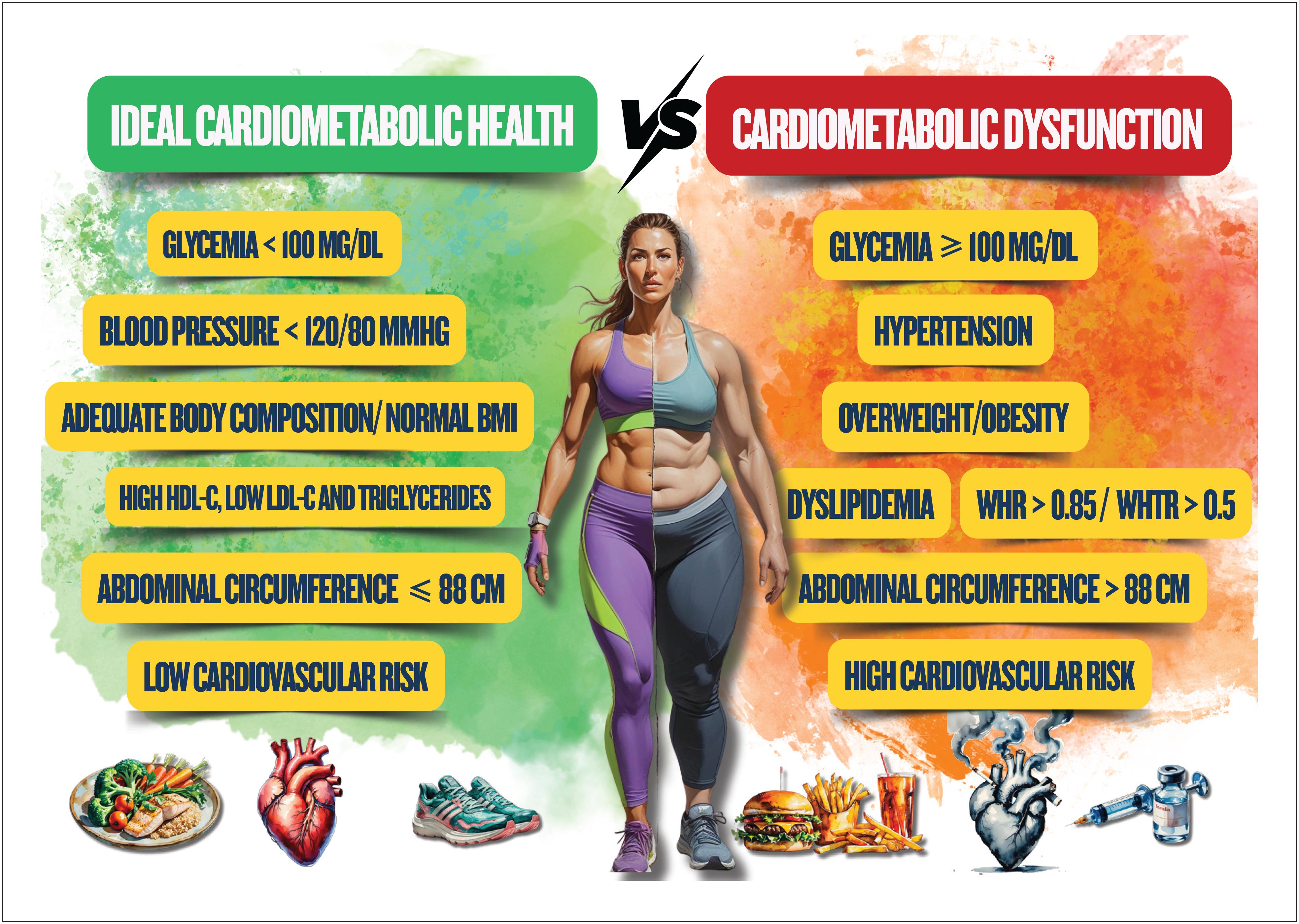
Cardiometabolic health can be characterized by ideal levels of serum glucose and lipids and of blood pressure (BP) in association with low adiposity and cardiovascular risk (CVR). Usually, cardiometabolic health is the absence of metabolic dysfunction characteristic of some diseases, such as cardiovascular diseases (CVD), type 2 diabetes mellitus (T2DM), and metabolic syndrome (MS). Poor metabolic health is responsible for a substantial population burden of disability, diseases, as well as cardiovascular, neoplastic, and all-cause deaths. Different characteristics related to reproduction have been increasingly associated with metabolic diseases based on life course epidemiology, which postulates that biological, behavioral, and social factors during sensitive stages of life, mediated by hormonal fluctuations, act independently, cumulatively and interactively to influence the posterior risk for health and disease1 , 2 ( Figure 1.1 ).

Spectrum of cardiometabolic health. BMI: body mass index; WHR: waist to hip ratio; WHtR: waist to height ratio.
The prevalence of cardiometabolic disorders is increasing worldwide among women and men and has been associated with higher rates of obesity and its risk factors (RFs), such as hypertension and T2DM. In addition, there is increasing evidence suggesting that sexual hormones, and sex- and gender-specific molecular mechanisms can influence the metabolism of glucose and lipids, thus impacting on cardiometabolic RFs. Moreover, there is an emerging predominance of common types of cardiometabolic disorders, such as heart failure (HF), atrial fibrillation, and ischemic heart disease (IHD), that differ between females and males. Significant sex-specific variations have been reported in risk profiles, with an especial emphasis on women, whose risk scores are inadequate to predict both atherothrombotic diseases and their related outcomes, such as cardiovascular death, myocardial infarction (MI), stroke, HF, ventricular and supraventricular arrhythmias, as well as need for revascularization and hospitalization.2
The ranking of CVD deaths and DALYs (disability-adjusted life years, a measure that represents the burden of disease on a population) rates per 100,000 inhabitants, attributed to RFs in women and men in Brazil in 2021, shows that metabolic RFs represent the first five RFs for women's death and the first four RFs for women's DALYs. It is worth noting that the increase in systolic BP, low-density lipoprotein cholesterol (LDL-c), body mass, and serum glucose, in that order, is frequently associated with CVD death and burden in the Brazilian female population. In addition, kidney dysfunction is an important metabolic RF for women and men. It is worth noting the increase in body mass and serum glucose in the past 21 years, representing a significant increase of obesity and diabetes mellitus (DM) in women and men.3( Figures 1.2 and 1.3 )

Ranking of mortality rates per 100,000 inhabitants due to cardiovascular disease attributed to risk factors, in women and men, in Brazil, in 2021.3
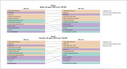
Ranking of DALYs rates per 100,000 inhabitants due to cardiovascular disease attributed to risk factors, in women and men, in Brazil, in 2021.3
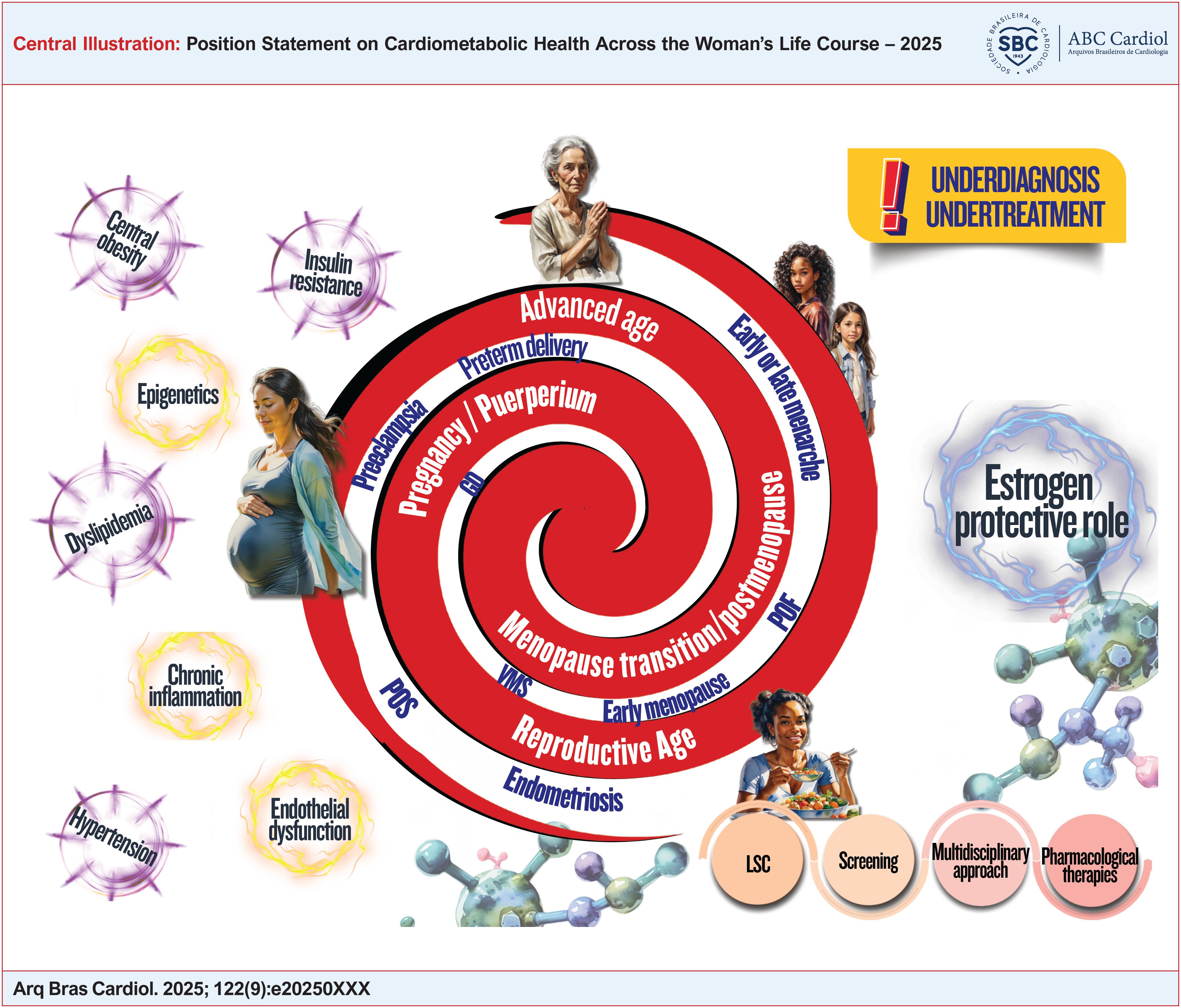
Recent evidence suggests that female reproductive characteristics can be related to RFs that contribute to later metabolic dysfunction that culminates in CVD in menopause. These reproductive characteristics are as follows: age of menarche, menstrual irregularity of endocrine origin, development of polycystic ovary syndrome (POS), excessive weight gain in pregnancy, gestational dysglycemia and dyslipidemia, hypertensive disorders of pregnancy, severity and timing of menopausal symptoms, and effect of hypoestrogenism on the cardiovascular system. Those RFs can be markers of future dysfunction or be explained by shared underlying etiologies that promote disease in the long run. Identifying potentially modifiable characteristics has a significant influence in strategies to implement a healthy lifestyle, as well as drug and surgical therapies that can relieve metabolic burden in the long run.4 - 6
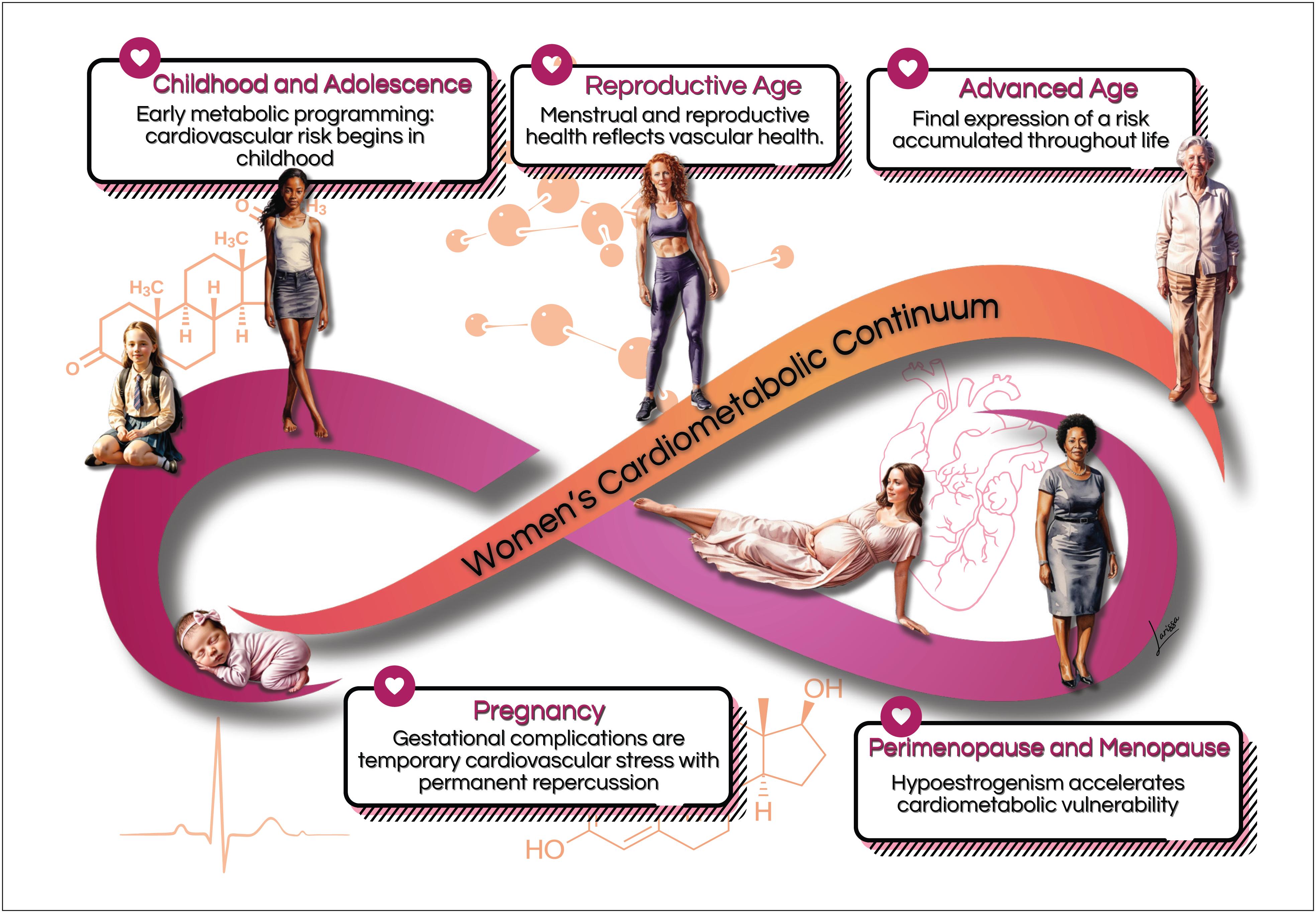
The cardiometabolic continuum , sequence of cardiovascular events resulting from gene-environmental interactions, influences of unhealthy lifestyles, and metabolic diseases, such as DM and systemic arterial hypertension (SAH), occurs predominantly over the course of a woman's life ( Figure 1.4 ). In a recent study, the cardiometabolic continuum was analyzed to assess differences between sexes and populations in two distinct cohorts: the UK Biobank (17,700 participants) and the Brazilian Longitudinal Study of Adult Health (in Portuguese, ELSA-Brasil - Estudo Longitudinal de Saúde do Adulto ) with 7,162 participants. The authors have studied the cardiometabolic continuum using machine learning and identified five patterns. They reported female disadvantage regarding the time of appearance of the cardiometabolic continuum . In the UK Biobank cohort, when SAH was the first disease of the cardiometabolic continuum and diagnosed in isolation, it occurred faster in women. In the ELSA-Brasil cohort, not only DM was more frequently the first disease in the female cardiometabolic continuum but also diagnosed faster when followed by SAH. In addition, women had a greater incidence of isolated SAH and DM, and a smaller percentage of them was classified as healthy. The authors have emphasized the unequal access to proper treatment and diagnosis of the female cardiometabolic continuum and stressed the need for sex-differentiated health policies in Brazil to reduce inequities.7

The cardiovascular system, kidneys and liver share RFs, such as dyslipidemia, hypertension, tobacco use, DM, and central/truncal obesity. Shared metabolic and functional disorders result in damage to those organs via overlapping pathophysiological pathways, to which hormonal influences, derived from the different cycles of a woman's life, are added. The increase in metabolic RFs over the years supports the need to improve the identification and treatment of women's cardiometabolic disorders, mainly because they are underdiagnosed and undertreated. This is compounded by the women's small participation in clinical trials that guide the currently available therapeutic strategies, which include lifestyle changes, drug therapy, and surgery. Moreover, the inflammatory nature, notably higher in women with cardiometabolic disorders, as well as the new pharmacological and surgical therapeutic options that provide management based on mechanisms that can reach multiple pathophysiological pathways influenced by hormones, require further studies in the female sex. Specific social programs to encourage healthy diets and physical activity, as well as better access to public health policies aimed at women with cardiometabolic disorders, are required.4 , 8
This Position Statement on Cardiometabolic Health Over the Course of a Woman's Life is a joint effort of the Women's Cardiology Department of the Brazilian Society of Cardiology (DCM/SBC), Brazilian Federation of the Societies of Gynecology and Obstetrics (FEBRASGO), and Brazilian Society of Endocrinology and Metabolism Study (SBEM) to close the knowledge gap on women's cardiometabolic disorders.
In what follows, we present the highlights of this position statement according to their respective chapters.
2. Highlights
Chapter 3
-
In women, menopause and pregnancy complications (mainly preeclampsia/eclampsia) activate inflammation and accelerate coronary atherosclerosis, causing vascular stiffness, endothelial dysfunction, and microvascular ischemia;
-
Biological variations between women and men result from differences in gene expression of sex chromosomes modulated by hormonal and environmental influences, which result in cardiovascular conditions associated with autonomic regulation and vascular and cardiac remodeling;
-
Women with menstrual irregularities of endocrine origin frequently exhibit a pro-inflammatory status with elevation of inflammatory markers that intermediate atherosclerotic genesis;
-
The increased prevalence of risk factors in women, such as obesity, diabetes, and hypertension, associated with the effects of menopause, seems to explain the higher prevalence of heart failure with preserved ejection fraction;
-
The association of chronic inflammation and adiposity in heart failure with preserved ejection fraction seems to be related to pericardial and epicardial adipokines, leading to microcirculatory inflammation, cardiac fibrosis, and physiological diastolic filling changes;
-
Postmenopausal hypoestrogenism promotes metabolic changes, such as increased central adiposity, worse glucose metabolism, and increased levels of total cholesterol, LDL-c, triglycerides, apolipoproteins, and lipoprotein a.
Chapter 4
-
Sex steroid hormones bind to hormonal receptors in several tissues and have multiple biological effects;
-
The different sex steroid hormones are frequently indicated to treat conditions, such as polycystic ovary syndrome, as well as for contraception and menopausal hormone therapy, which can have implications for cardiometabolic risk;
-
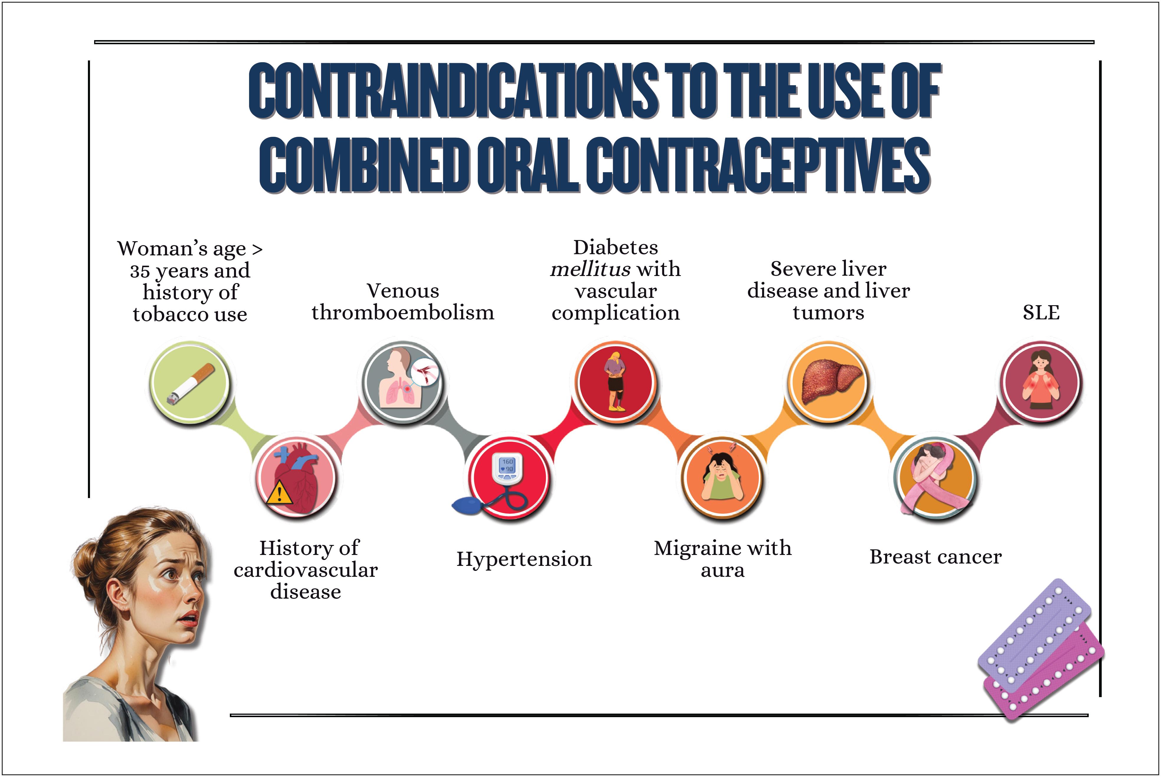
Combined oral contraceptives are not recommended for women with a history of tobacco use over the age of 35 years, cardiovascular diseases, venous thromboembolism, diabetes with vascular complication, migraine with neurological signs, severe liver disease, liver tumors, breast cancer, and systemic lupus erythematosus;
-
The cardiovascular effects of menopausal hormone therapy are influenced by hormone type and dose used, administration route, and timing of therapy initiation in relation to menopause onset ("window of opportunity"). More benefits and fewer adverse effects are believed to occur when therapy is initiated within 10 years from menopause onset;
-
The factors determining the type and dose of menopausal hormone therapy are as follows: patient's preference, uterus presence/absence, need for contraception, intensity of symptoms, and associated comorbidities.
Chapter 5
-
Menstrual cycle assessment can be used as additional data in the investigation of women's cardiometabolic health status;
-
Age of menarche (early or late), menstrual cycle irregularities, and polycystic ovary syndrome are associated with a higher future risk of cardiovascular diseases;
-
In pregnancy, abnormal weight gain and changes in lipid profile and glycemia can be associated with adverse pregnancy and postpartum maternal and infant outcomes;
-
Hyperlipidemia during pregnancy is associated with preeclampsia, preterm delivery, and gestational diabetes, and children are likely to develop fatty streaks and experience increased risk for progressive atherosclerosis;
-
Pregnancy is a condition of insulin resistance per se (40-50% increase), which can be aggravated in women with pre-pregnancy obesity, who are also at higher risk of developing gestational diabetes;
-
Women with glucose intolerance during pregnancy are at risk for adverse gestational outcomes, even when they do not develop diabetes;
-
In early menopause and in the presence of moderate to severe vasomotor symptoms, there is a higher likelihood of a more atherogenic lipid profile, insulin resistance, higher elevation of blood pressure, higher risk of metabolic syndrome, and worsening of endothelial function and inflammatory markers;
-
Anthropometric indicators, such as biomarkers (lipid and glycemic profiles, C-reactive protein, fibrinogen, homocysteine and adipokine), provide information on body fat composition and distribution, reflecting the cardiometabolic risk.
Chapter 6
-
Early or late menarche and menstrual irregularities of endocrine origin are important markers of metabolic and cardiovascular risk, whose early identification guides preventive measures, aimed at reducing metabolic risk and burden of cardiovascular diseases of the female population;
-
Obesity and eating disorders in childhood and adolescence involve genetic, metabolic, behavioral, and environmental aspects and significantly affect cardiovascular and mental health throughout adult life;
-
Early identification and continuous monitoring of obesity, in addition to implementation of therapeutic strategies since childhood are essential to interrupt the vicious cycle of obesity, eating disorders and their complications, minimizing the adverse outcomes in the long run.
Chapter 7
-
Women with polycystic ovary syndrome can have insulin resistance and dyslipidemia, which contribute to the development of cardiovascular diseases because of the excess of androgens, weight gain with visceral fat accumulation, and chronic inflammatory process;
-
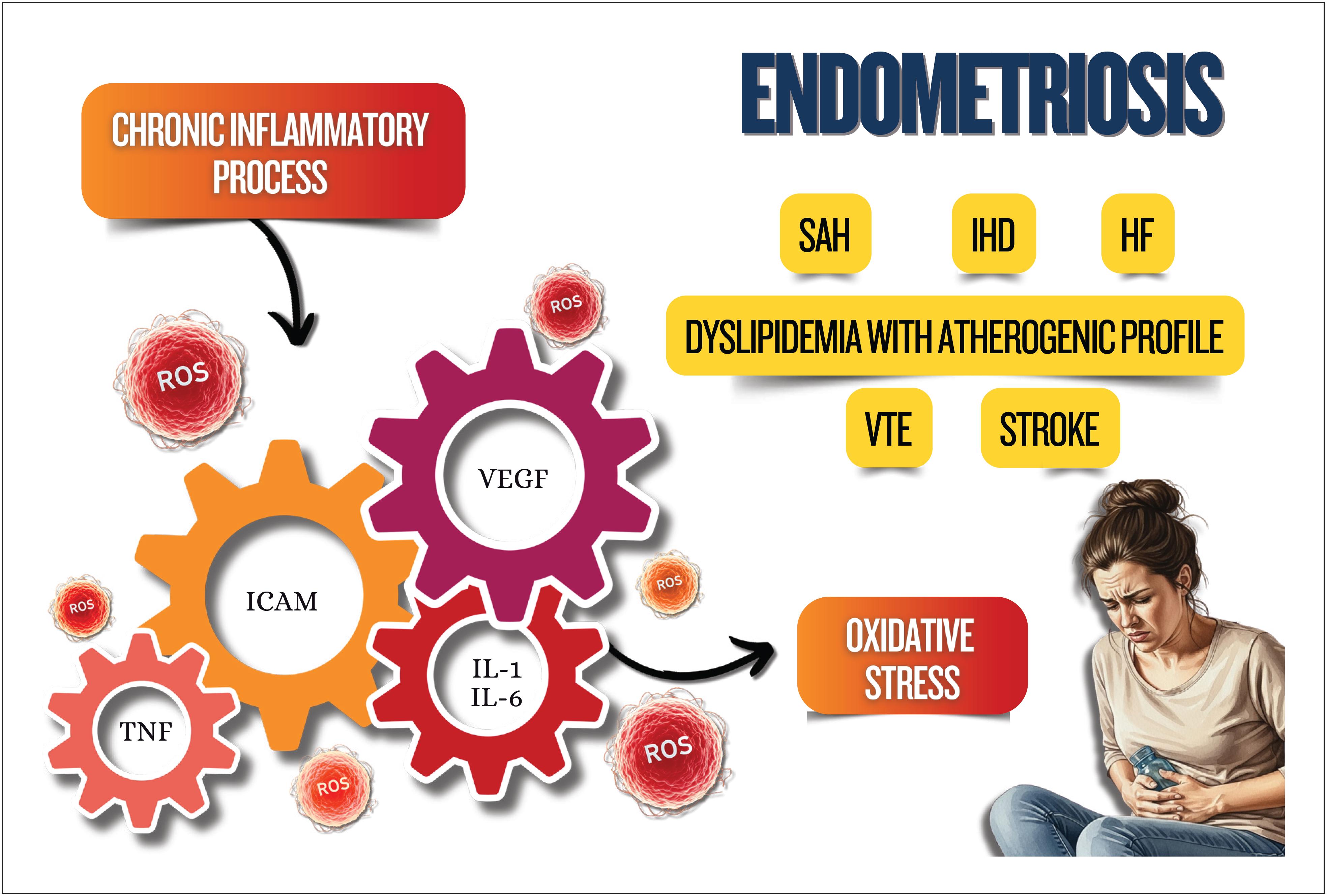
Endometriosis is associated with chronic inflammatory process with increased oxidative stress and elevation of cardiovascular risk factors, as well as an increased risk for venous thromboembolism, ischemic heart disease, heart failure, and stroke;
-
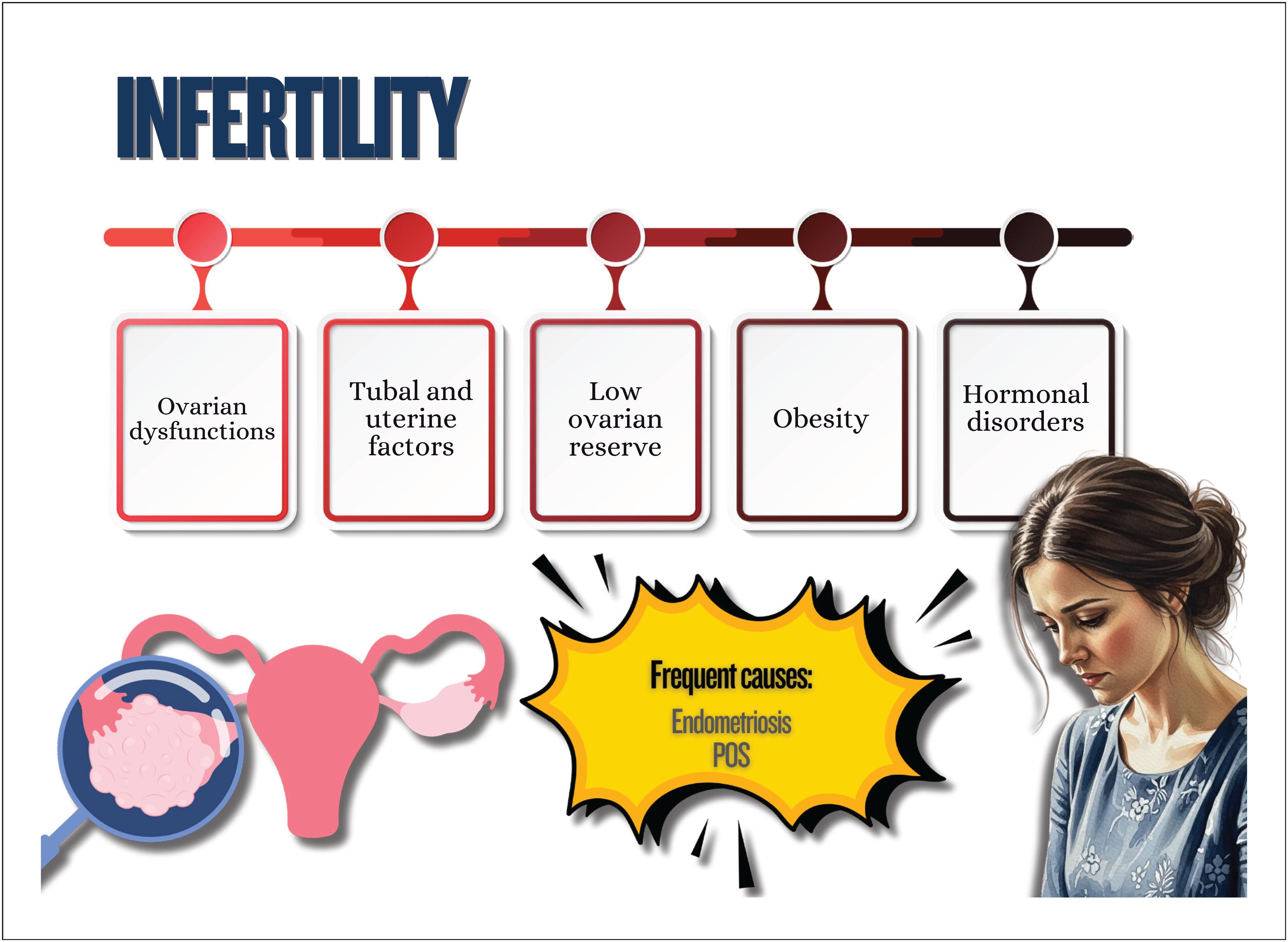
Treatment for infertility can have effects on cardiometabolism, such as ovarian hyperstimulation syndrome, with an increased risk for thromboembolic events, preeclampsia, gestational diabetes, and hypertensive disorders of pregnancy, increasing cardiovascular events in the long run;
-
Psoriasis is a systemic chronic inflammatory disease associated with obesity, metabolic syndrome, cardio- and cerebrovascular diseases, cardiac arrhythmias, sleep apnea, and others;
-
Preeclampsia is considered a risk marker of cardiovascular diseases throughout life, which seems to increase after menopause, with worsening of the cardiometabolic profile;
-
Postpartum excessive weight retention is associated with a higher risk of dyslipidemia and insulin resistance;
-
Women with history of gestational diabetes are more likely to experience metabolic disorders in the postpartum period and throughout life, independently of other traditional cardiovascular risk factors.
Chapter 8
-
The decrease in endogenous estradiol levels during menopause transition is associated with an increased risk of cardiometabolic disorders, such as abdominal adiposity, dyslipidemia, type 2 diabetes mellitus , and systemic arterial hypertension, which are related to renin-angiotensin-aldosterone system dysfunction, sympathetic activation, endothelial dysfunction, inflammation, and higher sodium sensitivity;
-
Women with premature ovarian failure and those with early menopause are at a higher risk of non-fatal cardiovascular events before the age of 60 years;
-
Menopausal hormone therapy is used to relieve menopause symptoms and not indicated for primary or secondary prevention of cardiometabolic disorders;
-
Testosterone replacement therapy is not indicated to improve cardiometabolic or musculoskeletal health, vasomotor symptoms, or mood changes;
-
Hormone implants for menopause, mainly of testosterone, are not recommended because their cardiometabolic effects and risks for breast and endometrial cancers are not known.
Chapter 9
-
Weight gain during pregnancy and its maintenance in the postpartum and polycystic ovary syndrome are risk factors for obesity and for the increase in insulin resistance and in other metabolic syndrome components;
-
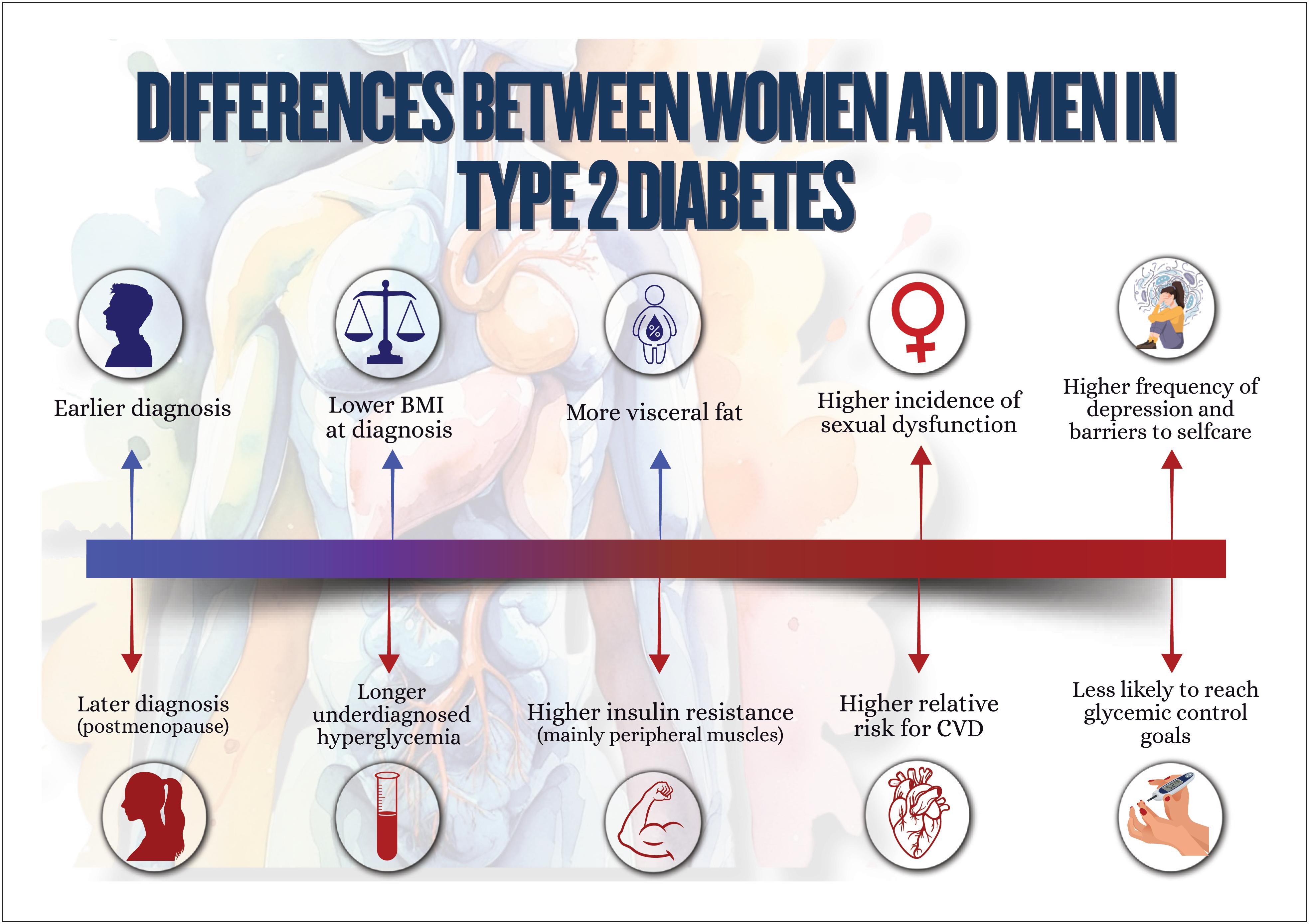
Women with type 2 diabetes mellitus are at higher risk for cardiovascular complications as compared to men;
-
Menopause, history of early menarche, and polycystic ovary syndrome are associated with an increase in women's susceptibility to metabolic dysfunction-associated steatotic liver disease, whose mortality rate due to cirrhosis is higher than that of men. It is the major cause of liver transplantation in women without hepatocellular carcinoma;
-
Regarding chronic kidney disease, women more often require dialysis and show faster glomerular filtration rate loss as compared to men, especially the elderly and postmenopausal ones.
Chapter 10
-
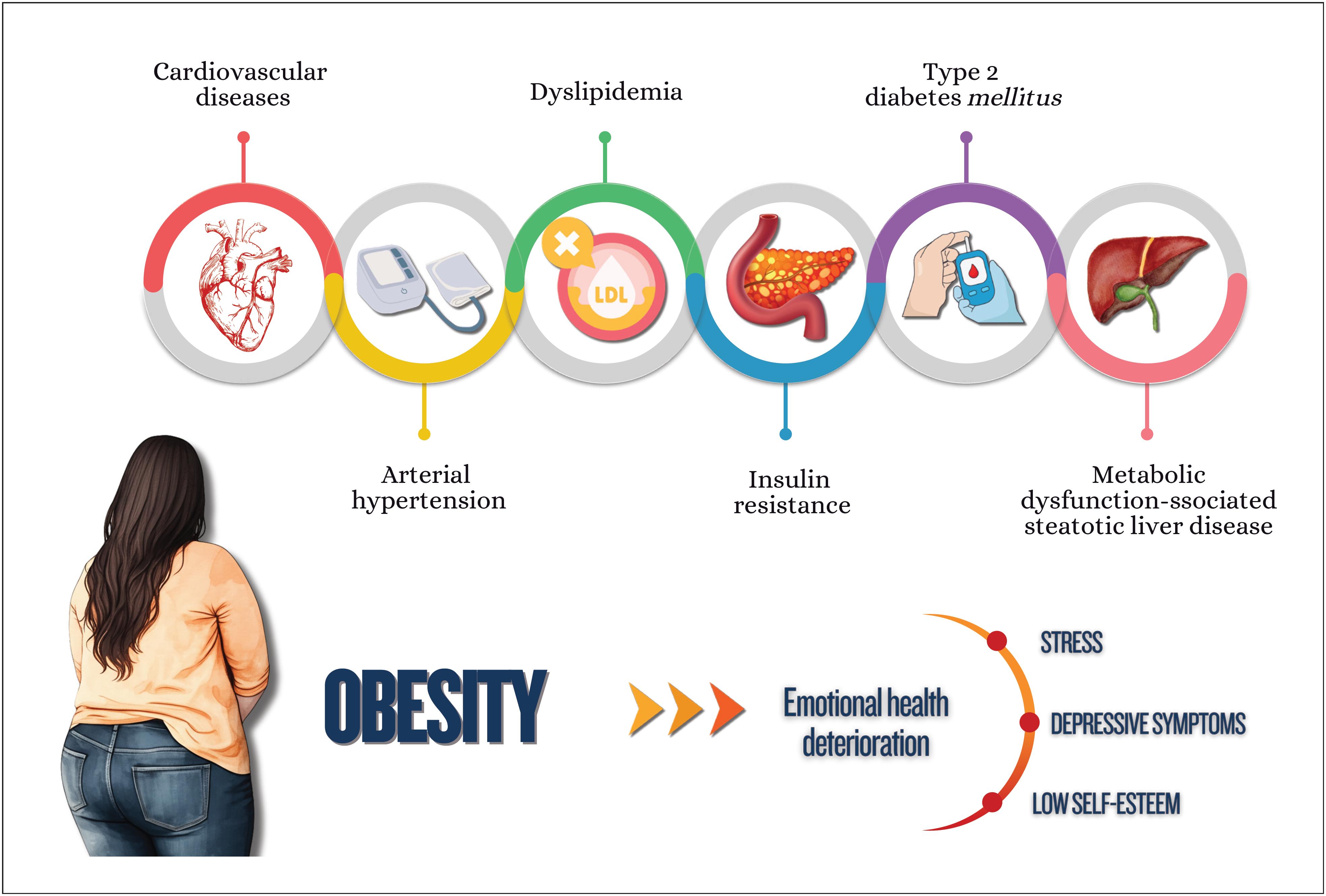
The increasing prevalence of cardiometabolic disorders in women represents one of the major challenges in public health because of the strong relation between obesity and cardiovascular diseases;
-
Nutritional education is crucial for preventing cardiometabolic disorders, and the association of nutritional plans with regular physical activity practice enhances beneficial effects, favoring cardiovascular function and decreasing morbidity and mortality among women;
-
Women who participate in integrated psychological support programs significantly improve their lifestyle and clinical outcomes, such as weight reduction, as well as glycemic and lipid profile control;
-
Tobacco use is a major chronic inflammatory factor, which is particularly compounded by menopause;
-
Multidisciplinary approach has shown to be effective in generating sustained lifestyle changes, reducing morbidity and mortality from cardiometabolic disorders;
-
Hypertension is the most prevalent risk factor for cardiovascular disease in all phases of a woman's life and its occurrence increases progressively with age. The choice and conduction of pharmacological treatment strategies should consider women's reproductive cycle phases, including peri- and postmenopause. The use of angiotensin-converting-enzyme inhibitors and angiotensin receptor blockers in women of reproductive age requires caution because of teratogenic risk;
-
Menopause is associated with significant elevations in total cholesterol, LDL-c, apolipoprotein B, triglycerides, and lipoprotein a, in addition to a possible reduction in the antiatherogenic protective effect of HDL-c. Although HDL-c levels above 50 mg/dL are desired in women, LDL-c reduction remains the therapeutic priority;
-
Women have a different progression of prediabetes to type 2 diabetes mellitus , frequently associated with higher obesity indices and increased risk of metabolic complications. Women can have higher risk of hypoglycemia with sulfonylureas and different response to glitazones, related to kidney function and body composition. Regarding treatment with GLP-1 analogues, women on oral contraception require especial attention;
-
There are several pharmacological treatments for obesity with different effects on body weight, but only more recent treatments are associated with a reduction in cardiovascular and metabolic outcomes;
-
The treatment of metabolic dysfunction-associated steatotic liver disease consists in lifestyle changes with focus on reducing at least 5% of body weight, because weight loss is the most effective measure to improve that disease's histological findings;
-
Pregnancy is one of the major causes of acute kidney injury in women of reproductive age and, along with preeclampsia, can lead to chronic kidney disease. Chronic kidney disease has a negative effect on pregnancy, even at the very beginning, and the risks increase with the progression of pregnancy and the concomitant presence of type 2 diabetes mellitus ;
-
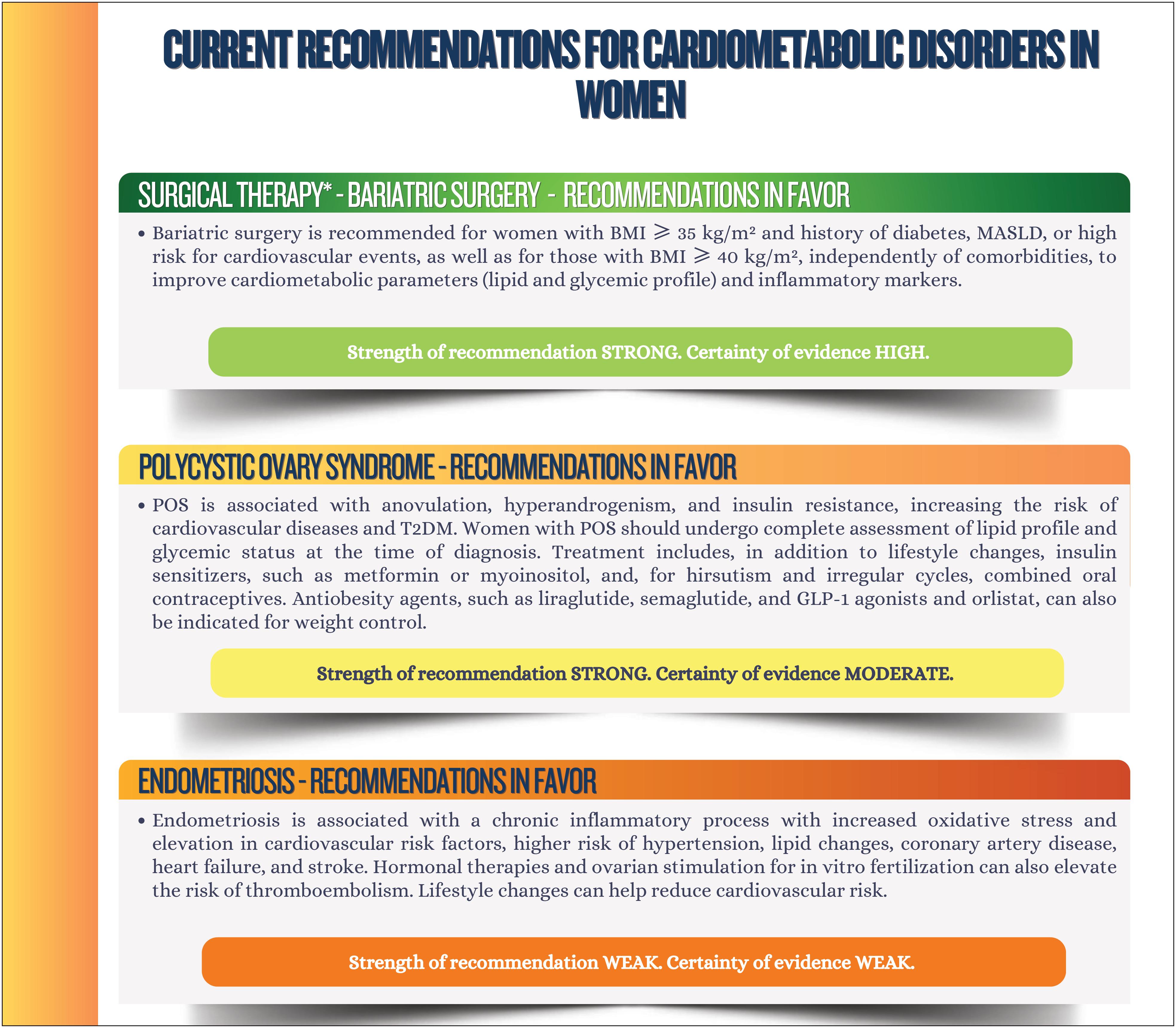
To improve cardiometabolic parameters (lipid and glycemic profiles) and inflammatory markers, bariatric surgery is recommended for women with body mass index ≥ 35 kg/m² and history of diabetes, metabolic dysfunction-associated steatotic liver disease, or at high risk for cardiovascular events, as well as for those with body mass index ≥ 40 kg/m², independently of comorbidities.
Chapter 11

Chapter 11

Chapter 11

3. Inflammation and Implications for the Cardiovascular System
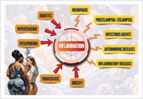
Inflammation is a process of defense activated by infectious agents, autoimmune and inflammatory diseases, influenced by traditional RFs, such as DM, hypertension, dyslipidemia, obesity, and tobacco use. In association with these metabolic cardiorenal factors, there are physiological changes of ageing, called immunosenescence or immune-aging.
In women, menopause and pregnancy, in particular preeclampsia/eclampsia, activate inflammation and accelerate coronary atherosclerosis, promoting vascular stiffness, endothelial dysfunction, and microvascular ischemia. Some relevant examples of the role played by inflammation in women's CVDs are as follows: heart failure with preserved ejection fraction (HFpEF) with its inflammatory phenotype; and ischemia/myocardial infarction with no obstructive coronary arteries (INOCA/MINOCA).
3.1. Inflammation and Atherosclerosis
Cardiovascular diseases, particularly IHD, are the major cause of death in women, mainly between the ages of 45 years and 75 years.9 , 10 Although sex-related differences in CVR have been well established, they are not completely understood.11Thus, better knowledge of the mechanisms that contribute to worsen women's risk profiles is mandatory to reduce morbidity and mortality from those diseases.
Several factors contribute to differences between sexes regarding the diagnosis and treatment of CVDs. It is a consensus that women are under-represented in large clinical trials, as evidenced in the VIGO Study, which has assessed 2349 women and 1152 men with acute myocardial infarction (AMI) under the age of 55 years, in the presence of three or more RFs. That study showed that women are less likely to receive guidance on cardiovascular prevention.12 , 13 This scenario can jeopardize the ability to accurately assess the efficacy and safety of different therapies in women, thus hindering specific strategies to prevent and treat CVDs.13
In addition to traditional RFs, several emerging conditions should be considered in female CVR, such as depression, domestic violence, socioeconomic and cultural profiles, obstetric and gynecologic history, gestational hypertension, gestational diabetes (GD), preterm delivery, premature menopause, POS, breast cancer, etc.10 , 14
In IHD, those factors reflect significant differences between sexes regarding pathophysiology, clinical presentation, and outcomes. INOCA is more frequent in women, specifically between the ages of 45 years and 65 years,15and so as MINOCA.16Although epicardial coronary vasospasm is more common among men, up to 70% of the cases of coronary microcirculation dysfunction occur in women.17Spontaneous coronary dissection, although rare, is a cause of acute coronary syndrome in women under the age of 50 years.18
The atherosclerotic process has been known for decades and was initially associated with inflammation by Rudolph Virchow, who showed that inflammation is a core element in the formation and progression of atherosclerotic plaque in coronary syndromes, for both chronic obstruction and plaque rupture.19
Atherosclerosis begins early with the accumulation of modified lipoproteins on vascular endothelium and worsens with age. In women, in addition to the metabolic effects of menopause, ageing contributes to endothelial dysfunction, mainly by increasing reactive oxygen species (ROS) production associated with senescence. Cytokines and inflammatory cells have pro-atherogenic effects by disrupting the endothelial barrier, reducing vasodilation, inducing the expression of adhesion molecules and chemokines, and facilitating the recruitment of leukocytes to atherosclerotic lesions.19 - 21
Biological variations between women and men result from differences in gene expression of sex chromosomes, modulated by hormonal and environmental influences, that result in cardiovascular conditions associated with autonomic regulation, as well as vascular and cardiac remodeling.18
In addition, epigenetic factors play a relevant role, especially in women, whose expression is widely modulated by menopause. These changes can influence gene transcription and translation via mechanisms, such as DNA methylation, histone modification, and regulation of non-coding RNAs. A related phenomenon, clonal hematopoiesis of indeterminate potential (CHIP), refers to the presence of somatic mutations in blood cells with no progression to hematological malignancies.22 , 23 These mutations increase with age, are detected in approximately 10% of individuals older than 70 years, and are associated with the concept of inflamm-aging, a term proposed by Franceschi et al . to explain the inflammatory process increase by immunosenescence.22 , 23
Thus, biological differences and underlying female-sex-specific pathophysiology of CVD have not been completely elucidated, and further research is required for the development of more effective preventive and therapeutic strategies.
3.2. Inflammation and Heart Failure in Women
Local or systemic inflammatory process participates in HF pathogenesis, with cardiac remodeling and fibrosis, in addition to macro- and microcirculation abnormalities. Low-intensity chronic inflammation is associated with different RFs, such as obesity, insulin resistance (IR), T2DM, dyslipidemia, chronic kidney disease (CKD), and ageing, and leads to a reduction in nitric oxide (NO) production, changing the ventricular diastolic physiology, promoting left atrial myopathy.24 , 25 The increased prevalence of those RFs in women, associated with the effects of menopause, seem to explain the higher prevalence of HFpEF. The association of chronic inflammation and adiposity in HFpEF results from evidence pointing to the role of the release of substances produced by the visceral adipose tissue (adipokines) particularly located around the pericardium and epicardium, leading to microcirculation inflammation, cardiac fibrosis, and physiological diastolic filling changes.26 , 27
In women, menopause induces cardiac function changes, resulting from the decrease in estrogen production and consequent reduction in NO production. This hormonal deficiency causes changes in the coronary circulation diastolic function and physiology. Clinically, HFpEF can show different phenotypes, two of which are common in women: the cardiometabolic phenotype, in which obesity and DM are associated with diastolic dysfunction; and atrial fibrillation and myopathy, present in elderly women.28 , 29 In addition, conditions such as obesity, DM, and hypertensive disorders of pregnancy increase the risk of HFpEF in women, probably because of associated inflammatory mechanisms. Despite the higher prevalence of those conditions in women, they are not included in most clinical trials, which leads to poor knowledge about treatment efficacy in the female sex.30
Biological markers of inflammation, such as ultrasensitive C-reactive protein (us-CRP) over 2mg/L, have been proven useful to characterize individuals with HFpEF of cardiometabolic phenotype, usually associated with worse prognosis.31
Preeclampsia has been recognized as a condition that leads to changes in immune and systemic inflammation activation, with consequent endothelial and cardiac dysfunction, and it can manifest over the following decades and increase the risk for HF.32 , 33
In women, autoimmune diseases, mainly systemic lupus erythematosus, can cause inflammation of the myocardium, pericardium, and valvular endocardium, as well as of the coronary macro- and microcirculation. Thus, pathophysiological abnormalities and clinical aspects of the disease must be understood so that preventive measures can be established and cardiovascular complications be early detected.34
The important role of HF has been evidenced in women. Studies, such as the PURSUIT-HFpEF and the analysis from TOPCAT study, have shown that women with HFpEF have more significant diastolic dysfunction and worse clinical outcomes as compared to men. Understanding the sex-specific mechanisms is crucial to advance the management of HFpEF.30
The post-hoc analysis from TOPCAT study has shown significant differences in the baseline characteristics between women and men with HFpEF.35Women (55.5% of the cohort) had fewer cardiovascular comorbidities (previous infarction: 18.7% vs. 30.9% in men, p < 0.001), worse cardiac function (NYHA class III/IV: 38.5% vs. 31.5%, p = 0.003), and higher ejection fraction (63.3% vs. 58.9%, p = 0.001). Women were older, had higher prevalence of obesity, atrial fibrillation, and SAH, while men more often had coronary artery disease and history of tobacco use. Despite these differences, both groups had similar HF symptoms and comparable use of cardioprotective drugs.35The results showed that women had lower cardiovascular mortality (8.9% vs. 14.8%, p < 0.001) and fewer hospitalizations due to HF (13.4% vs. 18.2%, p = 0.008). After adjustment, the female sex was a protective factor against cardiovascular mortality (HR: 0.53; 95%CI: 0.40–0.73). However, women had worse health-related quality of life and higher functional limitation. Regarding treatment with spironolactone, there was a reduction in all-cause mortality in women (HR: 0.68; 95%CI: 0.48–0.96), but not in men ( P for the interaction = 0.190), suggesting possible sex-specific benefit.
The PURSUIT-HFpEF study, a multicenter prospective registry from East Asia, has investigated sex differences in diastolic dysfunction and clinical outcomes in patients with HFpEF. Women represented 55.2% of the cohort (481 out of 871 patients).36The study showed that women had a higher prevalence of diastolic dysfunction (52.8% vs. 32.0% in men, p < 0.001), independently of comorbidities, such as SAH and T2DM. Anemia and obesity were factors associated with diastolic dysfunction only in women. Despite the worse diastolic function, women had a similar rate of combined events (death/hospitalization due to HF) in a non-adjusted analysis. However, after multivariable adjustment, female sex was independently associated with a higher risk of clinical events (HR: 1.54; 95%CI: 1.14–2.07), mainly for hospitalizations due to HF.30 , 36
Both studies emphasize that, from the metabolic viewpoint, HFpEF is a different condition in women, with:
-
Higher diastolic dysfunction associated with factors, such as obesity and anemia; thus, screening is important.
-
Better global survival despite worse symptomatology, possibly because of fewer ischemic comorbidities.
-
Different response to therapies, such as spironolactone, which can be more effective in women.
3.3. Hormonal Aspects in Inflammation
Women's cardiovascular health is influenced by a complex interaction of hormonal and inflammatory factors that manifest in a particular way in different phases of life, such as puberty, pregnancy, and menopause. Hormonal fluctuations, especially estrogen and progesterone levels, play an important role in maintaining vascular integrity and cardiac function. Therefore, understanding how such changes relate to the increase in CVD risk is crucial.
In postmenopausal women, hypoestrogenism promotes important metabolic changes, such as increase in central adiposity, worsening of glucose metabolism, and elevation in the levels of total cholesterol, LDL-c, triglycerides, apolipoproteins, and lipoprotein a (Lp(a)).37 , 38 In addition, endothelial dysfunction occurs, with consequent increase in IHD risk.
Estrogen acts by interacting with different receptors, such as ERα, Erβ, and GPER, activating genomic and non-genomic signaling pathways.39This results in inflammatory response modulation, reducing the production of pro-inflammatory cytokines, such as interleukin 6 (IL-6),40and increasing the expression of anti-inflammatory cytokines.41A key mechanism involves inhibition of nuclear factor kappa B (NF-κB) activity, reducing the transcription of inflammatory genes.41In addition, estrogen increases the production of NO in endothelial cells, contributing to vasodilation, and inhibits the expression of adhesion molecules, reducing leukocyte infiltration in the vascular wall.42
Endothelial function is benefited by estrogen that induces the expression of the enzyme endothelial nitric oxide synthase (eNOS), increasing NO production and promoting vasodilation. In addition, estrogen stimulates the regeneration of endothelial cells and inhibits the proliferation of vascular smooth muscle cells, preventing atherosclerosis progression.42
Chronic inflammatory processes represent another crucial underlying mechanism. Women with irregular cycles frequently have a pro-inflammatory status characterized by elevation in markers, such as C-reactive protein (CRP), IL-6, and tumor necrosis factor alpha (TNF-α). This systemic inflammation acts as an intermediate between menstrual dysfunction and atherogenesis.18
3.4. Inflammation in Cardio-Oncology
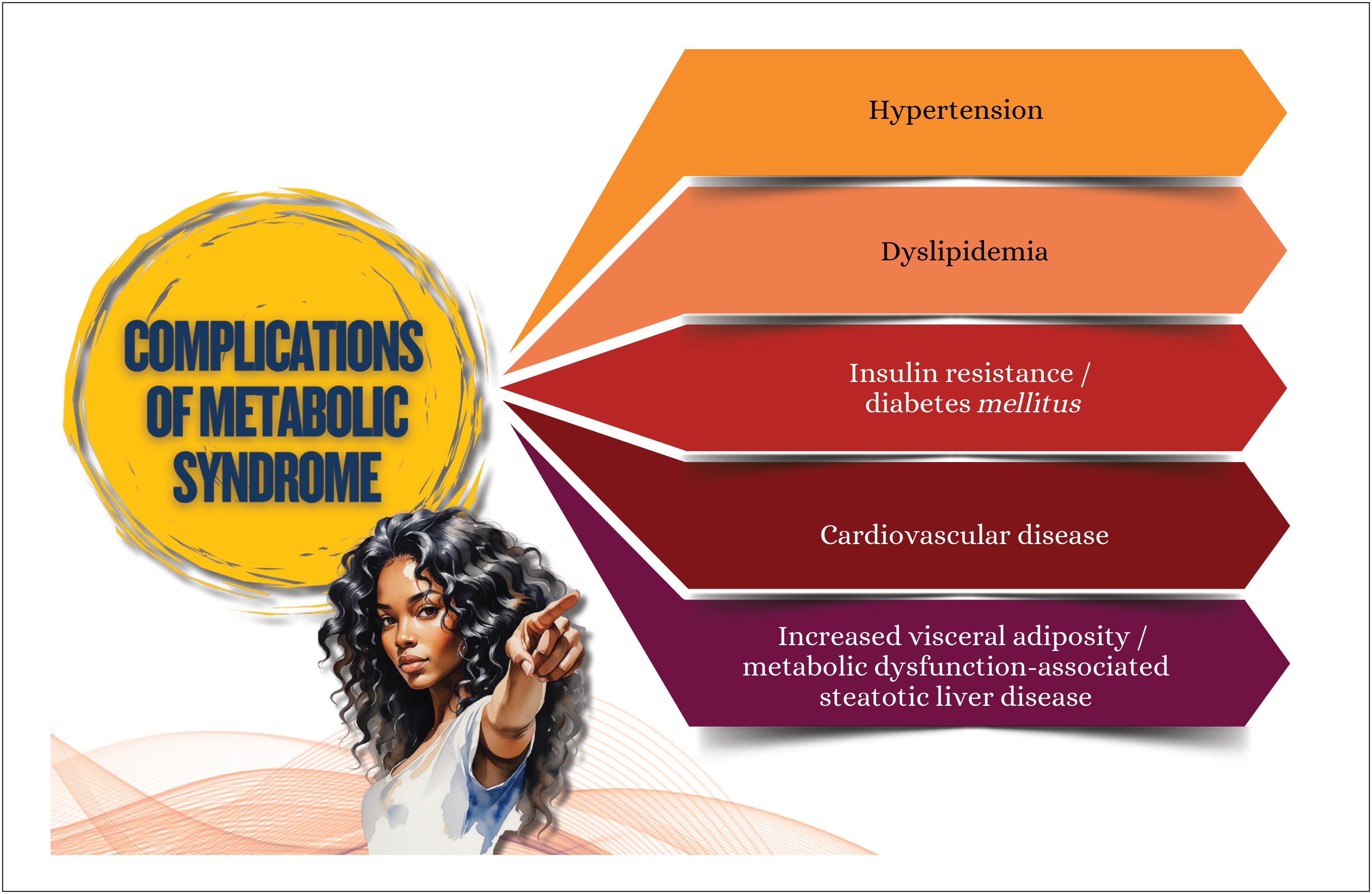
In addition to traditional RFs and MS ( Figure 3.1 ), women with cancer have an increased CVR because of the cardiotoxic effects of oncological therapies, such as those with anthracycline and trastuzumab. These therapies can lead to complications, such as HF, AMI, and stroke, even years after the end of oncological treatment.

Systemic chronic inflammation plays a significant role in the intersection of cancer and CVD. The tumoral inflammatory microenvironment and the side effects from oncological treatment contribute synergically to endothelial dysfunction, vascular remodeling, and atherosclerosis progression, especially in women. For example, thoracic radiotherapy has been associated with coronary inflammatory changes detected by biomarkers and the perivascular fat attenuation index (FAI), reflecting persistent vascular inflammatory activity. In addition, the prolonged use of aromatase inhibitors in breast cancer survivors has been associated with endothelial function worsening and higher risk of cardiovascular events.
In women undergoing oncological treatment, a multidisciplinary approach is essential, including baseline assessment of cardiac risk prior to the beginning of cancer therapy, which should be optimized to minimize cardiotoxicity. Interventions in lifestyle and maintenance of cardiovascular surveillance in the long run are also required. In addition, it is important to address racial and ethnic health disparities. Black and Hispanic women have a higher risk of cardiotoxicity and worse cardiovascular outcomes as compared to White women because of socioeconomic factors and inequalities in access to health care.43 , 44
3.5 Inflammation in Pregnancy and Preeclampsia
Preeclampsia is a multisystemic disease of pregnancy, whose pathophysiology has not been completely understood. Reduced placental perfusion, resulting from deficient trophoblastic invasion of maternal uterine wall, associated with the secretion of inflammatory cytokines and angiogenic factors, is believed to play a role in the disease. These factors contribute to endothelial dysfunction, vascular inflammation, and poor maternal perfusion. Endothelial dysfunction has been indicated as a major phenomenon responsible for preeclampsia and gestational hypertension.
Placental ischemia is related to the increased production of circulating antiangiogenic factors, such as soluble Fms-like tyrosine kinase-1 (sFlt-1) and soluble endoglin (sEng), that cause generalized endothelial dysfunction, NO pathway impairment, oxidative stress, excessive inflammation, imbalance in angiogenic factors, and loss of endogenous protective regulators.45Pregnant women with RFs associated with chronic inflammation (autoimmune diseases, obesity, pregestational hypertension, DM, and dyslipidemia) are more likely to develop preeclampsia. Inflammatory mediators might have local autocrine or paracrine effects, in addition to amplifying the effects of antiangiogenic factors.46
Pregnancy, even in normal conditions, is a state of oxidative stress because of increased maternal metabolism and placental activity. However, in preeclampsia, compensatory mechanisms fail, leading to the increased production of pathogenic factors and subsequent vascular dysfunction.47Observational and experimental studies have shown the association between inflammation and endothelial dysfunction. An important characteristic of systemic inflammation in preeclampsia is the predominance of Th1-type immunity and no Th2 biased immune response. However, normal pregnancy is characterized by a change to Th2-type immunity. In addition, the circulating levels of pro-inflammatory cytokines, such as IL-6, TNF-α, and chemokines IL-8, IP-10 (interferon-gamma-induced protein 10) and MCP-1 (monocyte chemoattractant protein 1) are elevated in preeclampsia.45 , 46
McCarthy et al . have investigated the role of mitochondrial dysfunction as a facilitator of oxidative stress, inflammation, apoptosis, and metabolic changes — all of which are crucial pathogenic intermediates in preeclampsia.47
Obesity increases the risk of preeclampsia. White adipose tissue secrets pro-inflammatory mediators that contribute to the chronic inflammatory state and metabolic complications of obesity. In addition, visceral adiposity is associated with metabolic RFs and complications, such as GD and preeclampsia.48The sFlt-1, the soluble form of the vascular endothelial growth factor (VEGF) receptor, is known to be secreted by adipocytes in non-pregnant women. Huda et al .48have shown that dysregulation of inflammatory pathways occurs predominantly in the visceral adipose tissue, with activation of macrophages and increased TNF-α and IL-6 expressions in that tissue, but not in the subcutaneous fat, emphasizing that, in preeclampsia, dysregulation of inflammatory pathways occurs predominantly in the visceral adipose tissue.
Metanalysis conducted by Guan et al .49has assessed the relation between pro- and anti-inflammatory biomarkers and their dynamic changes throughout preeclampsia progression. Women with preeclampsia had significantly higher levels of CRP, IL-4, IL-6, IL-8, IL-10, and TNF-α. The levels of pro-inflammatory cytokines were higher than those of the anti-inflammatory ones. Women with gestational age over 34 weeks had elevated levels of IL-6 and TNF-α. Higher systolic BP was associated with higher levels of IL-8, IL-10, and CRP. Such findings suggest that inflammatory imbalance is an independent RF for preeclampsia, and failure in anti-inflammatory autoregulation leads to disease progression.
Finally, recent studies have shown that neuroinflammation with participation of the autonomic nervous system can be present in the induction and evolution of inflammatory reactions associated with preeclampsia. In addition, the reduced autonomic regulation in patients with preeclampsia can be involved in the late fetal neurological maturation, with cognitive deficits and mental disorders. These findings show a link between maternal inflammation in preeclampsia and its impacts on fetal health and neurodevelopment.50
The factors associated with inflammation in women are summarized in Figure 3.2.
4. Implications of Sex Steroids for Cardiometabolic Health
4.1. Origin and Metabolism of Sex Steroids
The major sex steroids acting in the female body are estrogens, progesterone, and testosterone. These hormones are produced in the ovaries, adrenal cortex, and through peripheral conversion. Sex steroids have in common a cyclopentanophenanthrene ring derived from cholesterol. They are divided into three major groups according to the number of their carbon atoms as follows: the 21-carbon series includes corticosteroids and progestagens, being based on pregnane nucleus; the 19-carbon series includes all androgens, being based on androstane nucleus; and estrogens are 18-carbon steroids based on estrane nucleus.51
From cholesterol, which is obtained directly from blood stream, steroidogenesis originates natural sex steroids.51In mitochondria, an enzymatic hydroxylation system and a desmolase continue the process, forming the first steroid, pregnenolone. From that phase, steroidogenesis can progress via delta 5 pathway, which predominates in the follicle, to the synthesis of estrogens, or via delta 4 pathway, which predominates in the corpus luteum, to the synthesis of progesterone. Pregnenolone and progesterone are converted into androgens, androstenedione, and testosterone, in theca cells. Androstenedione, by the action of 17β-dehydrogenases, can be converted into testosterone and, through aromatization in granulosa cells, results in the formation of estrone and estradiol.52
Once synthesized, in the plasma those hormones are transported bound to SHBG (sexual-hormone-binding-globulin), CBG (cortisol-binding-globulin), or albumin, while a small fraction remains free. Then, sex steroids bind to hormone receptors present in several tissues, exerting their different biological effects.53These steroids are metabolized in the liver and excreted through the urine or bile.51
In addition, sex steroids can be extracted from plants in the pharmaceutical industry that manufactures products for clinical use. Their molecular structure is identical to that of hormones naturally produced by the female body (isomolecular), being, thus, called bioidentical hormones, whose effects are equal to those of endogenous hormones.
Synthetic sex steroids are produced in laboratories through a process involving several chemical steps and pharmaceutical engineering, originating molecules with different characteristics and biological potencies.51 - 53
4.2. Chemical Classification of Estrogens
4.2.1. Natural Estrogens
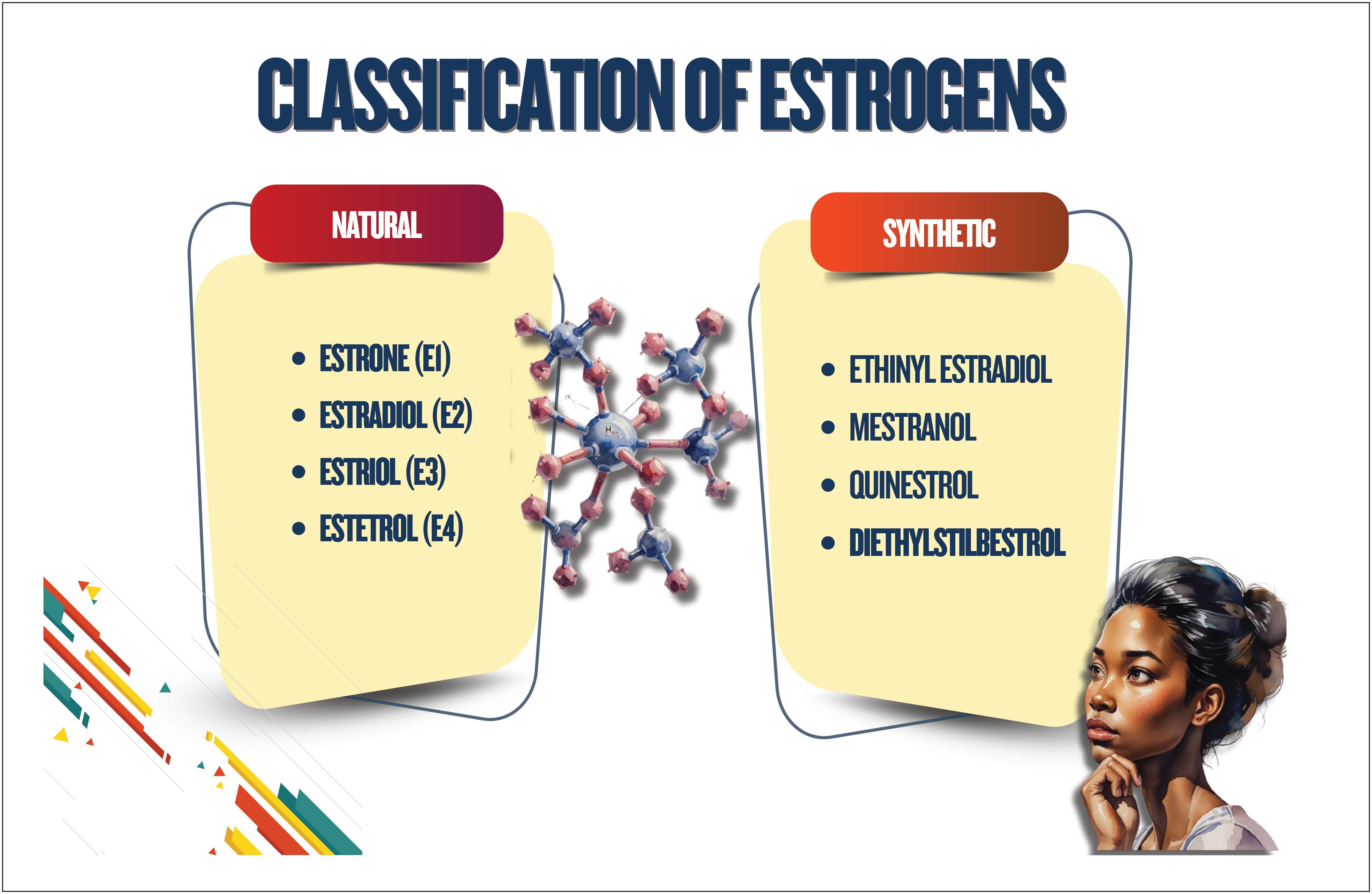
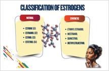
The natural estrogens present in bloodstream are estradiol, estrone, estriol, and estetrol. Estradiol is the major and most potent estrogen secreted by the human ovary, originating mostly from androstenedione. Estrone is secreted by the ovaries in significant daily amounts. Estriol is the peripheral metabolite of estrone and estradiol, rather than secreted by the ovary. Estriol is formed through general metabolic "detoxification", which means the conversion of biologically active material into less active forms. Estetrol is produced only during intrauterine life by the fetal liver.51 - 53
4.2.2. Synthetic Estrogens
Synthetic estrogens are classified as steroidal and non-steroidal, and their major examples are as follows:
-
Steroidal - ethinyl estradiol, mestranol, estradiol valerate
-
Non-steroidal - diethylstilbestrol, hexestrol, dienestrol Of those, estradiol valerate and ethinyl estradiol are currently the most clinically important.
Estradiol valerate is an estradiol ester, a prodrug that is rapidly metabolized to 17β-estradiol and valeric acid. The esterification of estradiol is aimed at improving its absorption and bioavailability after oral administration. After absorption, the esters are cleaved, releasing endogenous estradiol or 17β-estradiol. Thus, estradiol esters are considered bioidentical forms of estradiol.54 , 55 Their use in isolation or combination with progestagens is mainly related to menopausal hormone therapy (MHT) and contraception.
Ethinyl estradiol, an agonist of estrogen receptors, is more resistant to metabolism as compared to estradiol and has better bioavailability when used orally. These differences favor ethinyl estradiol use in combined contraceptive pills, although they also increase the risk of thromboembolism and other rare adverse effects.55( Figure 4.1 ).
4.3. Chemical Classification of Progestagens
4.3.1. Natural Progesterone
Progesterone, originally secreted by the ovaries, acts on the hormone-dependent target organs, especially endometrium and breast tissue. Its effects, however, go beyond, because they act on immunity, central nervous system, cardiovascular system, etc. From the physiological viewpoint, progesterone acts on tissues previously impregnated with estradiol. In addition, progesterone has a stereoisomer, dydrogesterone, available for oral use.56
4.3.2. Progestagens
Progestagens are a large group of synthetic molecules that differ regarding their chemical structure and the molecule they are created from, which bestow them a different effect profile based on affinity with and potency in different steroidal receptors. There are three large groups: testosterone derivatives (norethindrone, levonorgestrel, norgestimate, desogestrel, etonogestrel, gestodene, dienogest), progesterone derivatives (medroxyprogesterone, cyproterone, megestrol), and spironolactone derivatives (drospirenone). ( Figure 4.2 ).

Progestagens are characterized by high affinity with progesterone receptor; however, according to their origin, they can have affinity with other receptors, leading to effects, such as androgenic/antiandrogenic, antiestrogenic/estrogenic, antimineralocorticoid, and glucocorticoid activity. This pharmacodynamic profile of progestagens should guide their selection based on their expected benefits, safety profile, and unwanted effects.51 , 52 , 57 ( Figure 4.3 ).

Biological effects of endogenous progesterone and other progestagens. Adapted from Kuhl et al.55MPA: medroxyprogesterone acetate; NETA: norethisterone acetate; LNG: levonorgestrel.
Progestagens have different progestational potencies, determined by their capacity to change the animal endometrium. Considering progesterone to have progestational potency of 1, progestagens are as follows in decreasing progestational potency order: desogestrel, levonorgestrel, nomegestrol, medroxyprogesterone, norethisterone, and drospirenone.58
From the pharmacokinetic viewpoint, progesterone and progestagens differ regarding their pharmaceutical forms and administration routes. For example, micronized progesterone can be administered via oral and vaginal routes. Vaginal administration offers good absorption, with lower fluctuations in plasma concentration than the oral route.52
In addition, progestagens differ in their binding to plasma proteins. Micronized progesterone binds to albumin and CBG, but not to SHBG. Norethindrone, desogestrel and norgestrel bind to albumin and SHBG, while medroxyprogesterone acetate binds mainly to albumin.59
Differences in binding to plasma proteins in addition to clearance and association or not with estrogens determine differences in the plasma half-life of progestagens. Most progestagens are administered daily.
4.4. Common Indications of Therapies with Estrogens, Progestagens, and Testosterone in Women
4.4.1. Polycystic Ovary Syndrome
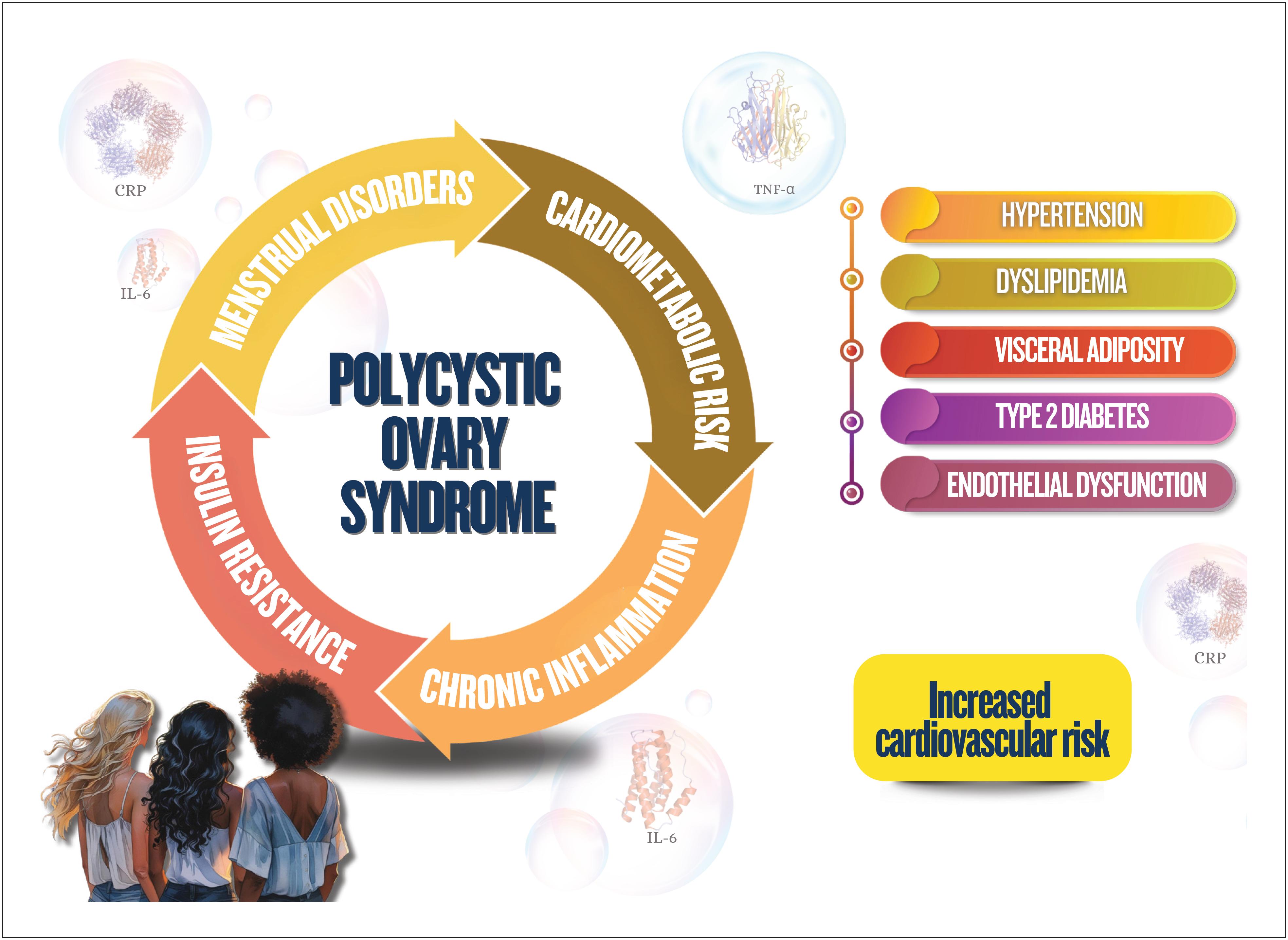
Polycystic ovary syndrome is a frequent indication of therapy with steroid hormones. It is a gynecological disorder associated with frequent endocrine disorders in women of reproductive age. In addition, metabolic disorders are present, increasing the CVR.60
Women with com POS can develop IR and dyslipidemia, which contribute to the development of CVD, such as atherosclerosis, SAH, and AMI.61These metabolic changes are boosted by the excess of androgens and weight gain, which can promote visceral fat accumulation and chronic inflammation. These factors combined create a metabolic environment that favors CVD development even in young women.62 - 64
The management of CVR in POS requires multidisciplinary approaches, including lifestyle changes, such as balanced diet and physical exercises, in addition to pharmacological interventions when necessary. Combined oral contraceptives (COCs) are often used in POS treatment to control menstrual changes and hirsutism, being effective to reduce the circulating levels of androgens, regulate the cycles, and protect the endometrium. However, that treatment does not improve IR and can even worsen it, depending on the type of progestagen used.60Insulin-sensitizing drugs, such as metformin or alternatives, such as pioglitazone65and myoinositol,61 , 66 reduce IR, improving ovarian function. Statins can be indicated to control dyslipidemia, and anti-hypertensive drugs are essential for women with SAH. Treatments for obesity, such as glucagon-like peptide 1 (GLP-1) analogues (liraglutide and semaglutide), can be used.67Early identification of those metabolic changes and their proper treatment are crucial to reduce CVR in women with POS.
4.4.2. Hormonal contraception
Sex steroids are often used for hormonal contraception. The COCs have two types of hormones (estrogen + progestagen) in different formulations.68The estrogen component offers cyclic endometrial stability and potentiates the suppressive effect of progestagen on the hypothalamic-pituitary-ovarian axis. The most used estrogen component is ethinyl estradiol, at current doses between 15μg and 30μg. New COCs with natural estrogens – estradiol (17β-estradiol and estradiol valerate) and more recently estetrol – appeared as alternatives with potential for lower hepatic and cardiovascular impact.69
In different COCs, those estrogens are combined with a progestagen. The major contraceptive effect of the progestagenic component is suppression of the luteinizing hormone (LH) secretion and of ovulation. In current formulations, several progestagens are used. They are synthetic progestagens that inhibit ovulation/pregnancy, and the most modern ones have been developed to cause fewer secondary/adverse effects. Pregnancy rates in the first year of COC use vary, being 0.3% with consistent and correct use. With typical use, it can reach 9%, showing the method's efficacy during real use70and emphasizing the importance of contraceptive counseling. The COCs are not recommended for women aged over 35 years with history of tobacco use, CVD, venous thromboembolism (VTE), SAH, DM with vascular complication, migraine with neurological symptoms, severe liver disease, liver tumors, breast cancer, and systemic lupus erythematosus. The contraceptive method safety has been well established by the Medical Eligibility Criteria for Contraceptive Use71( Figure 4.4 ).

Contraindications to the use of combined oral contraceptives. SLE: systemic lupus erythematosus.
Progestagen-only hormonal contraceptives contain only synthetic progestagen, such as desogestrel, norethisterone or drospirenone. Long-acting progestagen-only contraceptives can also be used as implants in the subdermal region of the forearm, and, in Brazil, the only implant approved for contraception is Implanon NXT®(etonogestrel), which has high contraceptive efficacy for 3 years.72The levonorgestrel intrauterine system (LNG-IUS) is a hormonal IUS that releases 20 µg of levonorgestrel per day with local intrauterine action and high contraceptive efficacy for 8 years. In addition, there is the IUS releasing 8 µg of levonorgestrel for 3 years. Long-acting progestagen-only contraceptives have the advantage of high efficacy for prolonged time and pregnancy rates lower than 1% per year, without depending on either correct use or user's motivation.72
4.4.3. Menopausal Hormone Therapy: Regimens, Doses, and Routes, Focused on Cardiovascular Health
In women, hormonal fluctuations that begin in menopause transition, being followed by a gradual and irreversible reduction in estrogen production in menopause, although physiological, creates a "window of vulnerability" and marks the inflection point of cardiometabolic risk, with negative impact on vascular health, glucose and lipid metabolism, and body fat distribution.73 , 74 Thus, MHT, especially estrogen, can interfere with that risk, as long as judiciously and individually prescribed. It should always be associated with integral health measures, with strategies for lifestyle, diet, and physical exercise practice.75
The MHT is the most effective treatment to relieve vasomotor symptoms (VMS) and genitourinary symptoms, as well as to prevent bone loss in peri- and postmenopausal women.76It should be initiated when menopausal symptoms begin to interfere with daily life. However, for women with premature ovarian failure (menopause < 40 years) or early menopause (between 40 years and 45 years), MHT is recommended to start as early as possible to reduce health risks in the long run.77Cardiovascular effects of MHT are strongly influenced by the type, dose, and route of administration used, as well as onset time of menopause ("window of opportunity"), with higher benefits and lower rates of adverse effects when MHT begins within 10 years of menopause onset.77Determining factors of MHT type and dose are: patient's preference, presence/absence of uterus, need for contraception, severity of symptoms, and comorbidities.75
Therapeutic regimens include estrogen-only therapy (for hysterectomized women) and combined therapy of estrogen and progestagen (women with uterus).76In the combined regimen, continuous use is indicated for women with postmenopausal amenorrhea for more than 12 months, while the sequential regimen is more indicated in perimenopause. MHT should be prescribed at the lowest effective dose to control symptoms.78The most used estrogen in MHT is 17β-estradiol, whose administration routes can be oral, transdermal (patch, gel, spray), or vaginal.75For proper relief of VMS and bone protection, low doses of estradiol (approximately 1 mg of oral estradiol or equivalent in other routes) are usually necessary.75 , 76 In healthy women without cardiovascular risk factors (CVRFs), any route, including the oral route, can be prescribed for MHT.75However, differently from the oral route, the transdermal route avoids the ‘hepatic first-pass effect’ in estrogen metabolism, resulting in lower impact on coagulation factors, triglycerides, and CRP, which can reduce the risk for VTE and cardiovascular events. Thus, transdermal route is preferred for women with RFs, such as obesity, MS, tobacco use, hypertension, as well as risk for VTE.79
The use of "compounded bioidentical" hormones for MHT is not recommended because of concerns regarding quality, regulation, safety, efficacy, and lack of standardization of the products used.75 , 76
The only indication for testosterone supplementation in postmenopause is to treat hypoactive sexual desire disorder, after excluding other causes. Although data on the effects of androgenic therapy on cardiovascular health of postmenopausal women with hypoactive sexual desire disorder are limited, evidence indicates that, in physiological doses, transdermal testosterone does not significantly increase CVR.80
An individualized approach with the best evidence-based decision should be used. Maintaining MHT and not starting MHT in women ≥ 60 years seem associated with a better risk-benefit profile for cardiovascular events.75Transdermal route for estrogen at low doses combined with progestagens of safe profile represents a favorable option regarding CVR.79
4.5. Risks: Metabolic Effects, Thromboembolism, and Breast Cancer
4.5.1. Metabolic Effects
MHT reduces visceral fat, IR, and the LDL-c to HDL-c ratio, but slightly increases triglycerides when used orally.81 , 82
A review has also confirmed lower levels of glycemia, insulinemia, and IR among MHT users.83The Women’s Health Initiative (WHI) study has found a lower significant risk of DM among MHT users as compared to those using placebo.84
There is evidence that the progestagen type used in MHT can interfere with metabolic effects. Medroxyprogesterone is known to have more glucocorticoid effects than norethisterone or progesterone.85Studies have suggested that progesterone and dydrogesterone interfere less with the benefits provided by estrogens,86 , 87 while norethisterone, depending on the dose, can lead to loss of some of those benefits.88
4.5.2. Thromboembolism
Ethinyl estradiol, a synthetic estrogen widely used in combination with progestagen in COCs, is associated with increased risk of VTE.89This risk is influenced by factors, such as dose of estrogen and type of progestagen present in the formulation. COCs containing levonorgestrel (second-generation progestagen) are associated with a lower risk of VTE as compared to those containing desogestrel, gestodene, drospirenone, or cyproterone (third- and fourth-generation progestagens).
When COC contains natural estrogens instead of ethinyl estradiol, the risk is slightly increased.89In addition, ethinyl estradiol of COCs can exacerbate the production of hepatic angiotensinogen, which increases BP via renin-angiotensin-aldosterone system.
When administered orally, they can increase the risk of venous thrombosis, odds ratio 1.58 (1.52–1.64), while the transdermal route is not associated with increased risk, odds ratio 0.93 (0.87–1.01).90In addition, a recent metanalysis has concluded that MHT combining conjugated equine estrogens plus progestagens increased systolic BP and the risk of hypertension, while other formulations, such as oral or transdermal estradiol plus progestagen, estradiol only, and tibolone, did not have significant effects on BP, showing that such effects can be influenced by different administration routes and formulations.91
4.5.3. Breast Cancer
Breast cancer is a hormone-dependent neoplasm, and the proliferative effects of sex steroids in breast tissue are well known.92
MHT can associate with an increased risk for breast cancer. The WHI study has found that MHT with conjugated estrogens plus medroxyprogesterone acetate associated with eight more breast cancer cases per 10,000 women-year.93
However, effects differ depending on the progestagen used in the formulation. Observational studies have shown no increase in the risk when the association in MHT was with micronized progesterone94or dydrogesterone.95
Thus, different MHT formulations and doses also seem to have different effects on breast cancer risk.
5. Indicators of Cardiometabolic Health
5.1. Assessment of Reproductive Risk Factors with Implications for Cardiometabolic Health
This chapter addresses the relevant RFs of women's cardiometabolic profile in puberty, pregestational, gestational, and postgestational periods, as well as menopause transition, which will be detailed in chapters 7 and 8.
5.1.1 Puberty and Pregestational Period
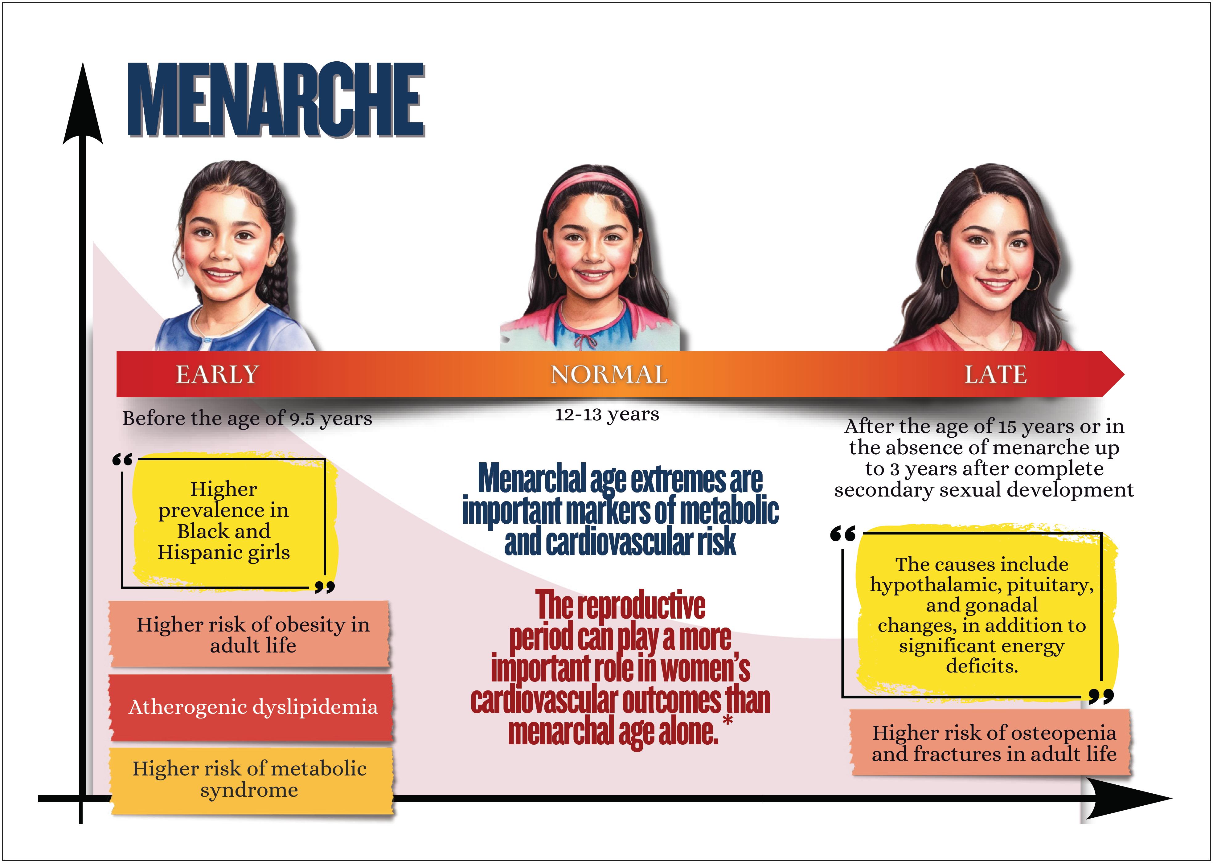
Menstrual cycle assessment has been suggested to be used as an additional vital sign in the investigation of women's general health.96In this scenario, age at menarche (early or late), menstrual cycle irregularities of endocrine origin, and POS have been associated with a higher risk of future CVD, especially atherosclerotic disease.5 , 97
5.1.1.1. Age at Menarche
The CARDIA ( Coronary Artery Risk Development in Young Adults ) study, which enrolled 2788 women, aged 18-30 years, initiating in 1985-1986, and followed them up for 35 years (ages between 50 years and 65 years), has observed that early menarche associated later with adverse levels of glucose and lipids, and each earlier year of menarche, in relation to the mean age of 12 years, associated with greater body mass index (BMI) and visceral adiposity.98
Other studies have shown that early menarche is associated with elevation in glucose, insulin, BP, and body fat, as well as with higher risk of future CVD.5
A cohort study of 648 women stratified according to age at menarche (≤10, 11, 12, 13, 14, ≥15 years) and using the mean age of 12 years as reference, has reported that early or late menarche associated with a higher risk of future cardiovascular events, represented by all-cause death, non-fatal AMI, non-fatal stroke, or hospitalization due to HF.99
In a cohort of 1.2 million women, with mean age of 56±5 years, no previous heart disease, and followed up for 12 years, those with early (≤ 10 years) and late (≥ 17 years) menarche had a higher risk of IHD, stroke, and SAH.100
However, a metanalysis of 12 cohort studies carried out up to 2018, with 2,341,769 participants and 79,363 deaths, has shown that, for each 1-year increase in the age at menarche, there was a reduction in the relative risk for all-cause mortality, cardiovascular mortality, IHD mortality, and stroke mortality,101evidencing the need for further studies on the impact of age at menarche on future CVD.
5.1.1.2. Menstrual Cycle Characteristics
A systematic review and metanalysis of observational studies performed up to 2022 has analyzed the association of oligomenorrhea and menstrual irregularity with CVR, observing the association of these changes with CVD, IHD, and AMI, but not with stroke, as documented in women with POS5 , 97 , 98 , 102 (see chapter 8).
5.1.2. Pregnancy and Postpartum Period
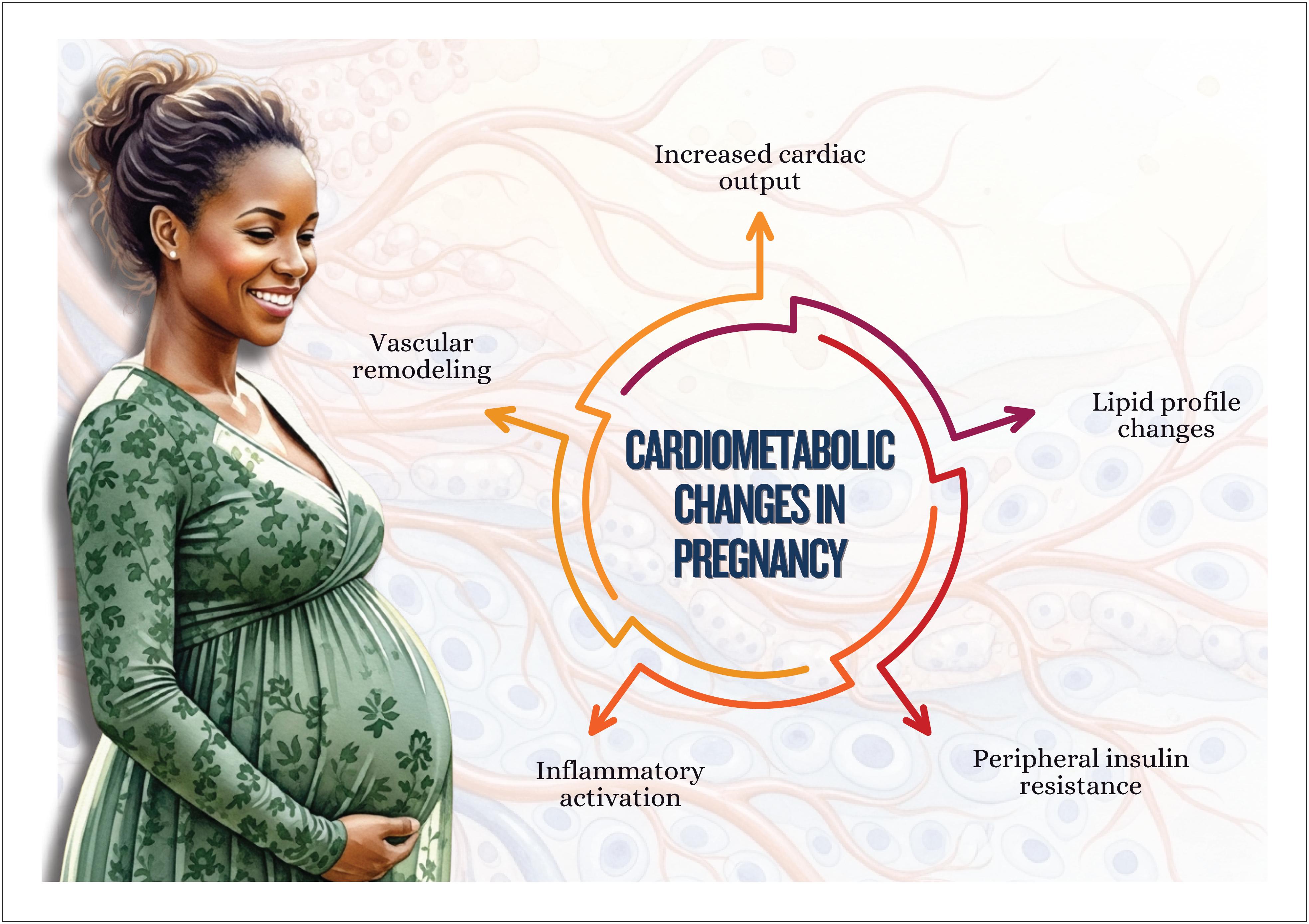
Pregnancy involves several physiological, hormonal, and metabolic transformations, fundamental to ensure proper fetal development and adaptation of the maternal body to new demands. Abnormal weight gain and changes in lipid and glycemic profiles, however, can be associated with adverse outcomes in pregnancy and postpartum for mother and infant.
5.1.2.1. Weight changes in pregnancy
There is evidence that both low and excessive gestational weight gain (GWG) are associated with negative fetal and neonatal outcomes.103
Pregestational obesity favors the risk for gestational hypertension and GD, cesarean birth, and high birth weight, being recognized as a significant RF for spontaneous abortion, preterm delivery, metabolic disorders that complicate pregnancy, and higher rates of abnormal deliveries, stillbirths, and neonatal death. On the other hand, malnutrition can contribute to lower birth weight, placental abnormalities and complications, higher rates of surgical births, and higher fetal and neonatal mortality.103 , 104
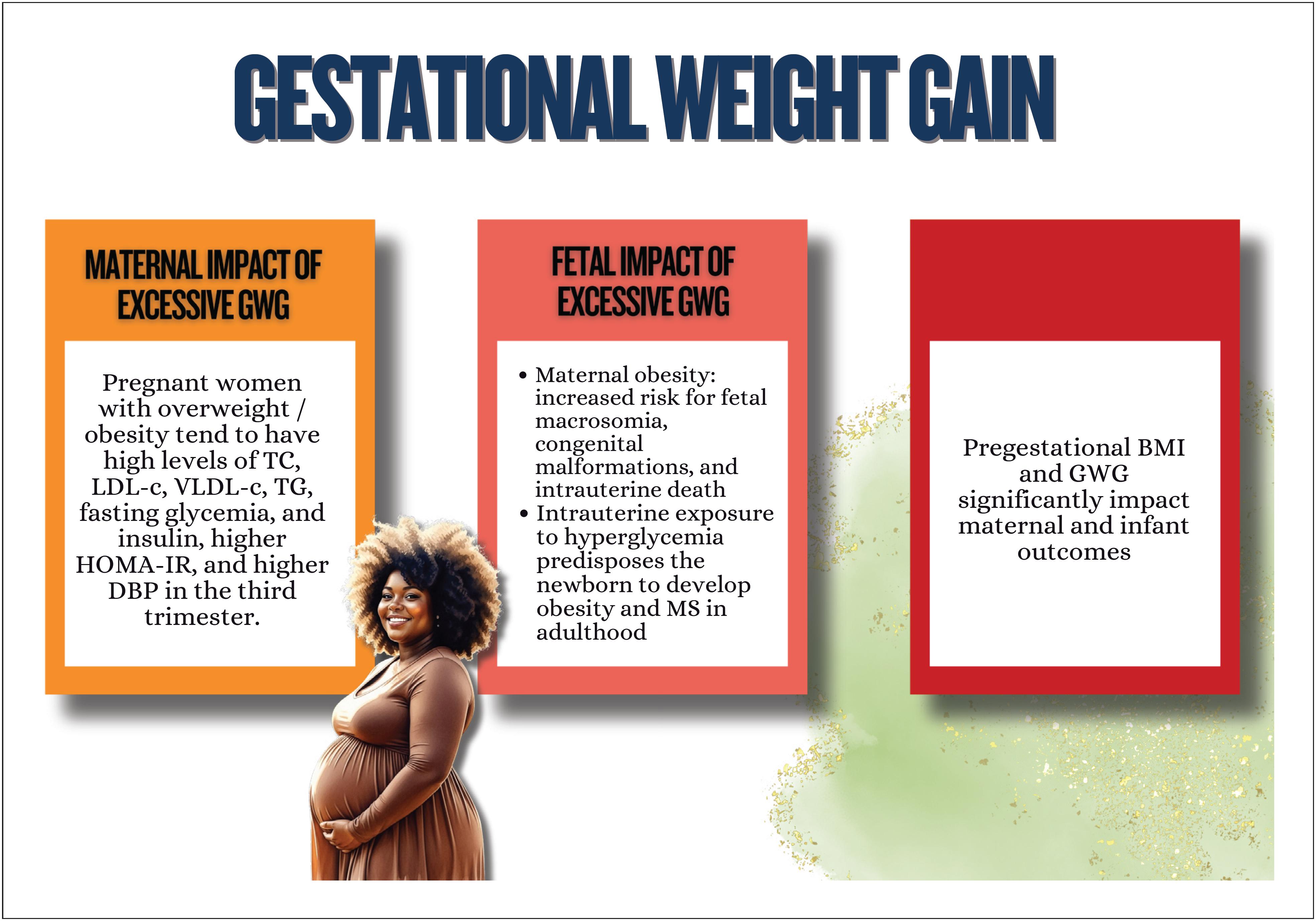
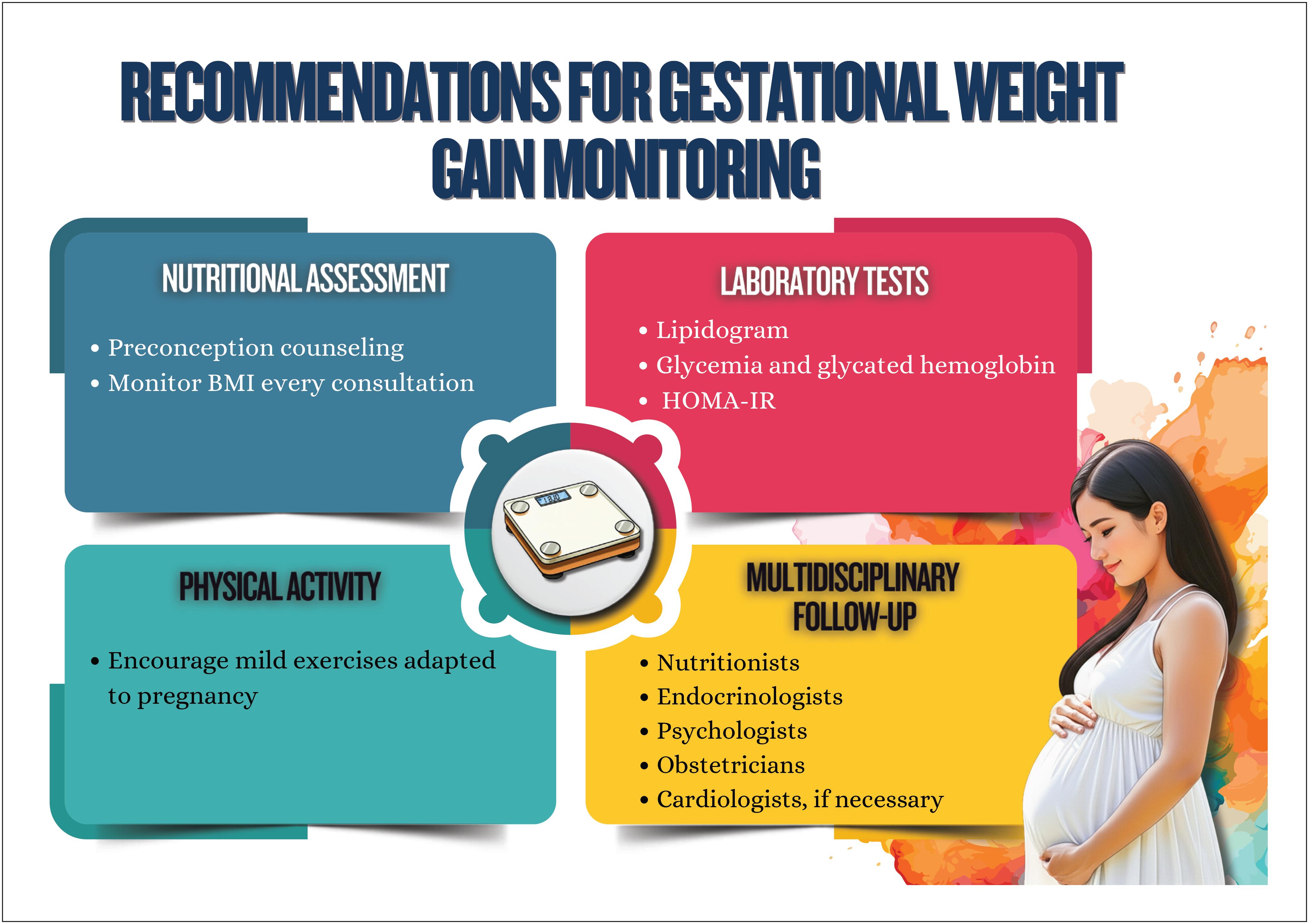
Guidelines recommend GWG values, but analyses of more recent studies have suggested that this gain should be personalized considering three classes of obesity.103( Table 5.1 and Figure 5.1 ) (details in chapter 7).
Weight gain in pregnancy according to the recommendation of the 2009 Institute of Medicine guidelines
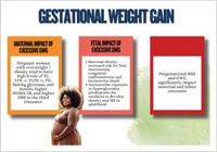
Implications of weight gain in pregnancy for cardiometabolic health. BMI: body mass index; DBP: diastolic blood pressure; GWG: gestational weight gain; HOMA-IR: homeostasis model assessment of insulin resistance; LDL-c: low-density lipoprotein cholesterol; MS: metabolic syndrome; TC: total cholesterol; TG: triglycerides; VLDL-c: very-low-density lipoprotein cholesterol.
5.1.2.2. Gestational dysglycemia
Pregnancy is a condition of IR per se, potentially aggravated by increased pregestational IR in women with obesity. During pregnancy, IR is increased by 40-50%, which increases the risk for GD.105
Pregnant women with glucose intolerance are at risk for adverse gestational outcomes, even in the absence of GD.
The primary underlying pathophysiology leading to hyperglycemia is related to the presence of IR, insulin deficiency, or mixed pathophysiology. Studies have shown that insulin-resistant glucose intolerance in pregnancy is a subtype with high risk for adverse results in pregnancy, such as high birth weight, admission to neonatal intensive care unit, gestational arterial hypertension, and cesarean delivery.106
Glucose intolerance in pregnancy increases the risk for future diabetes. Thus, recognizing glucose intolerance in pregnancy enables better DM screening and prevention (more details in chapter 7).
5.1.2.3. Gestational Dyslipidemia
Recent evidence shows that the increase in cholesterol, triglycerides, and metabolites associated with cardiometabolic dysfunction seems to have significant maternal and fetal vascular consequences.107
Historically, gestational dyslipidemia has been considered physiological and, thus, without clinical relevance. This physiological increase plays an essential role during pregnancy. However, high lipid levels in women who are predisposed to or have familial forms of hyperlipidemia can increase the risk for maternal-fetal complications. Hyperlipidemia during pregnancy is associated with preeclampsia, preterm delivery, and GD, and infants are more likely to develop vascular fatty streaks and experience increased risk for progressive atherosclerosis.108
5.1.2.4. Postpartum Behavior
In the postpartum period, weight loss is usually incomplete and full recovery of metabolic functions is not always obtained. Postgestational weight retention is an RF for SAH, dyslipidemia, and MS. In addition, studies have shown that women with preeclampsia have a worse cardiometabolic profile up to 1 year after delivery.
Although lactation is a factor of metabolic protection because it stimulates energy expenditure and better glycemic and lipid control, it depends on adequate emotional, social, and support factors. Lack of specialized guidance for postpartum lactation can favor the persistence of obesity, IR, and dyslipidemia, in addition to increasing the risk for MS and SAH.104
5.1.3. Menopause Transition
The lack of estradiol in menopause causes several metabolic, hormonal, inflammatory, and endothelial function changes, whose association favors the rapid progression of the atherosclerotic process in all arterial territories. Thus, from the eighth decade on, women have higher CVR than men do.109 , 110
Adequate management (primary prevention, early detection and proper treatment) of RFs that contribute to atherosclerosis development throughout life is crucial to delay CVD in peri- and postmenopausal women, as well as to primary and secondary prevention of events that lead to cardiovascular mortality.111This guidance on cardiovascular prevention should occur in all life stages prior to menopause, becoming more important from menopause on.109 - 111
5.1.3.1. Age at menopause
For the large majority of women (90%), natural menopause occurs around the age of 51 years (age range: from 45 years to 55 years).110 - 112 For 5% of women, menopause can occur spontaneously between the ages of 40 years and 45 years (early menopause), and, for 1%, before the age of 40 years, characterizing premature ovarian failure.110 , 112 Women submitted to bilateral oophorectomy and chemotherapy or radiotherapy can progress to menopause as a consequence of these treatments, independently of their age at treatment time.109 , 110 , 112
The CVR is even higher in women who experience menopause early (naturally or secondary to some treatment), in those with moderate to severe VMS, and in those on MHT.109 , 110 In early menopause, each year of earlier menopause onset increases by 3% the risk for CVD (IHD, stroke, and cardiovascular mortality).110 , 112
This higher risk seems to associate with a higher propension of this group to have a more atherogenic lipid profile, higher IR, SAH, higher risk of MS, as well as worse endothelial function and inflammatory markers, as compared to women who experience natural menopause.110 , 112
5.1.3.2. Vasomotor Symptoms
Menopausal VMS, characterized by hot flushes and night sweats, can be present in up to 80% of women and last 7-9 years. However, when they appear earlier, they can last longer.109 , 110 The intensity of VMS varies from mild to moderate and severe, and their diagnosis is associated with impairment of quality of life and increased use of health services.113 - 115
Several studies have shown that women with severe VMS, as compared to women with mild or no VMS, have a worse cardiometabolic profile, greater sympathetic hyperactivity, worse endothelial function, and higher incidence of subclinical atherosclerosis.113 , 115
5.1.3.3. Menopausal Hormone Therapy
Several clinical trials have shown that MHT with estrogens increases CVR (IHD, stroke, thromboembolism) in postmenopausal women, and that risk seems to be attenuated when MHT is used in younger women (50-59 years), who are at the beginning of menopause or within 10 years from its diagnosis. In addition, that risk is attenuated with the use of low doses of hormones and routes of administration other than the oral one, notably the transdermal route.109 , 110
Current guidelines establish that MHT has scientific evidence-based indication only to treat VMS in women without contraindication and should not be used in those with established CVD or high CVR.78 , 109 , 110
5.1.4. Postmenopausal Metabolic Health
Menopause is associated with hormonal changes that result in an imbalance between estrogen and testosterone. The ovaries, despite estrogen production cessation, produce testosterone at a lower rate, leading to its relative excess. Testosterone is associated with increased visceral fat and BP levels in postmenopausal women.116
The hormonal fluctuations and physiological changes related to ageing determine an impaired metabolic status, characterized by IR, increased total body fat, sarcopenia, and accumulation of abdominal fat.117
5.1.4.1. Menopausal metabolic changes
Insulin resistance is defined as an inadequate response to insulin in tissues (adipose, skeletal muscle), central nervous system, and liver, being one of the major factors leading to hyperglycemia and T2DM, along with impaired insulin secretion.
Epidemiological evidence has suggested significant protection against IR in premenopausal women, who are more sensitive to insulin as compared to men. This metabolic advantage, however, disappears gradually after menopause or when IR progresses to hyperglycemia and DM. Thus, menopause is associated with a higher risk of glucose intolerance, and an increase in BP and triglyceride levels, as well as in the risk for MS, with consequent acceleration of CVR.117 , 118
5.1.4.2. Diabetes mellitus
The changes described make women more likely to develop T2DM. Evidence suggests that women with menopausal VMS have higher risk of developing T2DM as compared to those who do not have such symptoms. In a data analysis from the WHI, the risk of developing T2DM was 18% higher in women with VMS, increasing in parallel with the severity of those symptoms.119
Women who develop DM before the age of 20 years tend to experience early menopause, while menopause is delayed in those with T2DM of late onset.119
A publication of the SWAN Study with a 20-year follow-up has concluded that women with multiple physical and psychological symptoms, as well as moderate to severe menopausal symptoms, had earlier onset of DM and MS.119
The MHT was associated with a reduction in the risk of developing T2DM in women without previous T2DM, and, in those with T2DM, MHT leads to better glycemic control.117
5.1.4.3. Dyslipidemia
Estrogen has a protective role in the cardiovascular system, and its ovarian production uses LDL-c as a substrate. In menopause, circulating LDL-c cannot be used to synthesize estrogen, resulting in the reduction of estrogen production and a mild increase in LDL-c serum levels. In addition, the size and density of LDL-c particles change in menopause. Studies have shown that the levels of small and dense LDL-c particles increase from 10-13% in premenopausal women to 30-49% after menopause.37
Menopausal estrogen deficiency impairs the hepatic and intestinal functions related to lipid metabolism and transportation, which can increase plasma lipid levels and accelerate atherosclerosis progression.120
The SWAN study has suggested that the antiatherogenic function of high-density lipoprotein cholesterol (HDL-c), which is its ability to promote cholesterol reverse transport, can decrease during menopause, when less elevated HDL-c levels are associated with atherosclerosis.109
5.2. Anthropometric Indicators
Cardiometabolic health is a pillar of well-being, encompassing integrity of the cardiovascular system and metabolism. In women, this is influenced by hormonal changes, reproductive events, and biological factors that manifest over the course of a woman's life, from childhood to old age.110
Anthropometric indicators are body measures that provide information on body fat composition and distribution, reflecting cardiometabolic risk. Simple and accessible, these measures are fundamental to large-scale populational screening.110 , 121 Therefore, they should be performed routinely, with attention to changes over the course of life.
5.2.1. Body Mass Index
The BMI, calculated as weight (kg) / height² (m), classifies the nutritional status as follows: < 18.5 (low weight); 18.5-24.9 (normal); 25-29.9 (overweight); and ≥ 30 (obesity). An elevated BMI is associated with SAH, dyslipidemia, and DM. However, it has limitations, such as not differentiating slim mass from fat and not assessing adipose distribution, which reduce its accuracy in elderly or athletic women.122 , 123
5.2.2. Waist circumference – Brazilian population
Waist circumference measures visceral fat, a more robust predictor of cardiometabolic risk than BMI. Visceral fat is associated with IR and chronic inflammation, central factors in CVD pathogenesis.124 , 125 In women, waist circumference > 88 cm indicates high risk, according to the World Health Organization guidelines.
5.2.3. Waist-To-Hip Ratio
Waist-to-hip ratio (WHR) compares waist circumference to hip circumference, reflecting fat distribution. A WHR > 0.85 in women suggests central fat accumulation, associated with higher risk of MS. Studies have indicated that WHR is superior to BMI in predicting cardiovascular events, especially after menopause.124 , 126
5.2.4. Body Fat Percentage
Body fat percentage is an important parameter to assess cardiometabolic risk, especially because women tend to have a higher subcutaneous fat proportion as compared to men. Hormonal changes, such as those of puberty, pregnancy, and menopause, have a direct impact on body fat distribution and accumulation, and can increase the risk for IR, dyslipidemias, and chronic inflammations.126 , 127
5.2.5. Waist-To-Height Ratio
Waist-to-height ratio (WHtR) is a predictive measure of central adiposity and cardiometabolic risk. Values greater than 0.5 are strongly associated with higher risk for SAH, T2DM, and cardiovascular events. This marker is particularly relevant in menopause transition, when there is a trend towards abdominal fat accumulation.128 , 129
5.3. Biomarkers
5.3.1. Lipid Profile (Total Cholesterol, LDL-c, HDL, Triglycerides)
LDL-c promotes vascular inflammation and cholesterol build-up in the arterial intima layer, while dysfunctional HDL-c (common in women with IR or MS) loses its antioxidant ability and of reverse transport, favoring plaque accumulation.130High triglyceride levels (>150 mg/dL) associates with small and dense LDL-c particles, as well as with particles similar to remnants, which intensify plaque instability.131As previously cited in this chapter, menopause promotes metabolic profile changes due to estrogen decline, increasing LDL-c (∼10-15%) and reducing HDL-c, as well as its protective function, worsening dyslipidemia, especially in women with visceral obesity or DM.73Thus, nonpharmacological strategies, such as Mediterranean diet and aerobic exercises, improve HDL-c functionality and modulate triglycerides.
5.3.2. Glycemia and Glycated Hemoglobin
Chronic hyperglycemia and elevated glycated hemoglobin (HbA1c) in women increase the CVR via mechanisms, such as glycation of vascular proteins, endothelial dysfunction, and activation of inflammatory pathways, which accelerate atherosclerosis and plaque instability.132Insulin resistance, common in conditions such as POS and previous GD, induces atherogenic dyslipidemia (reduced HDL-c, elevated triglyceride) and oxidative stress, exacerbating vascular injury.133
In postmenopause, estrogen decline reduces insulin sensitivity and increases metabolic dysfunction, correlating with higher arterial stiffness and systemic inflammation.134Based on findings from the SUSTAIN and EMPA-REG OUTCOME studies,135 , 136 guidelines recommend for most women with T2DM: HbA1c <7%; and the use of drugs with proven cardiovascular benefit for secondary prevention, such as GLP-1 agonists (ex.: semaglutide) and sodium-glucose cotransporter type 2 (SGLT2) inhibitors (ex.: empagliflozin), which reduce cardiovascular events and hospitalizations due to HF. Assessment of subclinical atherosclerosis (ex.: coronary artery calcium score) and aggressive management of comorbidities (SAH, dyslipidemia) are essential. Nonpharmacological strategies, such as Mediterranean diet and aerobic exercises, modulate inflammation and improve insulin sensitivity.
5.3.3. C-reactive Protein
Elevation of us-CRP in women is associated with increased CVR due to chronic vascular inflammation, which promotes endothelial dysfunction, atherosclerotic plaque instability, and thrombotic activation.137Hormonal factors modulate that relation, such as menopause, in which estrogen decline increases us-CRP (∼15-25%), which correlates with higher arterial stiffness and exacerbated inflammatory response.73
Comorbidities, such as autoimmune diseases (ex.: systemic lupus erythematosus), as well as oral MHT can increase us-CRP.138The use of statins (ex.: rosuvastatin) for patients with elevated us-CRP reduced cardiovascular events independently of LDL-c levels, as evidenced in the JUPITER study.137Adjuvant strategies include diet and aerobic exercise that modulate inflammation, especially in postmenopausal women.139Thus, us-CRP interpretation should be added to hormonal context, comorbidities, and lifestyle, guiding the treatment to mitigate CVR.
5.3.4. Fibrinogen
Fibrinogen is a soluble plasma glycoprotein, synthesized in the liver and involved in platelet aggregation, endothelial injury, and plasma viscosity. It plays a central role in the formation of thrombi. In addition, it is a protein of the acute phase of inflammation, induced mainly by IL-6.140 , 141
Women have higher circulating levels of fibrinogen independently of age, as well as higher levels of functional fibrinogen, as determined by using thromboelastography estimates of fibrinogen contribution to clot resistance.141
Differences in fibrinogen function and circulating levels between women and men define a behavior called sex dimorphism. Such differences are associated with sex hormones, and estradiol is an important mechanistic mediator in coagulation induction.141 - 143
Epidemiological data have shown the important predictive role of fibrinogen in IHD,140and a study published in 2025 with 5690 participants showed a relation between high fibrinogen levels and all-cause mortality, suggesting that fibrinogen is a potential biomarker of mortality risk.143 , 144
5.3.5. Homocysteine
Elevated homocysteine levels accelerate the development of atherosclerotic plaque in arteries via mechanisms, such as increased oxidative stress, LDL-c oxidation, NO depletion, endothelial dysfunction, inflammatory processes, epigenetic changes, and microRNA regulation.145
Changes in diet and lifestyle, regular physical activity, tobacco use cessation, and alcohol consumption reduction are essential to control hyperhomocysteinemia.145
5.3.6. Adipokines
Adipokines are peptides secreted by adipocytes, of which leptin and adiponectin are the most frequently known and studied. Normal physiological levels of adipokines are essential to maintain adequate cardiovascular function.
Leptin promotes satiety and regulates energy expenditure. Hyperleptinemia is present in obesity and T2DM, reflecting a state of leptin resistance associated with atherogenic processes, endothelial dysfunction, low-grade chronic inflammation, and vascular dysfunction. There are clear sex-related differences in circulating leptin levels, which are three to four times higher in women as compared to men.146 , 147
Adiponectin has anti-inflammatory, antiatherogenic, and insulinotropic effects. Its action includes improvement in insulin sensitivity, oxidative stress reduction, and modulation of endothelial inflammatory response. Adiponectin serum levels have been inversely associated with CVD risk and considered a biomarker of cardiovascular protection. Women have significantly higher total adiponectin circulating levels as compared to men in healthy populations. Sex-related differences in adiponectin levels have not been well characterized, but differences in regional fat distribution (subcutaneous vs. visceral) between women and men can contribute to the sex-related differences.146 , 147
The use of biomarkers for clinical assessment is summarized in Figure 5.2.
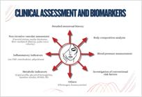
Use of biomarkers for clinical assessment. HOMA-IR: homeostasis model assessment of insulin resistance; us-CRP: ultrasensitive C-reactive protein.
6. Cardiometabolism in Childhood and Adolescence
The age of puberty onset has significant clinical and epidemiological implications, because variations in its timing are associated with several metabolic and cardiovascular outcomes. Menarche, defined as the first occurrence of menstruation, indicates the hypothalamic-pituitary-gonadal axis maturation, marking the onset of female reproductive ability. Studies have shown a secular trend towards a lower age of menarche onset, with a reduction of three months per decade in the past 50 years, a reflex of nutritional improvement and lifestyle changes.148Currently, the mean age of menarche onset is 12-13 years in developed countries, while, in low-income regions, that event occurs later.149Understanding the determinants of that variability and its metabolic impacts is crucial for cardiovascular health.150
6.1. Definitions and Diagnostic Criteria
When sex characteristics develop before the age of 8 years in girls, it is considered early puberty. Early menarche is defined as the first occurrence of menstruation before the age of 9.5 years. On average, menarche occurs two years after the development of breasts, and early menarche has higher prevalence in Black and Hispanic girls. However, the Brazilian consensus emphasizes the need for individual clinical assessment, considering each patient's context.151Late menarche is defined as the first occurrence of menstruation after the age of 15 years or the absence of menarche up to 3 years after complete secondary sexual development. Its causes include hypothalamic, pituitary, and gonadal changes, in addition to significant energy deficits.152
6.1.1 Pathophysiology
Puberty depends on the activation of the hypothalamic-pituitary-gonadal axis, regulated by the pulsatile release of gonadotrophin-releasing hormone (GnRH). This process stimulates the production of LH and follicle-stimulating hormone (FSH) by the pituitary, which stimulates the ovaries to secrete estrogens.
In early puberty, the activation occurs early, usually associated with high hypothalamic sensitivity to adipokines, particularly to leptin. This hormone, produced by the adipose tissue, signals to hypothalamus the existence of adequate energy reserves for reproduction. In obese girls, elevated leptin levels can exacerbate that trigger, accelerating puberty onset. In addition, the early exposure to endocrine disruptors, such as bisphenol A and phthalates, can interfere with the natural hormonal signals and precipitate the reproductive axis activation.
In late puberty, the reproductive axis activation is frequently delayed due to chronic energy deficits common in athletes or patients with eating disorders. Low energy storage reduces the secretion of leptin and other adipokines, impairing signaling to the hypothalamus. In some cases, there is resistance to the metabolic signals of insulin and leptin or changes in kisspeptin signaling, a hypothalamic neuropeptide essential for GnRH secretion.151
These mechanisms illustrate how nutritional status and body composition directly influence menarche timing and its metabolic repercussions. For example, obesity can induce metabolic dysfunctions that accelerate sexual maturation, while severe calorie deficits, observed in athletes or girls with eating disorders, such as anorexia nervosa, delay the process.
6.1.2. Early Menarche, Late Menarche, and Cardiometabolic Risk
Early menarche is strongly associated with adverse metabolic outcomes. One of the major impacts is the increase in BMI and central adiposity, with increased risk for adulthood obesity by approximately 30-60% as compared to women with normal age of menarche.150( Figure 6.1 ).
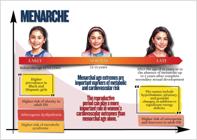
Association of cardiovascular risk with age of menarche. The extremes of menarchal age associate with a future risk of cardiometabolic disorder and cardiovascular disease and should be considered enhancers of cardiovascular risk. Source: Dastmalchi and Gulati.154
Other relevant consequence is IR, frequently identified in that profile and characterized by elevated levels of fasting insulin and early changes in glucose metabolism, often regardless of BMI.
In addition, unfavorable lipid profile changes, such as elevation of triglycerides and LDL-c, and HDL-c reduction, are common. These are findings of atherogenic dyslipidemia, which increases CVR.
In addition, there is a 1.5- to 2.5-fold increase in the risk for MS, associated with the combination of central obesity, SAH, IR, and dyslipidemia.100Even after adjusting for confounding factors, such as socioeconomic level, the metabolic impact of early menarche remains clinically important.
In late menarche, metabolic outcomes are more heterogeneous. Usually, patients have lower BMI and less adiposity, which reflects a more favorable metabolic profile. However, in cases related to energy deficits, such as eating disorders, malnutrition becomes a critical factor, influencing negatively metabolism and bone health152( Figure 6.1 ).
Insulin sensitivity is better in women with late menarche as compared to those with early menarche.
In late menarche, bone health is frequently impaired, with increased risk for osteopenia and fractures in adulthood. The reduced bone mineral density in those cases emphasizes the need for preventive interventions and continuous monitoring.100 , 152
Finally, the extremes of menarchal age correlate with a "U" shaped pattern of CVR. In early menarche, there is an increase in systolic and diastolic BP, carotid intimal thickening, and higher predisposition to subclinical atherosclerosis. In contrast, in late menarche, hormonal and nutritional disorders elevate the likelihood of late complications.153
6.2. Menstrual irregularities and cardiometabolic risk
Excluding structural causes, such as uterine myomatosis and polyps, the menstrual disorders related to hormonal causes are common manifestations during female reproductive life, affecting 14-25% of women at childbearing age, with peak incidence in reproductive life extremes, such as menarche and perimenopause.155These changes, classified as oligomenorrhea, polymenorrhea, amenorrhea, menorrhagia, and metrorrhagia, go beyond gynecological changes and emerge as important indicators of cardiometabolic vulnerability. Contemporary investigations have evidenced solid correlations between menstrual cycle anomalies and the development of CVD, emphasizing the need for an integrative approach for those patients’ assessment.156
6.2.1. Pathophysiological Mechanisms
The interrelation between irregular menstrual cycles and cardiometabolic risks involves multiple interconnected pathophysiological processes. The hypothalamic-pituitary-ovarian axis plays an essential role in that association, because hormonal imbalances impact reproductive and cardiovascular functions simultaneously. Oscillations in estrogen and progesterone levels directly modulate insulin sensitivity, lipid metabolism, endothelial function, and BP homeostasis.44 , 155
Insulin resistance is the central element in the pathophysiology of certain conditions, such as POS, promoting both menstrual irregularities and dyslipidemia, SAH, and visceral adiposity. Longitudinal follow-up studies have shown that women with IR have a substantially elevated risk for T2DM and cardiovascular events over the course of their life cycles.156
6.2.2. Clinical Assessment and Biomarkers
The assessment of women with menstrual irregularities requires a comprehensive approach of CVRFs. Detailed menstrual history is an essential component of risk stratification that needs to be complemented with body composition analysis, BP measurement, and investigation of conventional RFs.153 , 157 , 158
6.2.3. Therapeutic Interventions
The management of adolescents with menstrual irregularities at high cardiometabolic risk requires multidimensional strategies. Lifestyle changes are the basis of treatment, which includes regular physical activity, cardioprotective eating patterns, body weight control, and tobacco use cessation.157Pharmacological interventions require customization according to individual risk profile.
Despite significant advances, there are still important knowledge gaps of the relation between menstrual irregularities and cardiometabolic risks.
In clinical practice, the following measures are recommended: incorporation of detailed menstrual assessment into CVR stratification; early cardiometabolic screening in adolescents with persistent menstrual irregularities; adoption of a multidisciplinary approach in the management and early diagnosis of conditions, such as POS, frequently diagnosed at puberty onset;109 , 159 - 161 and development of educational programs focused on the interrelation of reproductive and cardiovascular health.44 , 162 , 163 These changes are detailed in chapter 7.
6.3. Obesity, Eating Disorders, and Cardiovascular Risk: Long-Term Impacts
Obesity in childhood and adolescence is associated with a series of adverse physical and psychological outcomes. From the physical viewpoint, there is a significant increase in the risk of developing chronic comorbidities, such as CVD, SAH, dyslipidemia, IR, T2DM, and metabolic dysfunction-associated steatotic liver disease (MASLD).164From the psychological viewpoint, obesity relates to deterioration of emotional health, including higher prevalence of stress, depressive symptoms, and low self-esteem.165
6.3.1. Eating Disorders and Obesity
The relation between obesity and eating disorders is known to be bidirectional. Children and adolescents with obesity are at higher risk of developing eating disorders, especially bulimia nervosa and periodic compulsive eating disorder. These disorders can aggravate the physical and psychological consequences already associated with obesity, establishing a pathological cycle of weight gain and eating-disordered behaviors.165
The American Academy of Pediatrics stresses the importance of early screening for eating-disordered behaviors and implementation of timely interventions. Such measures can promote significant improvement in clinical outcomes.164Dysfunctional eating behaviors identified during middle childhood correlate with the appearance of obesity and future cardiometabolic complications, justifying the need for early and longitudinal monitoring.165
6.3.2. Hereditary Factors and Prognosis of Eating Disorders
Several genetic and hereditary factors influence the risk and prognosis of eating disorders in childhood and adolescence.167The major ones include:
-
Genetic loci and heritability167
Studies have shown specific loci associated with anorexia nervosa (1p33–36 region) and bulimia nervosa (10p14), with estimated heritability between 48% and 74%.
-
Specific genes167
Mutations and genes, such as ESRRA, HDAC4 , AGRP, GHRL, and BDNF, have been associated with appetite regulation and increased risk of developing eating disorders.
-
Family history168
Family history of eating disorders or psychiatric disorders significantly increases individual risk, as evidenced in studies with monozygotic twins.
-
Comorbid psychiatric disorders167
There is genetic overlapping of eating disorders and other psychiatric conditions, such as obsessive-compulsive disorder and depression, worsening the clinical course.
-
Autoimmune and autoinflammatory diseases
There is evidence that the presence of autoimmune diseases in the family history can contribute for the risk of developing eating disorders, suggesting possible immune system involvement.
6.3.3. Future Consequences of Eating Disorders
Eating disorders that begin in childhood have long-lasting repercussions. Longitudinal studies have shown that inadequate early eating patterns are associated with disorders diagnosed in adolescence.169For example, childhood hyperphagia is related to increased risk for eating compulsion and periodic compulsive eating disorder, while extreme food selectivity and persistent malnutrition increase the risk for anorexia nervosa.170
The consequences extend beyond adolescence. Individuals affected by eating disorders in their youth have an elevated risk of developing psychiatric disorders, such as depression, anxiety disorders, and abuse of illicit substances in young adulthood. In addition, self-harm behaviors are more prevalent in that group. Moreover, early eating symptoms are associated with adverse weight outcomes, such as obesity or extreme thinness, and persistence of eating-disordered behaviors up to adulthood.
Early intervention associated with therapeutic strategies, such as cognitive-behavioral therapy, family interventions, and treatment of psychiatric comorbidities, is strongly advised.171
6.3.4. Childhood Obesity and Cardiovascular Outcomes
According to the American Heart Association, children with severe obesity frequently have multiple and simultaneous CVRFs, such as hypertension, dyslipidemia, and IR, since young age.171
Childhood obesity has substantial implications for cardiovascular health in adulthood. It is strongly associated with the development of hypertension, dyslipidemia, IR, T2DM, and MASLD. These factors persist and evolve, increasing cardiovascular morbidity and mortality.171
The systematic review by Sommer and Twig has shown increased incidence of CVD, such as MI and stroke, among individuals who were obese in childhood and adolescence.172Maintaining obesity from childhood to adulthood was associated with a significantly higher CVR as compared to individuals whose weight was normalized.172
The classical Bogalusa Heart Study has shown that such physiological changes are related to increased carotid intima-media thickness and arterial stiffness, which are markers of subclinical atherosclerosis.173
In addition, cardiac structural changes, such as increased left ventricular mass and myocardial hypertrophy, are common in those individuals and correlate with worse future cardiovascular outcomes.
6.3.5. Eating Disorders and Cardiovascular Risk
Eating disorders initiating in childhood, such as anorexia nervosa, result in persistent cardiovascular changes. Even after clinical recovery, individuals can have increased carotid artery stiffness, reduced aortic distensibility, endothelial dysfunction, and vagal hyperactivity.173
In addition, maladaptive eating behaviors during childhood are associated with the development of obesity and SAH during adolescence, both of which are CVRFs.171
The American Heart Association emphasizes that obese children with eating-disordered behaviors tend to have increased left ventricular mass, arterial stiffness, and elevated BP, factors that persist up to adulthood and contribute to the development of atherosclerosis and other chronic CVDs.171
7. Cardiometabolic Continuum and Reproductive Age
The cardiometabolic continuum refers to a process of The cardiometabolic continuum refers to a progressive process of interconnected metabolic and cardiovascular changes, often beginning in childhood. In women, the phases of reproductive life — menarche, pregnancy, puerperium, and menopause — directly influence this risk. Pregnancy is a physiological state with deep cardiometabolic changes, necessary to sustain fetal growth and adapt the maternal organism to new demands. Of such changes, increased cardiac output, expansion of plasma volume, reduction in systemic vascular resistance, lipid profile changes, higher peripheral insulin resistance, inflammatory activation, and vascular remodeling stand out. These adaptations can decompensate previous clinical conditions or reveal latent cardiometabolic vulnerabilities, with implications for immediate maternal health and future CVR.174 - 178
Recognizing this relation in the different phases of a woman's life is essential to develop preventive strategies and screening throughout life.
Figure 7.1 illustrates cardiometabolic changes in pregnancy.
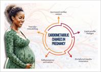
7.1. Polycystic Ovary Syndrome
The POS is the most common reproductive endocrinopathy, present in 6-10% of women. It is strongly associated with central obesity, IR, glucose intolerance, and dyslipidemia (increased LDL-c and triglycerides, decreased HDL-c), as well as with higher risk for SAH and early atherosclerosis,159hepatic steatosis, sleep respiratory disorders with higher incidence of obstructive apnea,5 , 97 , 98 and depression.
The pathophysiology of POS is complex and involves dysregulation of the hypothalamic-pituitary-ovarian axis, reduced hepatic synthesis of SHBG, and functional ovarian hyperandrogenism, a primary dysfunction of ovarian theca cells, in addition to elevated anti-müllerian hormone levels, which exacerbates even more ovarian dysfunction.160
Currently, there are four recognized phenotypes of POS, each one with different long-term implications for metabolism and health: 1) hyperandrogenism + oligoanovulation + polycystic ovary morphology; 2) hyperandrogenism + oligoanovulation; 3) hyperandrogenism + polycystic ovary morphology; and 4) oligoanovulation + polycystic ovary morphology.161The phenotypes with hyperandrogenism have worse metabolic profile.62
The major metabolic changes related to POS are as follows: obesity, in approximately 50% of cases; IR, in 60-95% of cases, leading to glucose intolerance in 31-35% of cases; and T2DM, in 7.5-20% of cases.
Insulin resistance plays a central role in metabolic and cardiovascular complications. The resulting hyperinsulinemia stimulates the hepatic production of triglycerides and reduces HDL-c levels, favoring the accumulation of atherosclerotic plaques. In addition, IR promotes neoglucogenesis, increasing the amount of glucose available (dysglycemia), reducing sex-steroid-binding proteins and insulin-like growth factors, worsening the clinical findings and increasing the chronic inflammatory process.63This leads to higher levels of CRP and pro-inflammatory cytokines, which also accelerate vascular injury and endothelial dysfunction.64
Dyslipidemia is the most frequent abnormality in POS, presenting with low HDL-c levels and high concentrations of triglycerides, and increased LDL-c levels can occur.179
Overweight and obesity, present in many women with POS, worsen the risk because the visceral adipose tissue releases free fatty acids and inflammatory adipokines, exacerbating IR and dyslipidemia. In addition, hyperandrogenemia in POS is associated with an atherogenic lipid profile, with increased oxidized LDL-c and reduced HDL-c, increasing the risk of early cardiovascular events.62
Analysis of 39 systematic reviews and metanalyses published up to 2019 has shown that women with POS are at higher risk for CVD.102This risk remained when stroke and IHD were assessed separately, but there was no association with HF. In addition, the risk of cardiovascular events was higher in young women of reproductive age with POS as compared to normal controls, but no association was observed in postmenopausal women with POS.102Currently, most of the therapy is centered on the patient's major complaint, reducing hyperandrogenism symptoms, restoring menstrual regularity, and obtaining conception in women.
The up-dating of the 2023 International Evidence-based Guideline for the Assessment and Management of Polycystic Ovary Syndrome reintegrated the 2018 International Evidence-Based Guideline for the Assessment and Management of Polycystic Ovary Syndrome , encompassing a wide synthesis of evidence and recommendations for POS.162The major up-dates were as follows:
-
strengthening of the recognition of broader characteristics of POS, including metabolic RFs, CVD, sleep apnea, prevalence of psychological characteristics, and high risk for adverse outcomes during pregnancy;
-
emphasis on the diverse and rarely recognized burden of the disease and on the need for improving health professional education;
-
maintained emphasis on a healthy lifestyle, emotional well-being, and better quality of life;
-
emphasis on evidence-based medical therapy.
Weight loss should be prioritized early.163
The POS changes associated with increased CVR are summarized in Figure 7.2.

7.2. Infertility and its Treatment
Infertility, defined as the absence of clinical pregnancy after 12 months of regular and unprotected sexual intercourse, affects 8-12% of reproductive-age couples.175 , 176 Female causes include ovulatory dysfunctions, tubal and uterine factors, low ovarian reserve, obesity, and hormonal disorders.177The most frequent causes are endometriosis, which is an inflammatory pathology, and POS, which is known to cause increase in androgen hormones and MS.178 , 179 Mulder et al. , assessing women with and without infertility in the same age group, have shown that infertile ones had a greater tendency to have specific cardiometabolic RFs, with an increase in metabolic disorders, such as obesity and dyslipidemia (total cholesterol, LDL-c, and triglycerides), but they found no change in fasting glycemia, IR, and BP.180( Figure 7.3 )

Endometriosis has a prevalence of up to 10% of the female population, is a frequent cause of infertility, and will be addressed separately.181 , 182
The treatment for infertility has cardiometabolic repercussions. Ovarian stimulation with clomiphene, letrozole, and gonadotrophins can induce ovarian hyperstimulation syndrome with risk for thromboembolic events.183 In vitro fertilization leads to higher risk of VTE, preeclampsia, and cardiovascular events in the long run. In addition, women getting pregnant after infertility treatment have higher risk for GD and hypertensive disorders of pregnancy.182
Interventions in lifestyle, such as balanced diet, physical activity practice, and behavioral changes, can not only improve cardiovascular health, but increase infertility treatment success as well. Weight loss in obese women is recommended to improve reproductive health and reduce complications during pregnancy. There is still limited evidence that weight loss improves in vitro fertilization outcomes. Weight control, however, is considered beneficial to fertility and cardiometabolism.184
7.3. Weight changes in pregnancy
Weight changes during pregnancy are intrinsically related to physiological and metabolic adaptations to meet fetal demands. Monitoring GWG is an important part of prenatal consultation and deserves multidisciplinary attention. Pregestational BMI and GWG significantly impact maternal and infant outcomes, as described in chapter 5. Both excessive and insufficient weight are associated with maternal and fetal cardiometabolic complications, such as preterm delivery and small for gestational age newborns.165
Brazilian researchers have developed curves and recommendations of specific GWG for the Brazilian population adopted by the Ministry of Health from 2022 on.186
Chart 7.1 shows the recommendation for GWG adjusted to pregestational BMI in single and twin pregnancies, as well as physiological weight gain distribution in women with normal BMI.186 , 187
7.3.1. Maternal Impact of Excessive Gestational Weight Gain
Almost 50% of women initiate pregnancy with overweight or obesity, and 51% gain weight above the recommended amount. In postpartum, on average, women retain 0.5-3 kg per pregnancy. Preconception overweight and obesity are associated with reduced fertility and a delay in conception. In addition, they increase the risk of maternal mortality and complications, such as excessive GWG, GD, hypertensive disorders of pregnancy, emergency cesarean delivery, congenital diseases, preterm delivery, fetal death, and future risk of DM and CVD. Studies have shown that pregnant women with overweight or obesity tend to have elevated levels of leptin, total cholesterol, LDL-c, very-low-density lipoprotein cholesterol (VLDL-c), triglycerides, fasting glycemia, and insulin, in addition to higher HOMA-IR (homeostasis model assessment of insulin resistance) and diastolic BP in the third trimester.188
7.3.2. Fetal Impact of Excessive Gestational Weight Gain
Maternal obesity is associated with increased risk of fetal macrosomia, congenital malformations, and intrauterine death. In addition, intrauterine exposure to a hyperglycemic environment can predispose the newborn to develop obesity and MS in adulthood, because of changes in fetal metabolic programming.189
In a study with 16 million births in the USA, pregnant women with healthy BMI had lower infant morbidity and mortality rates, while those with class 3 obesity had higher risks of those adverse outcomes, independently of GWG.190 Figure 7.4 shows the recommendations for GWG monitoring.
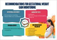
Recommendations for gestational weight gain monitoring. BMI: body mass index; HOMA-IR: homeostasis model assessment of insulin resistance.
7.4. Gestational Dysglycemia
Hyperglycemia is the most common metabolic change in pregnancy, driven by the obesity epidemic, higher prevalence of type 1 diabetes mellitus (T1DM) and T2DM in women of reproductive age, and late pregnancies.191 , 192 In pregnancy, there is a reduction in fasting glycemia because of fetal and placental glucose uptake, as well as mild postprandial hyperglycemia due to diabetogenic placental hormones, even in women with normal metabolism.192
Dysglycemia in pregnancy is classified as pregestational diabetes, diagnosed in pregnancy or GD. Prenatal consultations offer the opportunity to diagnose previously unidentified DM, based on established criteria of glycemia, oral glucose tolerance test (OGTT), or HbA1c.191 , 192 The most prevalent form of gestational hyperglycemia is GD, which affects up to 25% of the pregnancies and is defined as intolerance to carbohydrates initiated in pregnancy, without meeting the diagnostic criteria for DM outside this period.191 , 192 ( Chart 7.2 ).
Although less sensitive to diagnose GD, HbA1c can identify increased risk when ≥ 5.7% in the first trimester, indicating the need for early screening.192HbA1c is a reliable test to guide women with diabetes who want to get pregnant, and measures < 6% reduce the risk of fetal malformations.193The major RFs for GD are advanced maternal age, overweight, obesity, family history of DM, IR, hypertriglyceridemia, hypertension, excessive GWG, and previous GD.191
Gestational diabetes increases the risk of obstetric and neonatal complications, such as abortion, preeclampsia, prematurity, macrosomia, fetal anomalies and death, hypoglycemia, and neonatal respiratory distress. Adequate intervention improves outcomes.191 , 192 Its effects last throughout maternal life, increasing by four-fold the risk of T2DM and by two-fold the risk of cardiovascular events already in the first postpartum decade, and that increase persists throughout life and seems to be independent of T2DM development.194Regarding the fetus, there is higher risk of MS, obesity, DM, and SAH over the course of life.191 - 193
The initial treatment is based on nutritional guidance and physical activity practice. If glycemia is not controlled in up to 14 days, pharmacological therapy should be initiated, and that is mandatory for pregnant women with pregestational T1DM or T2DM.202 , 205 In such cases, aspirin (100–150 mg/day) is recommended from the 12th-16th week on to prevent preeclampsia.192Insulin is the drug of choice due to its efficacy, safety, and low placental transfer.192 , 195
Glycemic self-monitoring, especially in the post-prandial period, is fundamental for therapeutic adjustment, hypoglycemia prevention, and lower risk of preeclampsia.192 , 195 Glycemic goals include pre-prandial values of 65-95 mg/dL, 1st post-prandial hour < 140 mg/dL, and 2ndpost-prandial hour < 120 mg/dL.195
Prevention of GD should be initiated before conception, with weight control, healthy diet, and regular practice of physical activity.
7.5. Gestational dyslipidemia
During pregnancy, the maternal organism undergoes adaptations to adjust fetal growth and development. Thus, the increase in lipid and lipoprotein levels in this phase is important. These changes are stimulated by placental lactogen hormone, estrogen, and progesterone, as well as by elevations in leptin and insulin levels.196Cholesterol is crucial for embryonic and fetal development, because it is an essential component of cell membranes and responsible for several intracellular signaling functions. In addition, this cholesterol increase is necessary to meet the elevated demand for maternal and placental steroids, which accumulate in the maternal body since the 7th week, peaking in the second and third trimesters. By the end of pregnancy, stored lipids serve as a reservoir for the synthesis of fatty acids in the placenta.196The adaptations of lipids to pregnancy consist in an increase in total cholesterol and LDL-c by approximately 30-50% and in HDL-c, by 20-40%.197Triglycerides increase more significantly, reaching 2 to 4 times their pregestational levels in the third trimester, because, along with glucose, they are one of the major sources of fetal energy. LDL-c changes not only its levels, but increases small and dense, more atherogenic, particles, generating higher impact on patients with familial hypercholesterolemia. This unfavorable effect is attenuated by elevated levels of HDL-c and apolipoprotein A-I, which peak during the second trimester and can offer protection against atherogenic lipid fractions.196
In pregnancy, the levels of Lp(a) almost double and the mechanisms responsible for that are not clearly understood. The hypotheses for that increase include estrogen influence in Lp(a) synthesis and clearance, Lp(a) action as acute phase protein in endothelial injury, and Lp(a) possible role in placental development.198
Lipid levels in pregnancy and the magnitude of their changes during pregnancy are influenced by several factors, such as pregestational lipid levels, BMI, age, diet, and ethnicity.
Breast-feeding improves the lipid profile, with a higher reduction in LDL-c and triglycerides than in HDL-c.196 , 199
7.5.1. Impact of Dyslipidemia in Pregnancy
Lipid assessment in the first trimester can provide valuable information regarding short- and long-term results for the mother and newborn, in addition to identifying specific risk groups.197Dyslipidemia (elevation of apolipoprotein B, total cholesterol, LDL-c, and triglycerides), especially at the beginning of pregnancy, is associated with adverse outcomes for the mother and newborn. It has been shown that an atherogenic lipid profile increases the risk of endothelial injury via oxidative stress mechanisms in arterial wall. Maternal risks include preterm delivery, preeclampsia, SAH, GD, MS, and unfavorable lipid profiles. The risks for newborns include premature birth, macrosomia, development of pre-atherosclerotic lesions, and unfavorable lipid profiles. Women with preterm deliveries have a two-fold increased risk of developing CVD later. The placenta shows changes, such as atherosclerosis, infarction of villi, and thrombosis. Addressing lipid abnormalities in pregnancy might help reduce the risk of prematurity.200
In addition, the lipid profile in pregnancy has been related to the development of MS years later and can be used as an early marker of a woman's cardiovascular health, as described in chapter 5. Moreover, it can predict the lipid profile of children and be used as a predictor of children's health in the long run. Monitoring this phase can be a window of opportunity to initiate an early intervention and possibly reduce future CVR. Increased lipid levels have been associated with fatty streaks in the aorta and rapid progression of atherosclerosis in childhood.201
Elevation in Lp(a), an inflammatory protein, can negatively influence gestational outcomes, increasing the risk of complications, such as preeclampsia, DM, preterm delivery, and low birth weight. These conditions represent short-term risk of maternal and fetal morbidity and mortality, and are associated with increased CVR in the long run, such as MI, stroke, and HF.198
Therefore, lipid screening in the first trimester can provide valuable information about short- and long-term outcomes of the mother and newborn, in addition to identifying specific risk groups.
7.5.2. Impact of Pregnancy on Patients with Familial Hypercholesterolemia
Familial hypercholesterolemia is an autosomal semidominant condition, caused by mutations in genes related to lipid metabolism, resulting in elevated LDL-c levels and risk of early IHD.
Women with heterozygous familial hypercholesterolemia have total cholesterol and LDL-c levels approximately twice higher than those without the condition. A Norwegian study has reported that, although the relative increase in total cholesterol and LDL-c between the 17th and 36th gestational weeks was similar in the groups, the absolute increase was significantly higher in women with familial hypercholesterolemia. Triglycerides were also more elevated in that group although still within the normal range and with a similar relative elevation pattern. HDL-c levels, however, remained unchanged in both groups. Despite speculation about possible epigenetic effects of fetal exposure to high cholesterol levels, the mechanisms involved remain uncertain because of conflicting literature data.196
Despite the association of gestational dyslipidemia and adverse maternal-fetal outcomes, the European Society of Cardiology (ESC) guidelines only briefly mention the use of statins and discourage it in women of reproductive age who want to get pregnant. In addition, the United States Centers for Disease Control and Prevention (CDC) offers no specific guidance. They recommend that lipid-lowering drugs should be avoided during pregnancy and lactation, except in severe cases, such as familial hypercholesterolemia, for which bile acid sequestrants (non-absorbed) or LDL-c apheresis are considered.196The scarcity of data about the treatment is mostly due to the systematic exclusion of pregnant women from clinical trials, which limits the knowledge on the safety of lipid-lowering drugs. Metanalyses and systematic reviews about the subject have provided controversial and biased data.196
7.6. Endometriosis and Cardiovascular Risk
The association of endometriosis with RFs for CVDs, such as SAH and dyslipidemia with atherogenic profile, has been shown, as well as the increased risk for VTE, IHD, HF, and stroke.202Endometriosis, an estrogen-dependent chronic inflammatory disease, is the major cause of chronic pelvic pain in young women and one of the major causes of infertility. It is associated with a chronic inflammatory process mediated by substances, such as intercellular adhesion molecule, IL-1 and IL-6, TNF-α, and VEGF, which induce an increase in oxidative stress.181A systematic review with 254,929 participants has revealed that the condition is associated with higher risk of IHD (HR 1.50) and cerebrovascular disease (HR 1.17).203
The complete spectrum of pathogenesis and pathophysiology of endometriosis is recognized as a multifactorial condition involving hormonal, pro-inflammatory, pro-angiogenic, immunological, and genetic processes. Genetic studies have identified variants associated with complex diseases, such as IHD, enlarging the knowledge of its pathophysiology and suggesting new therapeutic targets, especially in lipid metabolism. In addition to hormonal and inflammatory factors, there is a relevant genetic component, with dysregulation of inflammasome, promoting cell proliferation and chronic inflammation. This inflammatory process contributes to a pro-thrombotic state, favoring atherosclerosis and supporting the hypothesis that endometriosis is a CVRF.182 , 204
7.6.1. Risk factors
In endometriosis, there is an elevation in well-known RFs for CVD, such as hypertension and dyslipidemia with major atherogenic profile, in addition to increased risk for VTE, IHD, HF, and stroke.182Population studies have shown an association between endometriosis and SAH, with relative risk (RR) of 1.14 for SAH in women with endometriosis and RR of 1.29 for endometriosis in women with SAH, suggesting common inflammatory mediation. Regarding dyslipidemia, data from the Nurses’ Health Study II 205have shown a 25%-higher risk of hypercholesterolemia in women with endometriosis, in addition to higher prevalence of endometriosis in women with an atherogenic lipid profile. Changes in the metabolism of phospholipids and sphingolipids play a significant role in endometriosis pathophysiology.
Although data on tobacco use, diabetes, and pollution are inconclusive, a recent metanalysis has shown increased risk of 23% for CVD and of 13% for hypertension in women with endometriosis. In addition, association with coronary events (RR 1.62) has been observed, despite the methodological heterogeneity of the studies. Hysterectomy before the age of 50 years, with or without oophorectomy, has been correlated to higher risk of IHD, possibly because of an adverse cardiometabolic profile. In addition, hormonal treatment for endometriosis can negatively impact CVR because of effects on weight and lipid metabolism. Lifestyle and behavioral factors, such as sedentary lifestyle and inadequate diet, contribute to the interrelation between endometriosis and CVD.206
Recognizing endometriosis as a potential RF for CVD is crucial, and so are the implementation of strategies for lifestyle changes and early intervention to prevent and minimize CVR in such women.182 , 206 Figure 7.5 depicts inflammatory markers and CVDs associated with endometriosis.
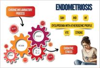
Inflammatory markers and cardiovascular diseases associated with endometriosis. HF: heart failure; ICAM: intercellular adhesion molecule; IHD: ischemic heart disease; IL-1: interleukin-1; IL-6: interleukin-6; ROS: reactive oxygen species; SAH: systemic arterial hypertension; TNF: tumor necrosis factor; VEGF: vascular endothelial growth factor; VTE: venous thromboembolism.
7.7. Psoriasis
Psoriasis is traditionally known to cause inflammatory plaques on the skin. However, there is increasing evidence that it is a systemic chronic inflammatory disease associated with cardiovascular comorbidities, such as obesity, MS, and CVD, cerebrovascular diseases, cardiac arrhythmias, sleep apnea, etc. Although its pathophysiology has not been totally clarified, psoriasis’ systemic inflammation is believed to relate to a pro-inflammatory state with the participation of cytokines, such as TNF-α, IL-6, leptin, and other adipokines.
The study of US female nurses has shown an increased risk of developing DM (RR 1.63) and SAH (RR 1.17) among female nurses with psoriasis.207Recent systematic review and metanalysis have shown the association of psoriasis with IHD.208
7.8. Hypertensive Disorders of Pregnancy and Endothelial Dysfunction after Menopause
Hypertensive disorders of pregnancy affect up to 10% of pregnancies and represent important predictors of CVR over the course of life, being identified as exclusive RFs of the female sex. Increasing evidence has suggested that women with history of hypertensive disorders of pregnancy have persistent changes in endothelial function even decades after pregnancy, which contributes to higher risk of CVD in postmenopause.219Other sessions of this position statement have already addressed the large impact on future cardiometabolic health of women with hypertensive disorders of pregnancy.
Endothelial dysfunction, characterized by a reduction in NO bioavailability and increased inflammatory and pro-thrombotic markers, is an early event in atherogenesis. Longitudinal studies have shown that women with previous preeclampsia had elevated endothelin-1 levels, endothelium-dependent vasodilation dysfunction, and carotid intimal thickening years after delivery. This impact seems to increase after menopause, with worsening of the cardiometabolic profile.210 , 211
In addition, biomarkers of endothelial activation, such as VCAM-1 (vascular cell adhesion molecule) and E-selectin, remain elevated in late postpartum, suggesting low-grade chronic inflammation. Early interruption of estrogen exposure, common in women with preterm delivery due to preeclampsia, may also lead to an earlier loss of hormonally-mediated vasculogenic protection. Inclusion of obstetric history in CVR stratification is essential to the elaboration of individualized preventive strategies, especially in postmenopause.212
Thus, preeclampsia should not be considered only an isolated gestational complication, but rather a marker of female vascular susceptibility over the course of life.213The recent ESC guideline indicates that sex-specific factors are potential reclassifiers of CVR to a higher risk category. Hypertensive disorders of pregnancy should be considered in the individualized CVR stratification, although only a few disorders have shown to improve risk prediction or discrimination beyond traditional factors.214
For such women, it is essential to intensify, as early as from postpartum on, the approach using the eight pillars of cardiovascular prevention, such as control of BP, glycemia, dyslipidemia, and weight, physical activity, sleep quality, tobacco use cessation and healthy eating.213 , 214
7.9. Metabolic Changes in Postpartum
In recent decades, there has been a significant advance in protocols for pregnancy and delivery care. However, the postpartum period continues to be neglected, despite its critical importance to long-term maternal health. In that period, women face significant changes in their reproductive system recovery, as well as in metabolic, endocrine, and nutritional aspects.
One of the most evident changes is body weight variation. On average, weight loss occurs at a rate of 0.6-0.8 kg/month in the first six months after childbirth.215However, some women retain or even gain weight, which can lead to obesity and increase the risk of cardiometabolic complications. Weight retention after childbirth has significant implications for cardiovascular health, as shown in Figure 7.4. 216
7.9.1. Cardiometabolic Risk Factors in Postpartum
Recent studies have shown that factors such as race/ethnicity, socioeconomic level, and GD history, are strong predictors of cardiometabolic risk in postpartum.217 , 218 To minimize these risks, some strategies should be considered:
-
Lactation: breastfeeding is a protective factor against maternal obesity, improving lipid metabolism via hepatic lipolysis and combined action of prolactin and insulin;219
-
Diet: postpartum diet should focus on calorie reduction and nutritional quality. Addition of 500 kcal/day is recommended in the first six months, and 400 kcal/day in subsequent months to ensure adequate metabolic balance;220
-
Physical activity: physical exercise improves cardiorespiratory fitness, preserves lean mass, and helps lose weight, reducing metabolic risk;221
-
Sleep: sleep deprivation (<5 hours/day) has a negative impact on glucose metabolism and favors obesity and IR.222
7.9.2. Impact of gestational diabetes and dyslipidemia in postpartum
Excessive weight retention in postpartum is associated with a higher risk of dyslipidemia and IR. Women with history of GD are more likely to have metabolic disorders in postpartum, such as hyperinsulinemia and hypertriglyceridemia. These effects seem to be influenced by BMI and the gestational age at which the metabolic changes appeared. Studies have shown that, even years after childbirth, women with GD maintain elevated total cholesterol and LDL-C levels, independently of other traditional CVRFs.223These women tend to have elevated levels of triglycerides and LDL-c, independently of BMI, up to months after giving birth. Hyperglycemia and IR in pregnancy contribute to dyslipidemia persistence in postpartum, impacting cardiovascular health in the long run.224 , 225
7.9.3. Prolactin and Metabolism in Postpartum
In addition to its function in lactation, prolactin plays a relevant role in maternal metabolism. Although the relation of prolactin levels and GD has not been totally clarified, evidence suggests that, in postpartum, especially during lactation, elevated prolactin levels are associated with lower circulating insulin levels, higher function of beta cells, and increased insulin sensitivity.226
Maternal overweight and obesity, excessive GWG, and weight retention in postpartum are well established RFs for cardiometabolic complications. Therefore, the postpartum period should be seen as a window of opportunity for preventive interventions, with special attention to balanced and quality diet, regular physical exercise, body weight control, adequate sleep, and breastfeeding. The adoption of such strategies can contribute to preserve maternal cardiovascular health and reduce the risk for cardiometabolic disorders throughout life.
8. Cardiometabolic Health in Menopause Transition, Menopause and Postmenopause
Cardiometabolic disorders, such as T2DM and CVDs with their associated RFs, such as obesity, IR, hypertension, and dyslipidemia, are the major causes of mortality and burden of diseases in both sexes.73 , 109 Women and men, however, experience different trajectories of cardiometabolic risk throughout life influenced by hormonal fluctuations. Women's reproductive phase is a window of opportunity for prevention and approach of CVRFs and cardiometabolic disorders that are magnified in menopause transition, during menopause, and in postmenopause.73 , 109 Reduction in endogenous estradiol levels during menopause transition has been associated with an increased risk of cardiometabolic health issues, such as abdominal adiposity, dyslipidemia, T2DM, and hypertension.73 , 109
Usually, the last menstrual cycle occurs between 45 years and 55 years of age in 90% of women, and, in parallel, the CVR increases in the fifth decade of life, 10 years later than in the male sex. The age of menopause onset seems to be a marker of not only reproductive ageing with the decline in estrogen levels, but also of an increase in cardiometabolic complications.73 , 109
Menopause before age 40 years is considered premature, currently named premature ovarian failure, and has a prevalence of 1%. When menopause occurs between 40 years and 45 years, it is classified as early menopause and affects 7.3% of women. Evidence has shown that women with natural or surgical premature menopause are more likely to develop CVD.73 , 109 ( Figure 8.1 ) In the study by Honigberg et al ., assessing 144,260 women, natural premature menopause was independently associated with aortic stenosis, VTE, ischemic stroke, IHD, and atrial fibrillation. Surgical menopause, however, has been related mainly to mitral regurgitation, pulmonary thromboembolism, HF, and IHD. Menopause at a younger age remained independently associated with time until the first diagnosis of incident CVD (HR, 1.02/year of earlier age of menopause onset [95%CI, 1.01-1.03]; p < 0.001).157

Concept of menopause and increased cardiovascular risk. CVR: cardiovascular risk; POF: premature ovarian failure.
In addition, women with premature and early menopause have a substantially higher risk of a non-fatal cardiovascular event, especially IHD and stroke, before the age of 60 years, possibly associated with low estrogens levels that are related to subclinical atherosclerosis progression. A study with 301,438 women in five countries has reported a 30%-higher risk of CVD in women with early menopause as compared to those experiencing menopause at the age of 50-51 years.227 , 228
The literature has reported a statistically significant association of early age of natural menopause onset with risk of mortality due to general and cardiovascular causes, while more advanced age of menopausal onset results in longer life expectancy, higher bone mineral density, and lower risk of fractures.229 , 230
8.1. Severity of Symptoms and their Implications for Cardiovascular Health
The severity of the clinical manifestations of menopause transition are also related to women's cardiovascular health. They are mainly characterized by VMS, night sweats, neurological complaints, such as sleep, mood, cognition, and memory disorders, in addition to changes in genitourinary and sexual functions. They impair women's quality of life, productivity, and physical and mental health.73 , 109
Vasomotor symptoms are the major complaints, affecting 80% of women during menopause transition, peaking around the final menstrual cycle.231They have been associated with BP elevation and increased risk of CVD, such as stroke and IHD. The major causing factors include autonomic dysregulation with exacerbation of the sympathetic nervous system, sleep interruption, endothelial dysfunction, and more severe subclinical atherosclerosis.232 , 233 Evidence has suggested the association of early VMS onset, duration, and severity with risk for CVD.232Sleep disorders are reported by 40% to 60% of menopausal women and interfere negatively with their quality of life, as well as mental and physical health.234
Identifying premature ovarian failure and early or premature menopause, as well as severity of symptoms, is fundamental to recognize the increased CVR in middle-aged women. This approach enables assessing the therapeutic window for hormonal replacement and intensification of lifestyle changes, optimizing the control of other important factors to reduce cardiovascular events, such as BP, cholesterol, glycemia, body weight, quality of sleep, mental stress, cardiorespiratory fitness, and tobacco use cessation.
8.2. Cardiovascular Risk Factors and Menopause
Hypertension is one of the major CVRFs in women, in whom the association with CVD risk is higher than in men.235Globally, the rate of BP control is estimated to be 23% in women with SAH.236With ageing and after menopause, SAH prevalence increases and the BP control rate decreases.235Some factors, such as unfavorable lifestyle and diet, obesity, and ageing, as well as characteristics related to menarche, reproduction, and menopause contribute to increase BP.
Concomitantly with menopause, there is a decrease in the circulating levels of estrogen and androgens. In addition, there are renin-angiotensin-aldosterone system dysregulation, sympathetic activation, endothelial dysfunction, inflammation, and higher sodium sensitivity.237 , 328 This leads to a rapid and accentuated increase in BP239and high SAH prevalence in postmenopause, with consequent elevation in the risk of cardiovascular events, such as AMI, HFpEF, stroke, cognitive deficit, and peripheral artery disease, usually with systolic BP 10 mm Hg lower than in men.240Moreover, women with SAH more frequently have CKD, left ventricular hypertrophy, and coronary microcirculation dysfunction.235
In managing SAH, BP monitoring outside the medical office is important because of the high incidence of white coat hypertension and masked hypertension. Of nonpharmacological measures, salt restriction stands out. Physical exercise reduces BP and arterial stiffness.241Regarding pharmacological treatment, there is no specific recommendation regarding classes of antihypertensive drugs for menopausal women. Adverse events can be more frequent in women, probably related to pharmacokinetic and pharmacodynamic properties of antihypertensive drugs.235 , 238
Regarding lipids, studies have shown that, in postmenopausal women, total cholesterol and LDL-c levels are higher, and dyslipidemia treatment and control rates are lower than those in men of the same age.242 , 243 HDL-c levels are lower.231Studies have shown that the antiatherogenic function of HDL-c can be impaired and, in such case, elevated HDL-c levels can be associated with atherosclerosis.244In addition, Lp(a) concentration tends to increase during menopause, and elevated Lp(a) levels are more common in women than in men after the age of 50 years, increasing the risk of CVD.235 , 243
These particularities in BP behavior and in lipid disorders suggest that current guidelines for the management of such conditions might be inadequate to meet women's specific needs. Thus, targeted interventions, such as non-medicamentous and medicamentous measures, are essential to improve menopausal women's cardiovascular care. It is worth noting that women are under-represented in clinical trials, which determines scarcity of solid scientific information on women.233 , 235 , 238 , 244
In menopause, the decline in ovarian function results in a significant impact on not only BP, but also other cardiometabolic RFs, such as weight gain, central adiposity, atherogenic lipid profile, and increased glycemia associated with IR, a significantly threatening set of factors to women's cardiovascular health.235 , 238 , 243
Factors, such as race/ethnicity, reproductive factors, body composition, lifestyle, genetics, and premenopausal cardiovascular health, can affect natural menopause and are associated with cardiometabolic risk. In a cohort study including 3639 Dutch women with natural menopause, those who had experienced early natural menopause had a 2.4-higher risk of T2DM as compared to those with late menopause onset, and lived less and fewer years without T2DM as compared to women who had experienced normal or late menopause.245
However, another study involving 177,131 women from four different countries has shown that those with a cardiovascular event before the age of 35 years, as compared to women without CVD, had a twice higher risk of early menopause, raising the hypothesis that the associations can be bidirectional. Thus, a worse premenopausal cardiovascular health profile can influence natural menopause onset.227
Menopause is accompanied by several metabolic adaptations related to IR, increased total body fat mass, and accumulation of central abdominal fat, predisposing women to develop T2DM. Metabolic syndrome has high prevalence in menopausal women, indicating loss of estrogen protection in metabolic and cardiovascular health. In addition, early menopause has been related to increased risk of T2DM.73 , 109
Moreover, high estrogen exposure in premenopausal women, such as pregnancy, has been associated with adverse metabolic changes (hyperglycemia or increased BP) that could impact the risk of developing T2DM and SAH later in life.246
Environmental factors and those related to lifestyle, such as diet, alcohol consumption, physical activity, overweight and obesity, tobacco use, and exposure to environmental toxins, as well as sociodemographic, psychological, and social cognitive factors, are associated with the risk of both early natural menopause and cardiometabolic disease. Environmental factors and those related to lifestyle can trigger underlying mechanisms of cardiometabolic disease associated with menopause, including its influence on DNA methylation and gene expression, induction of low-grade inflammation and oxidative stress, probably interfering with hormone-related signaling in menopause.247
8.3. Menopausal Hormone Therapy, Hormone Implants, and Cardiometabolic Impact
Menopausal hormone therapy is used as the most effective intervention to relieve menopausal symptoms. It offers significant benefits, although it cannot be used for primary or secondary prevention of cardiometabolic disease. With the administration of estrogen and, in some cases, progesterone, MHT is aimed at relieving symptoms and improving quality of life. There are several MHT administration forms, such as pills, patches, gels, and spray. Factors, such as MHT onset and time, duration, dose, and administration route, determine its benefits and drawbacks. The choice of MHT type depends on several factors, such as associated risks, indications, and contraindications.73 , 109
Beginning MHT within the first ten years from menopause onset leads to higher safety in relation to the risk of cardiometabolic disorders, resulting in lower absolute risk of VTE, CVD, and stroke in the first years of menopause. MHT should be taken for the shortest period of time needed and at the lowest dose, and transdermal route has fewer metabolic effects. Oral MHT in women with baseline thromboembolic risk can increase the risk for VTE and stroke on a dose-dependent way; however, transdermal estradiol (only or combined with micronized progesterone) is considered safer. The vaginal route can be used, indicated only for the treatment of genitourinary symptoms.73 , 109
Randomized and controlled clinical trials have suggested that MHT could reduce IR and T2DM incidence, improve glucose metabolism, and increase the glucose hepatic production suppression, and oral preparations have a higher potential to improve glucose metabolism and reduce the risk of developing T2DM.155 , 248 Usually, oral 17β-estradiol is preferred in women with T2DM, because that route has more beneficial effects in glucose metabolism. Oral estrogens are also suggested to women with low risk of CVD in perimenopause or menopause of recent onset. Oral micronized progesterone or dydrogesterone and oral or transdermal norethisterone are the most used progestagens for postmenopausal women with T2DM and intact uterus. The administration of MHT to postmenopausal women with T2DM can be safe and effective if the therapeutic regimen is properly selected. However, MHT is not recommended for primary prevention of T2DM or cardiometabolic disorders.73 , 109 , 117
Testosterone replacement therapy is not indicated to improve cardiometabolic or musculoskeletal health, VMS, or mood changes. Sufficient solid studies on the impact of androgens on cardiometabolic health are not available. In addition, there are few clinical studies on hormone implants for MHT, and most of them address testosterone implants. These studies have small case series and/or low level of evidence (retrospective or observational studies). This impedes the understanding of the cardiometabolic effects of such implants, as well as of their risks for breast and endometrial cancers; therefore, such implants are not recommended for MHT.73,109,1173
8.4. Stratification of Cardiometabolic Risk in Menopause
In menopause and postmenopause, there is an abrupt drop in estrogen levels, an essential hormone for female cardiovascular protection. Some of the changes resulting from this hormone deficiency are as follows: increase in LDL-c, total cholesterol, apolipoprotein B, and triglyceride levels; dysfunctional HDL-c, whose ability to promote cholesterol efflux is reduced, leading to partial loss of its cardiovascular protection; glucose intolerance and increased risk for T2DM; accumulation of visceral fat and ectopic fat deposits in the liver and heart, exacerbating subclinical inflammation; and simultaneous increase of abdominal obesity, hypertension, dyslipidemia, and hyperglycemia.2 , 249
In the United States, between 2013 and 2017, there was a 7% increase in CVD among middle-aged women, attributed mainly to the increase in obesity and prevalence of cardiometabolic RFs. It is worth noting that women with history of T2DM, SAH, and tobacco use have significantly higher relative risk for CVDs as compared to men with the same CVRFs. For example, T2DM increases CVR by 3 to 7 times in women as compared to 2 to 3 times in men, possibly due to higher BMI, higher systemic inflammation, more deficient glycemic control, and higher burden of RFs at the time of female diagnosis.2 , 250
In addition, estrogen loss causes endothelial dysfunction and impairment of vascular integrity, resulting in: reduced flow-mediated dilation; increased arterial stiffness, identified as increased pulse-wave velocity; carotid intimal thickening, which is a marker of subclinical atherosclerosis; increased coronary artery calcium score (CAC); and presence of breast arterial calcification. The CAC obtained on computed tomography without contrast material is currently recognized as a strong marker of atherosclerotic burden and important predictor of risk for IHD, reclassifying the CVR obtained by use of traditional RFs. Studies have shown that, although women have fewer coronary calcifications than men of same age, the presence or increase of CAC is associated with higher relative risk in women. In addition, even in women considered of "low risk" according to Framingham score, a detectable CAC (>0) increases in up to five times the CVR. Breast arterial calcification incidentally detected on mammography also emerges as an independent risk marker of CVD, with 2.4-fold increased risk. In several cases, its detection justifies intensification of RF screening and complementary assessment with CAC.208 , 209
In addition, biomarkers have a relevant role in the cardiometabolic risk assessment of women in menopause transition, menopause, and postmenopause. The following biomarkers stand out: us-CRP, an independent predictor of CVR (usually >3 mg/L or 0.3 mg/dL) even in women with normal lipid levels, integrating Reynolds Risk Score; elevated serum levels of triglycerides (≥175 mg/dL), Lp(a) (≥50 mg/dL or ≥125 nmol/L) and apolipoprotein B (≥130 mg/dL), which are worsening factors; and high-sensitivity troponins and natriuretic peptides, which have prognostic value for future cardiovascular events, although not yet routinely recommended by the ACC/AHA guidelines for population screening of asymptomatic patients.2 , 117 , 249
Before initiating MHT, it is essential to consider the patient's total CVR. The initial assessment includes: complete lipid profile (total cholesterol, LDL-c, HDL-c, and triglycerides), fasting glycemia, HbA1c, and mammography.226In the absence of specific scores for perimenopause and postmenopause, traditional scores are used and can be refined by the identification of enhancing RFs and markers of subclinical atherosclerosis. Patients with T2DM, CKD, familial hypercholesterolemia, or severe SAH are automatically considered at high or very high risk.209 , 250 This initial assessment using the CVR score is important to define if MHT can be prescribed and its best administration route, because, in cases of moderate CVR, the transdermal route should be preferentially chosen.226
Factors, such as age of menopause onset, stress, anxiety, depression, and quality of sleep, need to be considered for CVR reclassification, although their measurement can be hindered by menopausal symptoms.109 , 226
In addition, a sedentary lifestyle in postmenopause leads to worse physical fitness and poorer cardiometabolic control, in addition to higher incidence of fractures and mortality. Tobacco use increases the risk of early menopause and the likelihood of CVD, stroke, osteoporosis, T2DM, and all-cause mortality.109 , 226
Stratification of CVR should be performed, followed by dietary and lifestyle counseling. The management of cardiometabolic RFs in menopause should always be individualized, with focus on hypertension, T2DM, and dyslipidemia. When prescribed to control menopausal symptoms or prevent osteoporosis, MHT can also have a beneficial, even though indirect, effect on cardiometabolic RFs.109 , 226
9. Cardiometabolic Disorders in Women
The incidence of T2DM and obesity has increased considerably in past decades. Projections indicate that there will be more than 600 million individuals with T2DM in 2045.251Obesity and T2DM are associated with several other cardiometabolic disorders. Although both sexes are affected by obesity and T2DM, some studies have indicated differences between men and women regarding the prevalence, diagnosis, treatment, and complications of obesity and T2DM. Evidence available on those differences does not imply sex-specific therapeutic and diagnostic recommendations. However, adequate understanding of those differences is crucial for women's clinical management.252 , 253
9.1. Obesity and Metabolic Syndrome
Obesity is the direct cause of or contributes to the development of several clinical conditions, such as T2DM, MASLD, sleep apnea, articular diseases, several types of cancer, SAH, and HF, in addition to increasing the risk of cardiovascular mortality.254The increase in cardiovascular mortality is independently associated with obesity, even in individuals without other metabolic changes.255Body weight is maintained by a balance between energy intake and energy expenditure,256 , 257 and, when there is excessive energy, it is stored as fat in adipose cells, usually in the subcutaneous tissue. There are factors that limit the physiological storage of fat, which then begins to accumulate ectopically in other tissues, such as liver, pancreas, kidneys, muscles, and epicardium.258Obesity has a strong genetic burden259 , 260 and is influenced by life habits; its prevalence, however, has been increasing in women and men across the world.261In Brazil, VIGITEL data from 2023 indicate a prevalence of obesity of 24.8% in women and 23.8% in men,262and, when overweight individuals are considered, 59.6% of women and 63.4% of men have that condition. ( Figure 9.1 ).
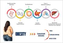
Women usually have a higher percentage of body fat and tendency towards fat accumulation in the subcutaneous tissue of lower limbs, while men have higher visceral fat amounts.263These differences in body fat distribution are associated with sex hormones and vary throughout a woman's reproductive life. Thus, after menopause, women experience body fat redistribution, with visceral fat increase.264The CVR accompanies these differences in hormones and body composition, and women's classic cardiovascular protection during menacme disappears in menopause.265 , 266 In addition, gestational weight gain and its maintenance in postpartum are important RFs for obesity.267The POS is associated with the presence of obesity, increased IR, and other MS components.
The World Health Organization's definition of obesity takes into consideration the BMI, independently of gender, and is subdivided into grade 1 (BMI: 30.0-34.99 kg/m²), grade 2 (BMI: 35.0-39.99 kg/m²), and grade 3 (BMI over 40.0 kg/m²). Recently, a new classification for obesity diagnosis and staging has been proposed,268dividing it in pre-clinical and clinical and considering BMI an inadequate method for obesity diagnosis alone. However, this new classification has not been totally accepted by scientific societies. To our knowledge, BMI is adequate for the diagnosis of obesity in individuals with BMI over 30 kgm/m². For those with lower BMI (below 30 or even below 25 kg/m²), however, excessive adiposity and its metabolic consequences can occur. In such cases, it is better to use other tools for the identification of excessive fat, such as simple measurements of waist and waist-to-hip and waist-to-height ratios, as described in chapter 5. Waist measurements vary between women and men, 88 cm being the cut-off point of waist for Brazilian women. The cut-off point of WHR is 0.85 for women, while 0.5 is the cut-off point of WHtR for men and women.269Another more accurate way to measure adiposity is body composition assessment using bioimpedance or DEXA (dual-energy X-ray absorptiometry). However, further studies confirming that its systematized use changes management and has measurable benefits to the management of patients with obesity are required. Anthropometric measures addressed in chapter 5 are important to establish the diagnosis of excessive body adiposity and to assess the disease's severity, in addition to being parameters of response to treatment.
9.2. Type 2 Diabetes Mellitus
There are differences between the sexes regarding T2DM diagnosis and epidemiology, and they can reflect biological, social, and behavioral factors. Usually, men are diagnosed earlier and at lower BMI values, while women tend to develop the disease later, often after menopause, when the hormonal protection of estrogens decreases.270 - 277 In addition, there is evidence that women can remain longer with underdiagnosed hyperglycemia because of different patterns of symptoms and lower sensitivity of some diagnostic criteria, such as fasting glycemia.278
The prevalence of T2DM was estimated as 8.8% of the world population in 2017, slightly higher among men than among women (9.1% versus 8.4%).251Despite that difference, women with T2DM have higher risk of cardiovascular complications, such as cerebrovascular disease, and early mortality, which can be related to inequities in access to diagnosis and treatment.279In addition, socioeconomic and cultural factors influence those disparities, affecting women's screening and search for care in different regions. Therefore, understanding the sex-related differences in T2DM diagnosis and prevalence is essential for the implementation of more equitable and effective strategies of prevention and care.
Differences in T2DM pathophysiology involving hormonal, genetic, and metabolic factors influence susceptibility to T2DM and its progression. Usually, women have higher IR, especially in the musculoskeletal tissue, while men tend to have higher visceral fat deposition, which is strongly associated with metabolic dysfunction.276 , 280 After menopause, the decline in estrogen levels in women contributes to increase central adiposity and worsens insulin sensitivity, changing the inflammatory and lipid profiles.281In men, testosterone reduction also associates with IR, although with different mechanisms, involving smaller muscle mass and changes in glucose hepatic metabolism.282In addition, differences in gene expression related to glucose transportation, inflammation, and energy metabolism suggest a biological basis for such disparities.280
Systematic reviews with metanalysis have shown sex-related heterogeneity in the contribution of each RF for cardiovascular outcomes. While BP, cholesterol, and BMI seem to contribute equivalently to the risk of coronary and cerebrovascular diseases,278 , 283 , 284 T2DM contributes to higher risk in women.285The risk of coronary disease is usually lower in women, but, in the presence of T2DM, such differences disappear.285T2DM poses a relative risk of 44% for coronary disease and of 27% for cerebrovascular disease to women; however, women's absolute risk is similar to that of men with T2DM.286The higher burden of comorbidities, including RF clustering, as well as hormonal and behavioral issues might contribute to that difference.287 , 288
Studies have shown that women less frequently adhere to drug treatment and self-care, which can be related to psychosocial barriers, less social support, and higher prevalence of depression.276 , 289 There is evidence that women are less likely to achieve the goals of glycemic, BP, and lipid control, even with treatment similar to that of men.290In addition, pharmacokinetic and pharmacodynamic differences affect response to antidiabetic drugs.
It is worth noting the psychosocial and behavioral aspects of T2DM in the female sex. The cognitive capacity of all patients with T2DM should be monitored throughout life, and female sex is a RF for cognitive dysfunctions. Depression also is more frequent in individuals with T2DM, with predominance in the female sex.291 - 293 Women with T2DM have higher incidence of sexual dysfunction, whose occurrence is influenced by both organic (such as autonomic neuropathy) and behavioral factors.
Figure 9.2 summarizes some differences between the sexes regarding T2DM.

Differences in T2DM between women and men: earlier diagnosis in men and higher cardiovascular and psychosocial risks in women. BMI: body mass index; CVD: cardiovascular disease.
9.3. Metabolic Dysfunction-Associated Steatotic Liver Disease
The most prevalent liver disease worldwide is MASLD,294 - 296 characterized by excessive fat accumulation in hepatocytes. It consists in a spectrum of hepatic manifestations associated with metabolic and cardiovascular disorders, such as obesity and/or unfavorable fat distribution, IR, SAH, dyslipidemia, and T2DM.297MASLD is recognized as the hepatic manifestation of MS, being strongly associated with IHD, which is the major cause of mortality in the population with MASLD.297
The natural history of MASLD consists of stages, such as steatosis (when there is only excessive fat in the liver, exceeding 5% of the hepatic parenchyma, with minimum inflammation) and steatohepatitis (when there is lobular inflammation and hepatocyte ballooning, with or without fibrosis).298Independently of sex, individuals with metabolic dysfunction-associated steatohepatitis (MASH) can progress with different grades of fibrosis, cirrhosis, and complications, such as portal hypertension or hepatocellular carcinoma.295 , 298
MASLD is estimated to affect at least 30% of the Western population.299In individuals with overweight and obesity, the global prevalence of MASLD is 50.7%,300and, in those with T2DM, its estimated prevalence is 65.3%.299Although obesity is more common in women, MASLD is more prevalent in men, and the risk of MASLD in women increases after menopause.301It is estimated that, in the general population, women have a 19%-lower risk of hepatic steatosis as compared to men; steatohepatitis rates are similar in both sexes, and women have 37% more risk of advanced fibrosis.302This higher risk for progression occurs especially in women over the age of 50 years, and it is worth noting the possible participation of sex hormones in the etiopathogenesis of MASLD.302
Menopause, history of early menarche,303and POS are associated with increased female susceptibility to MASLD.304Women have a higher mortality rate due to cirrhosis as compared to men, and MASLD is a major cause of liver transplantation in women without hepatocellular carcinoma.305
One justification of the differences in MASLD prevalence over the course of a woman's life is the influence of female sex hormones, particularly estrogen, on hepatic metabolism and on body fat distribution pattern.301Activation of estrogen receptor alpha (ERα) in the liver reduces the synthesis, uptake, and storage of triglycerides, and simultaneously favors the catabolism and export of lipids, effects that together protect non-menopausal women against MASLD.306In addition, the gynoid fat distribution pattern, characterized by greater gluteofemoral fat concentration and smaller visceral fat accumulation, is the typical fat distribution of women in menacme301and is associated with a lower risk of MASLD and MS.295 , 301 However, after menopause, estrogen decline promotes fat redistribution, favoring centralization and, thus, a more android pattern,301known to be associated with MASLD and MS.295 , 301
The diagnosis of MASLD consists in the presence of hepatic steatosis associated with at least one criterion for MS, in the absence of secondary causes of steatosis.295 , 298 Steatosis can be inferred using traditional imaging methods, such as ultrasonography, computed tomography, and magnetic resonance, which can also evidence signs of cirrhosis and portal hypertension. However, it is worth noting that such methods can identify neither MASH nor fibrosis at early stages.297Liver biopsy differentiates precisely patients with steatosis from those with MASH, but, because it is an invasive method, it is mainly used in situations in which there are doubts about the liver disease etiology.295 , 297 , 298
The diagnosis of MASLD is based on the identification of fibrosis, for which some tools, such as clinical-laboratory risk scores for advanced fibrosis and elastographies, are useful. Of those scores, Fibrosis-4 (FIB-4) stands out, calculated based on age, platelet count, and serum level of aminotransferases; the interpretation of the result does not depend on sex.295 , 298 When applying the cut-off point of 1.3, from which patients at higher risk of advanced fibrosis are identified, the test has high sensitivity and excellent negative predictive value.298 ,907, 308 For example, for a woman with FIB-4 ≤ 1.3, the likelihood of advanced fibrosis is very low.
Both ultrasound and magnetic resonance elastographies can estimate liver stiffness and, thus, the presence and amount of fibrosis, including in its initial stages. Transient elastography using the Fibroscan®method is the most validated in the literature and recommended by national295 , 298 , 307 , 308 and international309guidelines. MASLD should be actively screened in individuals at higher risk of fat accumulation in the liver and progression to more severe forms of liver disease, aiming at identifying those with significant fibrosis. The groups of risk comprise postmenopausal women, individuals with glucose homeostasis changes (prediabetes or T2DM), excessive weight, MS, and positive family history for cirrhosis and hepatocellular carcinoma. One rational way to perform population screening is to calculate FIB-4 in individuals at risk and perform elastography in those with FIB-4 > 1.3.295 , 298 , 309
It is worth noting that the isolated assessment of serum levels of aminotransferases has low accuracy to identify patients with MASH and fibrosis. However, elevated levels of those enzymes indicate the need for screening other liver diseases, especially chronic viral hepatitis and alcoholic hepatopathy (excessive alcohol consumption: >20 g/day for women and >30 g/day for men).295 , 298 , 310 - 321
9.4. Chronic Kidney Disease
Chronic kidney disease, mainly diabetic kidney disease (DKD), is a worldwide public health problem that affects millions of individuals. We address briefly the potential differences between genders, and women are a population of special interest because of their biological and social characteristics.
Chronic kidney disease is defined as kidney injury or kidney function reduction for over 3 months, which can progress to need for renal replacement therapy. The progression of CKD is frequently insidious. It is estimated that 425 million individuals have DM, and approximately 30% of those with T1DM and 40% of those with T2DM will develop CKD at different stages.322The major global cause of renal replacement therapy is DM, followed by SAH,323and its incidence has been increasing.324North American data have shown higher prevalence of DKD in women (14.8% x 12.6% in men).325However, it seems that men are at higher risk for CKD relapse and progression to advanced stages of disease as compared to women.326 - 328 These data are heterogeneous and can be related to hormonal changes, such as menopause, T2DM duration and age of onset, and the criteria used for diagnosis.329Studies have shown higher velocity of estimated glomerular filtration rate loss in elderly and menopausal women, in addition to inconsistency of results related to MHT and renal function changes.330Several studies have shown that men are more likely to develop albuminuric DKD as compared to women, in whom the most common form is nonalbuminuric. In a large Italian cohort, moderate to severe albuminuria was present in 29.8% of men as compared to 18.3% of women.331
Despite the report of differences between men and women regarding kidney hemodynamics in DM and mechanisms associated, such as higher frequency of glomerular efferent arteriole vasoconstriction in the female sex, their clinical significance remains uncertain.332 - 337
Current literature data do not allow the use of different criteria for screening, follow-up, and treatment of DKD between men and women. Thus, the following recommendations should be used for both sexes:323
-
The first screening for DKD should be performed with a random urine sample to determine the albumin/creatinine ratio and with estimated glomerular filtration rate determined with serum creatinine, using the 2021 CKD-EPI equation or Schwartz equation for children. In T2DM, screening should begin at the time of diagnosis. In T1DM, screening should begin from puberty onset on or at the age of 10 years in patients with at least 5 years from diagnosis and repeated annually. Every abnormal albumin/creatinine ratio test should be confirmed at least in two out of three samples repeated within three to six months because of daily variability.
-
In individuals with T1DM or T2DM, the HbA1c goal of 6.5-7% should be pursued when estimated glomerular filtration rate >60 mL/min/1.73m² and albumin/creatinine ratio >30 mg/g, to reduce the progression of albuminuria and DKD in the long run. The HbA1c goal of 7-7.9% should be maintained in individuals with T1DM or T2DM when estimated glomerular filtration rate <45 mL/min/1.73m² or if the patient is on dialysis, to prevent excessive mortality.
In conclusion, considering the differences between women and men, as well as the interactions observed with the presence of DM with or without DKD, sex hormones might contribute to sex-related differences in the pathophysiology of DKD beginning and progression.
10. Strategies for Addressing Cardiometabolic Disorders in Women
10.1. Nonpharmacological Measures
The increasing prevalence of cardiometabolic disorders in women represents one of the major challenges in public health because of the strong link between obesity and CVD. Globally, obesity accounts for 4.7 million deaths annually.338 , 339 Abdominal fat accumulation contributes to metabolic changes, chronic inflammation, and IR, favoring atherosclerosis, MI, and stroke. According to the Brazilian Ministry of Health and World Health Organization, in Brazil, CVDs are responsible for approximately 28% of female deaths.340 , 341
In Brazil, 24.8% of women are obese and 38.7% are overweight, totaling 63% of women above their recommended weight.262The ELSA-Brasil study has revealed elevated rates of abdominal obesity among Black (62%) and Mixed-race (59.5%) women, with a general overweight prevalence of 61.8%.342 , 343 These data show the need for urgent investments and interventions to reverse these pathological processes.
10.2. Nutritional Interventions
Nutritional re-education is key for the prevention of cardiometabolic disorders, mainly when tailored to patients’ conditions and habits. Dietary protocols that reduce simple carbohydrates and saturated fats and increase fiber consumption have proven effective in improving glycemia, dyslipidemia, and IR. The Mediterranean diet is widely recognized for its protective effects.344
In Brazil, that diet can be adapted to local food, such as olive oil, local nuts, fruits, such as papaya and avocado, freshwater fish, and pulses, such as beans and chickpea. The dietary guide for the Brazilian population emphasizes the consumption of natural food and the respect for regional dietary culture.345Adhesion to nutritional plans supervised by specialized professionals results in weight loss and sustained improvement of metabolic parameters, contributing to CVD prevention.346A tailor-made approach allows that cultural and regional preferences be respected, because they are relevant factors, considering the wide range of eating habits in Brazil.109
10.3. Physical Activity
Regular physical activity practice plays an essential complementary role in the treatment of cardiometabolic disorders, mainly during menopause transition and menopause. The combination of aerobic exercises with resistance training promotes significant improvement in body composition, insulin sensitivity, and glycemic control.347In addition, it contributes to BP reduction, lipid profile improvement, and systemic inflammation reduction.348
Association of individualized nutritional plans with regular physical activity practice enhances the beneficial effects, favoring cardiovascular function and reducing morbidity and mortality.139Moreover, physical activity is associated with lower incidence of SAH, dyslipidemia, and T2DM, a reduction in the risk of developing depression and dementia, and improvement of bone mineral density and quality of sleep.139 , 149
The current recommendation is moderate aerobic activity for at least 150 minutes per week or vigorous activity for 75 minutes per week associated with muscle resistance exercises at least twice a week.139 , 350 The prescription should be individualized, considering physical fitness, functional limitations, and context of a woman's life, ensuring adhesion and sustained effects in the long run.109 , 209 A recent study has shown that, when women and men practiced equivalent doses of physical activity, women derived greater gains in all-cause and cardiovascular mortality reduction as compared to men.351
10.4. Psychosocial Interventions
Psychosocial stress, such as loneliness, significant losses, and mental disorders, contribute directly to CVR, impairing adhesion to treatment and favoring risk behaviors, such as tobacco use and sedentary lifestyle.352Women who participate in integrated psychological support programs have a significant improvement in lifestyle, which results in better clinical outcomes, such as weight reduction and glycemic and lipid profile control.353Strategies, such as cognitive-behavioral therapy, are especially effective in such context, and studies have shown their ability to reduce anxiety and depression symptoms, improve quality of life related to cardiovascular health, and increase adhesion to treatment.353
Cognitive-behavioral therapy is associated with a lower rate of hospital readmissions and improved self-perception of health in women with CVD.354Moreover, a cognitive-behavioral therapy applied to stress management was associated with sustained behavioral changes, higher frequency of physical activity, healthy diet, and smoking cessation.354
10.5. Tobacco Use and Alcohol Consumption
Tobacco use is an important chronic inflammatory factor, worsened in postmenopausal women.355Electronic cigarette, in particular, represents a new threat especially among young individuals and pregnant women, who often consider such devices less harmful.356Electronic cigarette use can lead to nicotine levels up to six times higher than those of conventional cigarettes, increasing the risk of dependence, oxidative stress, endothelial dysfunction, and vascular inflammation.356Similarly, alcohol consumption significantly affects female health and is associated with higher prevalence of MS, dyslipidemias, and hyperinsulinemia.357
Estrogen reduction in menopause increases inflammatory and atherosclerotic susceptibility, endothelial dysfunction, arterial stiffness, and lipid changes, contributing to the pro-atherogenic and procoagulant state.358Addition of the inflammatory factor of tobacco use in postmenopause accelerates the atherosclerotic process.
10.6. Specific Clinical Conditions
Endometriosis and POS are frequently associated with hormonal imbalances that exacerbate IR, increase the risk of developing T2DM, and promote subclinical inflammation.355In POS, hormonal dysregulation affects the metabolism of glycoproteins and intensifies fat accumulation, contributing to an unfavorable lipid profile and increasing CVD risk.359In endometriosis, the chronic inflammation associated with the ectopic presence of endometrial tissue worsens local symptoms and impacts negatively the systemic environment, affecting metabolic and vascular regulatory mechanisms.360
In conclusion, nonpharmacological interventions have shown clear efficacy in the management of CVRFs.139The combination of regular physical activity with nutritional interventions, in addition to psychosocial support, for weight control results in a significant improvement in vascular and metabolic parameters.361
The multidisciplinary approach has proven effective, allowing personalization and adaptation of strategies to individual needs, generating sustained lifestyle changes, and reducing morbidity and mortality due to cardiometabolic disorders.361
10.7. Pharmacological Strategies
10.7.1. Systemic Arterial Hypertension Treatment
Hypertension is one of the most prevalent RFs for CVD in women; its presence is observed in all life phases and increases progressively as age advances.362The choice and conduction of the pharmacological strategies should consider the phases of the reproductive cycle, including perimenopause and postmenopause. Certain conditions associated with secondary SAH, such as renovascular disease resulting from fibromuscular dysplasia, Cushing syndrome of endogenous origin, as well as thyroid and parathyroid disorders, are more prevalent among women.363
-
Angiotensin-converting-enzyme inhibitors and angiotensin receptor blockers: With proven efficacy to reduce BP levels, they offer additional benefits, such as nephroprotection and left ventricular hypertrophy regression. They are recommended as first line drugs, especially for women with diabetes, CKD with albuminuria, HF with reduced ejection fraction, or IHD. In addition, they can be safely used in patients without comorbidities.214 , 364 The use of angiotensin-converting-enzyme inhibitors (ACEI) and angiotensin receptor blockers II (ARB) in women of reproductive age requires caution because of their teratogenic risk, and it is fundamental not only to exclude the possibility of pregnancy before starting their use but to ensure contraception use during therapy as well.214
-
Calcium channel blockers: Widely recommended for SAH management in women, they are a therapeutic option in perimenopause and postmenopause for patients without significant comorbidities.214It is important to emphasize that calcium channel blockers can be associated with worsening of VMS, such as hot flushes and night sweats in menopausal women. These medications can intensify VMS, with negative impact on quality of life.214
-
Thiazide diuretics: Effective to control SAH in women with overweight or obesity, especially when edema is associated.214Of the major representatives, indapamide stands out due to its additional vasodilating effect and prolonged action, providing sustained BP control and lower metabolic impact as compared to hydrochlorothiazide.364Although widely available and accessible, hydrochlorothiazide has a shorter efficacy and higher risk for electrolytic and metabolic disorders.364The choice should be individualized, considering clinical profile and therapeutic objectives.
-
Spironolactone: This aldosterone antagonist is effective in resistant SAH, when there is higher activation of the renin-angiotensin-aldosterone system.214It is also indicated in primary hyperaldosteronism and POS, because of its antiandrogenic action.214 , 364 It has a favorable metabolic profile, with low impact on glycemia and lipids, which is relevant in women with increased CVR.214It is necessary to monitor renal function and potassium levels, mainly in elderly female patients or those with CKD, because of the risk of hyperkalemia.364
The hypertensive disorders of pregnancy were discussed in chapter 7, as was their therapeutic approach. Figure 10.1 shows the implications of these disorders over the course of a woman's life.

Implications of hypertensive disorders of pregnancy over the course of a woman's life. DM: diabetes mellitus; HF: heart failure; IHD: ischemic heart disease; SAH: systemic arterial hypertension. Source: Davis et al.365
10.7.2. Management of Dyslipidemias
Dyslipidemia is particularly important in menopause transition. The drop in estrogen levels is related to relevant metabolic changes, such as IR, body fat redistribution with predominant abdominal accumulation, and lipid profile changes.366Menopause associates with significant elevations in the levels of total cholesterol, LDL-c, apolipoprotein B, triglycerides, and Lp(a), in addition to a possible reduction in the protective antiatherogenic effect of HDL-c.109Levels of LDL-c ≥130 mg/dL are considered elevated, while HDL-c levels <50 mg/dL represent an additional RF for CVD, especially when associated with other MS components.109This CVR increase should be considered in the individualized therapeutic management, and dyslipidemia treatment should be based not only on the levels of LDL-c, HDL-c, and triglycerides, but also on the patient's global CVR profile.109 , 212
10.7.3. Oral Lipid-Lowering Drugs
Statins are the first-line therapy for dyslipidemia. They are effective to reduce cardiovascular events and atherosclerosis in women at high risk, with benefits comparable to those observed in men.367They should be indicated for both primary and secondary prevention, with LDL-c goals defined according to CVR: < 100 mg/dL for intermediate risk; < 70 mg/dL for high risk; and < 50 mg/dL for very high risk.367 , 368 In women of reproductive age, statin use should be individualized for those at high risk, being contraindicated during pregnancy and lactation.369
Ezetimibe, when associated with statins, enhances LDL-c reduction and contributes to atherosclerosis regression and endothelial function improvement, maintaining safety and efficacy similar in both sexes.367It is particularly useful in women with intolerance to elevated doses of statins or with insufficient response to monotherapy.
Although HDL-c levels over 50 mg/dL are desirable in women, LDL-c reduction remains the therapeutic priority.367Hypertriglyceridemia, which is frequent especially in menopause, should be treated when triglycerides exceed 200 mg/dL and must be treated when over 500 mg/dL.367Of fibrates, fenofibrate is the preferential option, because of its safety profile and additional metabolic benefits in women.109
PCSK9 inhibitors, such as alirocumab, evolocumab, and inclisiran, are indicated for patients with elevated LDL-c levels who do not meet their goals with statins or are intolerant to them, including cases of familial hypercholesterolemia.368Inclisiran, an RNA silencer, stands out due to its semestral posology after the induction dose, with good adhesion and sustained efficacy. These medications should not be used during pregnancy or lactation and have similar efficacy in women and men to reduce cardiovascular events.368
The maternal and fetal impacts of atherogenic lipid profiles during pregnancy were described in chapter 7 and are summarized in Figure 10.2. The recommendations for addressing dyslipidemias in pregnancy are shown in Figure 10.3. .
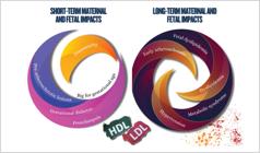
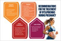
10.7.4. Diabetes Mellitus Control
Although there is not sufficient evidence available for female-sex-specific therapeutic recommendations, sex-specific hormonal, behavioral, and social factors should be considered in the management of T2DM and its complications. In addition, it is fundamental to raise awareness of these differences among health professionals and patients to ensure more equitable and effective care.
Therefore, women's T2DM management requires a comprehensive approach that contemplates clinical and metabolic heterogeneity. The key objective is to meet specific goals to reduce cardiovascular and metabolic morbidity and mortality. The most relevant goals are HbA1c <7% and control of body weight and abdominal circumference, which are factors closely related to IR and CVR.369
Women have a different progression of prediabetes to T2DM, frequently associated with higher indices of obesity and increased risk of metabolic complications. Thus, the management should contemplate not only glycemic control, but also specific strategies to reduce IR and prevent associated comorbidities.109 , 209
Regarding the drugs used to treat T2DM, some considerations are worth noting: women can have higher risk of hypoglycemia with sulfonylureas and different response to glitazones, related to kidney function and body composition.182However, men can have higher response to weight loss induced by SGLT2 inhibitors, partially due to the difference in body fat distribution. In addition, SGLT2 inhibitors are associated with a higher rate of genital fungal infections, especially in women.291Regarding treatment with GLP-1 analogues, special attention should be given to women on oral contraception. The American Diabetes Association recommends that women on oral contraception should choose a non-oral contraceptive method or add a barrier method of contraception during the first four weeks of tirzepatide use (GLP-1 analogue), because of its effects on gastric emptying, with potential consequences to the pharmacokinetics of oral contraceptives.292Without implying sex-specific therapeutic recommendations, the following considerations are worthy of note:
-
Metformin: remains the first choice for T2DM treatment, especially in asymptomatic cases, in POS, in the absence of established CVD or kidney disease, and in individuals at low global CVR.369
-
SGLT2 inhibitors: show important benefits in glycemia reduction and offer cardiovascular and renal protection in women with elevated CVR, HFpEF, HF with reduced ejection fraction, and albuminuric CKD.369
-
Pioglitazone: is a particularly useful thiazolidinedione for women with IR, such as POS, and prediabetes or T2DM with atherogenic metabolic profile. In addition to improving insulin sensitivity, it has beneficial effects on lipid profile and vascular inflammation. However, it should be carefully used, considering risks, such as weight gain, bone mass loss, and fluid retention, especially in women at high risk for HF.369
10.7.5. Management of Obesity
Obesity has a challenging and complex management and should ideally be addressed by a multiprofessional team. The treatment should consider minimizing the stigma related to obesity and requires special attention in women because of their higher vulnerability.214It is important to establish with the patient feasible therapeutic targets associated with clinical benefits, as well as to use strategies to approach weight loss, such as those to fight tobacco use (5 As: ask, assess, advice, agree, assist ). 271The Brazilian Society for the Study of Obesity and Metabolic Syndrome (ABESO) has proposed a classification of obesity based on body weight history and therapeutic goals derived from that assessment, thus aiming at a goal consisting in the proportion of weight loss in relation to maximum BMI in life. The non-realistic goal of BMI normalization to <25 kg/m2is not recommended.252Goals are shown in Table 10.1 .
As already mentioned, nonpharmacological measures, such as calorie restriction diet, regular physical exercise practice, and programs including psychosocial approach, are fundamental to manage metabolic diseases, notably obesity. Calorie restriction is more relevant for weight reduction, but physical exercise practice is essential to maintain weight loss and prevent muscle mass loss that usually accompanies weight loss.271However, the benefits of nonpharmacological measures tend to decrease with time,272and no benefit to long-term cardiovascular outcomes is found with that intervention for women and men. A subgroup analysis of the Look AHEAD clinical trial with patients who lost at least 10% of their body weight in the first year has shown a 20% reduction in the primary outcome (major adverse cardiovascular events and hospitalization due to angina).273
There are several pharmacological treatments for obesity with varied effects on body weight, but only most recent treatments associated with a reduction in outcomes. The SELECT trial was the first to show cardiovascular benefits in that scenario.274The use of semaglutide, a GLP-1 agonist, subcutaneously, at the weekly dose of 2.4 mg to patients with overweight and established CVD for approximately 3 years reduced body weight by 8.51% and resulted in a 28% decrease in the risk for major adverse cardiovascular events. The subgroup analysis of the women included (n=4872, 28% of the sample) showed a reduction of HR 0.84 (95%CI: 0.66-1.07), while men had 0.79 (95%CI: 0.70-0.90), with no report if there was a statistically significant difference in the results between the groups analyzed. The numerical differences found might be related to the lower power of the subgroup analysis of women because of its reduced sample size. Several other more potent antiobesity medications, leading to body weight reduction of up to 25%, are being studied.275However, the results of phase 3 studies with cardiovascular outcomes have not been made available.
10.7.6. Management of Metabolic Dysfunction-Associated Steatotic Liver Disease
The treatment of MASLD consists in lifestyle change with focus on body weight reduction of at least 5%, because weight loss is the most effective measure for histological improvement of MASLD.295 , 298 Alcohol consumption should be limited, as well as the intake of fructose used in ultra-processed foods and sugary drinks. In addition to lifestyle change, women with more advanced forms of MASLD can benefit from pharmacological therapies that act on MASH and/or liver fibrosis. Some examples of such drugs are as follows: resmetirom, semaglutide, pioglitazone, tirzepatide, vitamin E, and SGLT2 inhibitors.
Resmetirom, a selective agonist of thyroid hormone receptor β (THR- β), was the first drug to be approved by a regulatory agency for the treatment of non-cirrhotic MASH, with moderate to advanced fibrosis (compatible with stages F2 and F3). In the MAESTRO-NASH,310phase 3 study that enrolled 966 participants with MASH treated for 52 weeks (322 in the 80-mg resmetirom group, 323 in the 100-mg resmetirom group, and 321 in the placebo group), resmetirom was associated with fibrosis improvement as compared to placebo. There was no subgroup analysis regarding sex. Semaglutide, a GLP-1 receptor agonist, also improved histological results in a population with MASH and fibrosis F2-F3. In the interim analysis of ESSENCE,311a phase 3 study that enrolled 800 participants treated for 70 weeks (57.1% were women and 55.9% had T2DM), there was significant improvement of MASH without fibrosis worsening and significant reduction in fibrosis without MASH worsening (primary outcomes) with the use of semaglutide at the weekly dose of 2.4 mg versus placebo.
Pioglitazone, a selective agonist of the peroxisome proliferator-activated receptor gamma (PPARγ), is recommended to treat MASH and/or fibrosis in individuals with T2DM,295 , 298 , 308 because most studies comparing pioglitazone and placebo have shown benefits in inflammation and histological changes.312However, there was no sub-analysis regarding sex in most of them. It is worth noting that pioglitazone can worsen HF symptoms, because of fluid retention and, specifically in women, it is associated with increased risk of bone fractures and weight gain.312 , 313
Tirzepatide, a co-agonist of glucose-dependent insulinotropic polypeptide (GIP) and GLP-1 receptors already approved in some countries as an antidiabetic and antiobesity agent, has also been assessed to treat individuals with MASH and fibrosis F2-F3. In the SYNERGY-NASH,314a phase 2b placebo-controlled study, tirzepatide promoted MASH resolution without fibrosis worsening in 61% of the individuals assessed after 52 weeks of treatment (57% of patients were women, but no subanalysis was conducted in that group).
In the PIVENS study,315conducted in individuals with MASH and no T2DM, vitamin E use (800 IU/day) for two years improved liver disease activity, which was histologically estimated using the NAS score (Non-Alcoholic Steatohepatitis Activity Score), without fibrosis increase as compared to placebo (43% vs. 19%; p < 0.001).
Finally, although studies of histological outcomes related to MASLD with the use of SGLT2 inhibitors are scarce, there is evidence of reduction in liver enzymes, liver fat, and liver stiffness assessed with elastography as a result of the use of those drugs in a population with T2DM.316 - 318 Thus, some guidelines for the management of MASLD recommend that treatment with SGLT2 inhibitors be considered for individuals with T2DM and MASH and/or fibrosis, without sex-related difference regarding outcomes.295 , 298 , 308
When the combination of lifestyle change and pharmacotherapy fails, individuals with MASLD associated with fibrosis and obesity from class II on should be considered for bariatric surgery.319The benefits of that surgery for MASLD have been consistent in several studies assessing different surgical techniques and, although MASH reduction is evident by the end of the first postoperative year, significant benefits for fibrosis require longer, as shown in studies with follow-up of 5 to 6 years.320 , 321
10.7.7. Management of Chronic Kidney Disease
Current data in the literature do not allow the use of different criteria for screening, diagnosis, and treatment of CKD, especially DKD, and, thus, we recommend that the Brazilian Society of Diabetes criteria be used. Further research is necessary to identify clinically relevant physiological differences between sexes, aimed at identifying new therapies that change clinical outcomes.
The sex-related differences in albuminuria can influence treatment, with therapies targeted at reducing proteinuria, such as renin-angiotensin-aldosterone system blockade, SGLT2 inhibitors, and mineralocorticoid receptor antagonists.331
Large randomized clinical trials using SGLT2 inhibitors have shown that women and men seem to benefit equally, although only 28.5% to 36.9% of those studies’ participants were women. An analysis of the EMPA-REG OUTCOME, CANVAS, DECLARE TIMI-58, and CREDENCE trials has shown that there was no sex-related difference in the reduction of major adverse cardiovascular events or adverse events from SGLT2 inhibitors, such as amputation, fracture, and urinary tract infection, although women were more likely to have genital infection.370
Despite the equal benefit from SGLT2 inhibitors independently of sex, women are less likely to receive a prescription of an SGLT2 inhibitor, even when diagnosed with DKD, HF with reduced ejection fraction, or atherosclerotic CVD. This deprives many women from the cardiovascular and renal benefits of those drugs.
Table 10.2 shows a summary of the drug classes for treatment of DKD and sex-related differences.
10.7.8. Role of GLP-1 Analogues in Women's Cardiometabolic Treatment
The GLP-1 analogues promote glucose-dependent insulin secretion, reduce appetite, and delay gastric emptying. They contribute to effective glycemic control and improvement of CVR markers.369
10.7.9. Specificities in Women
Female hormonal factors and body fat distribution contribute to a more severe expression of cardiometabolic syndrome. Studies have shown that GLP-1 analogues have particularly beneficial effects, with higher impact on weight reduction and improvement of inflammatory and lipid parameters in women as compared to men. This can be partially explained by different hormonal responses and higher sensitivity to the anorexigenic effects of those drugs.371 , 372
10.7.10. Semaglutide
Semaglutide is a GLP-1 receptor agonist, with consolidated efficacy in glycemic control and weight reduction. In overweight or obese patients, semaglutide has shown significant reductions in body weight and HbA1c, even in the absence of T2DM.371In patients with T2DM, it reduced by 26% the risk of major cardiovascular adverse events.135In individuals with previous atherosclerotic CVD, BMI ≥ 27, and no history of T2DM, semagludite reduced by 20% the combined risk of major cardiovascular adverse events and all-cause mortality.274In addition, significant renal effects have been shown in patients with T2DM and CKD. There was a 24% reduction in the risk of severe kidney events and death due to renal or cardiovascular causes in the group treated with semaglutide, with smaller annual decline in estimated glomerular filtration rate.373Recent evidence has shown that its oral use in patients with diabetes also contributes to reduce cardiovascular events, being useful in clinical contexts where the injectable use is less feasible.374
10.7.11. Tirzepatide
Tirzepatide, a dual GIP and GLP-1 receptor agonist, represents an advance in cardiometabolic disease management. Its use has shown significant reductions in HbA1c levels (up to 2.4%) and body weight (up to 20%), in addition to lipid profile improvement and reduction in inflammatory markers.375 , 376 These characteristics make this dual GIP/GLP1 agonist a promising alternative, especially in women with central obesity and IR.
10.7.12. Future Perspectives
A recent study assessing the efficacy of the weekly use of 2.4 mg of semaglutide as compared to 5-15 mg of tirzepatide to treat obesity has shown the superiority of tirzepatide for both weight loss and metabolic control, with comparable cardiovascular effects and safety profile. However, the subgroup of women treated with semaglutide showed higher proportional weight loss as compared to men and improvement of cardiometabolic RFs after 72 weeks of treatment. Planning and final therapeutic decision should always be individual and based on each patient's physiological, hormonal, and metabolic characteristics.376
10.8. Specific pharmacological considerations
10.8.1. Drug interactions
Women with chronic conditions, such as osteoporosis, depression, and anxiety, are frequently exposed to polypharmacy, increasing the risk of relevant drug interactions, because of lower renal clearance and higher body fat proportion. Of the interactions with highest impact, selective serotonin reuptake inhibitors stand out. They can inhibit CYP3A4 and interfere with the metabolism of lipophilic statins, increasing the risk of myopathy.367
10.8.2. Importance of Weight Control
Interventions that reduce body weight have a direct impact on lipid profile improvement, BP control, and insulin sensitivity. Of such interventions, GLP-1 agonists stand out, showing consistent effects on weight reduction with additional benefits to the prevention of major cardiovascular adverse events. Management of obesity should be understood as a fundamental component of the therapeutic strategy for cardiometabolic disorders.117 , 274 , 373 - 376
10.8.3. Individualized Approach
The clinical heterogeneity among women requires personalized approaches for the management of cardiometabolic disorders that consider not only physiological parameters, such as age and presence of comorbidities, but also psychosocial factors, reproductive history, and individual preferences. Therapeutic individualization, thus, represents an essential pillar for woman-centered care, promoting more effective and safe interventions throughout a woman's healthcare journey.377 , 378
10.9. Surgical Treatment
10.9.1. Bariatric Surgery
Bariatric surgery is a widely acknowledged strategy across the globe, with perioperative mortality ranging from 0.03% to 0.2%.379In Brazil, approximately 70% of the patients undergoing the procedure are women.380Its benefits comprise a significant improvement of MS and associated comorbidities, such as T2DM, sleep obstructive apnea, SAH, dyslipidemia, Pickwick syndrome, MASLD, and gastroesophageal reflux disease.381The indication follows guidelines that recommend surgery to patients with BMI ≥ 35 kg/m² and associated comorbidities, or isolated BMI ≥ 40 kg/m², independently of the presence of other diseases.382
Bariatric surgery has been associated with a lower incidence of major cardiovascular adverse events in patients with CVD and obesity in a cohort of 2638 patients followed up for 4.6 years, with the highest benefit found in the groups with HF and IHD.383
After the procedure, women show a slightly smaller weight loss than men. The weight reduction can lead to an abrupt SHBG increase, testosterone decrease, and FSH elevation, improving ovulatory dysfunction and menstrual irregularity, thus favoring spontaneous conception at reproductive age.384
Contraindications to bariatric surgery are as follows: severe psychiatric disease without control; moderate to severe dementias; alcohol or illegal drug dependence; severe IHD or other severe heart diseases; and portal hypertension with esophageal varices. In patients with BMI > 50 kg/m², the surgical risk is high because of the higher incidence of comorbidities and anatomical complexity, which result in longer surgery duration, higher perioperative morbidity, and longer hospitalizations, according to some studies.382
Figure 10.4 summarizes the strategies to approach women's cardiometabolic disorders, emphasizing the importance of a woman-centered multidisciplinary approach, involving the promotion of healthy habits, individualized screening of RFs and their control, integrated clinical management, and inclusion of psychosocial aspects in the cardiometabolic assessment.
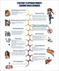
Strategies to approach women's cardiometabolic disorders. ACEI: angiotensin-converting-enzyme inhibitor; ARB: angiotensin receptor blocker; CCB: calcium channel blocker; CKD: chronic kidney disease; CV: cardiovascular; CVD: cardiovascular disease; CVR: cardiovascular risk; DLP: dyslipidemia; T2DM: type 2 diabetes mellitus; GLP-1: glucagon-like peptide 1; HFpEF: heart failure with preserved ejection fraction; HFrEF: heart failure with reduced ejection fraction; MACE: major adverse cardiovascular events; MASLD: metabolic dysfunction-associated steatotic liver disease; MRA: mineralocorticoid receptor antagonist; MS: metabolic syndrome; POS: polycystic ovary syndrome; RAAS: renin-angiotensin-aldosterone system; SAH: systemic arterial hypertension; SGLT2: sodium-glucose cotransporter type 2; VMS: vasomotor symptoms.
11. Recommendations for the Management of Cardiometabolic Disorders in Women
For the recommendations provided at the end of this chapter, a systematic review was conducted (Supplement 1) with ten PICO (Population, Intervention, Comparison, and Outcome) questions. This systematic review included systematic reviews, metanalyses, multicenter randomized controlled trials, and guidelines. The following databases were searched: PubMed/MEDLINE, Embase, Cochrane Library, LILACS, and BVS. The GRADE (Grading of Recommendations Assessment, Development and Evaluation) approach, a system for assessing the certainty of evidence and the strength of recommendation in systematic reviews and clinical practice guidelines was used. The GRADE approach classifies evidence into levels (high, moderate, low, or very low) and, based on that classification, determines the direction (AGAINST or IN FAVOR) and the strength of recommendation (STRONG or WEAK) as follows.
-
High: There is high confidence that effect estimates are close to the true effect.
-
Moderate: There is moderate confidence in effect estimates. Future studies are likely to impact confidence in effect estimates.
-
Low: Confidence in effect estimates is limited.
-
Very low: There is uncertainty regarding effect estimates.
Figure 11.1 summarizes the structure of the systematic review supporting this position statement.
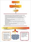
Although the importance of cardiometabolic disorders in women has been increasingly acknowledged, there is significant lack of evidence from multicenter randomized controlled trials specifically addressing the treatment of those conditions in the female sex. This limitation impairs the elaboration of clinical recommendations based on solid evidence adapted to women's particularities. It is worth noting that women continue to be under-represented in clinical trials of cardiovascular diseases and cardiometabolic disorders. In recent analyses, female participation in large multicenter randomized controlled trials has ranged from 27% to 38% of participants, a smaller proportion than the prevalence of those disorders in the female population.385 , 386 Only one third of studies has reported analyses of women-specific results, hindering the efficacy and safety assessment of treatments in that group. In addition, the scarcity of multicenter randomized controlled trials focused on women determines that many clinical recommendations be based on indirect evidence or evidence extrapolated from studies whose sample is mostly male, which might not reflect the best management of cardiometabolic disorders in women.387
There is strong correlation between cardiometabolic disorders and inflammatory conditions over the course of a woman's life, such as POS, GD, and preeclampsia, which predispose to increased risk for AMI, stroke, and IHD, as well as later repercussion on the mother-child dyad. Recent study with 12,480 mother-child dyads has shown that maternal cardiometabolic RFs were significantly associated with 4.88% and 1.90% increases in systolic and diastolic BP of the child, respectively. The combination of hypertensive disorders of pregnancy with pregestational obesity or GD was significantly associated with higher BP between the ages of 2 years and 18 years, emphasizing the importance of the early management of cardiometabolic disorders in women's childhood, adolescence, and reproductive period.388
Sex-related differences regarding cardiometabolic disorders and inflammatory diseases have suggested that sex hormones regulate women's inflammatory pathways. There are changes in women's inflammatory signaling that can account for differences in hypertension, atherosclerosis, obesity, CKD, and MASLD. Knowing the specific mechanisms that boost women's chronic inflammatory conditions, such as POS, GD, and postmenopausal hypertension, will support the multidisciplinary approach necessary to decrease the burden of CVDs in women.30
Several interventions have a combined effect on chronic diseases, especially the cardiometabolic ones. A recent study has assessed the associations between physical activity pattern and incidence of 678 conditions in 89,573 participants (mean age, 62 ± 8 years; 56% women) in the UK Biobank prospective cohort study, who used an accelerometer for one week from June 2013 to December 2015. Both physical activities concentrated over 1 or 2 days and more regular activity patterns were associated with a similarly lower risk for more than 200 diseases, particularly lower risk of cardiometabolic disorders.389
In what follows, we present the recommendations with relatively robust evidence for cardiometabolic disorders in women.
CURRENT RECOMMENDATIONS FOR CARDIOMETABOLIC DISORDERS IN WOMEN
ALCOHOL CONSUMPTION AND TOBACCO USE356 , 409 - 417
CURRENT RECOMMENDATIONS FOR CARDIOMETABOLIC DISORDERS IN WOMEN
DYSLIPIDEMIA366
ARTERIAL HYPERTENSION209 , 211 , 214
METABOLIC DYSFUNCTION-ASSOCIATED STEATOTIC LIVER DISEASE309 , 311 , 314 , 302 , 414 , 433 , 434
CURRENT RECOMMENDATIONS FOR CARDIOMETABOLIC DISORDERS IN WOMEN
CHRONIC KIDNEY DISEASE435 - 441
SURGICAL THERAPY * – BARIATRIC SURGERY442 - 448
* (Roux-en-Y gastric bypass, vertical gastrectomy, laparoscopic adjustable gastric banding, duodenal jejunal bypass liner/biliopancreatic diversion)
POLYCYSTIC OVARY SYNDROME43 , 65 , 97 , 141 , 156 , 159 , 172 , 265 , 304 , 444
SUPPLEMENT
SUPPLEMENT
SUPPLEMENT
SUPPLEMENT
SUPPLEMENT
SUPPLEMENT
Which drugs for the treatment of hypertension and dyslipidemia influence cardiometabolic disorders in women?
SUPPLEMENT
SUPPLEMENT
SUPPLEMENT
Which interventions improve cardiometabolic disorders in women Metabolic Dysfunction–Associated Steatotic Liver Disease (MASLD)?
SUPPLEMENT
-
Development:
Department of Women's Cardiology of the Brazilian Society of Cardiology (In Portuguese: Departamento de Cardiologia da Mulher da Sociedade Brasileira de Cardiologia – DCM/SBC)
-
SBC Clinical Practice Guidelines Committee:
Pedro Gabriel Melo de Barros e Silva (Coordenador), Helena Cramer Veiga Rey, Humberto Graner Moreira, José Augusto Soares Barreto Filho, Nadine Oliveira Clausell – Period 2025-2027.
-
Note:
These statements are for information purposes and should not replace the clinical judgment of a physician, who must ultimately determine the appropriate treatment for each patient.
-
List of Abbreviations and Acronyms
- –ACEI: angiotensin-converting-enzyme inhibitors
- –AMI: acute myocardial infarction
- –ARB: angiotensin receptor blocker
- –BMI: body mass index
- –BP: blood pressure
- –CAC: coronary artery calcium score
- –CBG: cortisol-binding-globulin
- –CHIP: clonal hematopoiesis of indeterminate potential
- –CKD: chronic kidney disease
- –COC: combined oral contraceptive
- –CRP: C-reactive protein
- –CVD: cardiovascular disease
- –CVR: cardiovascular risk
- –CVRF: cardiovascular risk factor
- –DALYs: Disability-Adjusted Life Years
- –DCM/SBC: Womens’ Cardiology Department of the Brazilian Society of Cardiology
- –DKD: diabetic kidney disease
- –DM: diabetes mellitus
- –eNOS: endothelial nitric oxide synthase
- –ESC: European Society of Cardiology
- –FAI: perivascular fat attenuation index
- –FEBRASGO: Brazilian Federation of the Societies of Gynecology and Obstetrics
- –FIB-4: Fibrosis-4
- –FSH: follicle-stimulating hormone
- –GD: gestational diabetes
- –GIP: glucose-dependent insulinotropic polypeptide
- –GLP-1: glucagon-like peptide-1
- –GnRH: gonadotrophin-releasing hormone
- –GWG: gestational weight gain
- –HbA1c: glycated hemoglobin
- –HDL-c: high-density lipoprotein cholesterol
- –HF: heart failure
- –HFpEF: heart failure with preserved ejection fraction
- –HOMA-IR: homeostasis model assessment of insulin resistance
- –IHD: ischemic heart disease
- –IL: interleukin
- –INOCA: ischemia with no obstructive coronary arteries
- –IP-10: interferon-gamma-induced protein 10
- –IR: insulin resistance
- –LDL-c: low-density lipoprotein cholesterol
- –LH: luteinizing hormone
- –LNG-IUS: levonorgestrel intrauterine system
- –Lp(a): lipoprotein a
- –MASH: metabolic dysfunction-associated steatohepatitis
- –MASLD: metabolic dysfunction-associated steatotic liver disease
- –MCP-1: monocyte chemoattractant protein 1
- –MHT: menopausal hormone therapy
- –MI: myocardial infarction
- –MINOCA: myocardial infarction with no obstructive coronary arteries
- –MS: metabolic syndrome
- –NF-κB: nuclear factor kappa B
- –NO: nitric oxide
- –OGTT: oral glucose tolerance test
- –POS: polycystic ovary syndrome
- –RF: risk factor
- –ROS: reactive oxygen species
- –RR: relative risk
- –SAH: systemic arterial hypertension
- –SBEM: Brazilian Society of Endocrinology and Metabolism Study
- –sEng: soluble endoglin
- –sFlt-1: soluble Fms-like tyrosine kinase-1
- –SGLT2: sodium-glucose cotransporter type 2
- –SHBG: sexual-hormone-binding-globulin
- –T1DM: type 1 diabetes mellitus
- –T2DM: type 2 diabetes mellitus
- –TNF-α: tumor necrosis factor alpha
- –us-CRP: ultrasensitive C-reactive protein
- –VCAM-1: vascular cell adhesion molecule
- –VEGF: vascular endothelial growth factor
- –VLDL-c: very-low-density lipoprotein cholesterol
- –VMS: vasomotor symptoms
- –VTE: venous thromboembolism
- –WHI: Women's Health Initiative
- –WHR: waist-to-hip ratio
- –WHtR: waist-to-height ratio
Acknowledgments
We would like to thank all those involved in the management of the journals of the Brazilian Society of Cardiology, as well as librarian Gesner Francisco Xavier Junior for his essential technical and scientific support in making this work possible.
References
-
1 Loh WJ, Watts GF. Cardiometabolic Risk Factors in Women: What's Sauce for the Goose is Not Sauce for the Gander. Curr Opin Endocrinol Diabetes Obes. 2025;32(2):59-65. doi: 10.1097/MED.0000000000000882.
» https://doi.org/10.1097/MED.0000000000000882 -
2 Rajendran A, Minhas AS, Kazzi B, Varma B, Choi E, Thakkar A, et al. Sex-Specific Differences in Cardiovascular Risk Factors and Implications for Cardiovascular Disease Prevention in Women. Atherosclerosis. 2023;384:117269. doi: 10.1016/j.atherosclerosis.2023.117269.
» https://doi.org/10.1016/j.atherosclerosis.2023.117269 -
3 Global Burden of Disease. GBD Compare: Brasil [Internet]. Seattle: Institute for Health Metrics and Evaluation; 2021 [cited 2025 May 22]. Available from: http://ihmeuw.org/6flc .
» http://ihmeuw.org/6flc -
4 Nichols AR, Chavarro JE, Oken E. Reproductive Risk Factors Across the Female Lifecourse and Later Metabolic Health. Cell Metab. 2024;36(2):240-62. doi: 10.1016/j.cmet.2024.01.002.
» https://doi.org/10.1016/j.cmet.2024.01.002 -
5 O’Kelly AC, Michos ED, Shufelt CL, Vermunt JV, Minissian MB, Quesada O, et al. Pregnancy and Reproductive Risk Factors for Cardiovascular Disease in Women. Circ Res. 2022;130(4):652-72. doi: 10.1161/CIRCRESAHA.121.319895.
» https://doi.org/10.1161/CIRCRESAHA.121.319895 -
6 Gerdts E, Regitz-Zagrosek V. Sex Differences in Cardiometabolic Disorders. Nat Med. 2019;25(11):1657-66. doi: 10.1038/s41591-019-0643-8.
» https://doi.org/10.1038/s41591-019-0643-8 -
7 Paula DP, Camacho M, Barbosa O, Marques L, Griep RH, Fonseca MJM, et al. Sex and Population Differences in the Cardiometabolic Continuum: A Machine Learning Study Using the UK Biobank and ELSA-Brasil Cohorts. BMC Public Health. 2024;24(1):2131. doi: 10.1186/s12889-024-19395-9.
» https://doi.org/10.1186/s12889-024-19395-9 -
8 Cosentino F, Verma S, Ambery P, Treppendahl MB, van Eickels M, Anker SD, et al. Cardiometabolic Risk Management: Insights from a European Society of Cardiology Cardiovascular Round Table. Eur Heart J. 2023;44(39):4141-56. doi: 10.1093/eurheartj/ehad445.
» https://doi.org/10.1093/eurheartj/ehad445 -
9 Global Burden of Disease Collaborative Network. Global Burden of Disease Study 2019 (GBD 2019) Results [Interne]. Seattle: Institute for Health Metrics and Evaluation, 2020 [cited 2025 May 22]. Available from: http://ghdx.healthdata.org/gbd-results-tool .
» http://ghdx.healthdata.org/gbd-results-tool -
10 Vogel B, Acevedo M, Appelman Y, Merz CNB, Chieffo A, Figtree GA, et al. The Lancet Women and Cardiovascular Disease Commission: Reducing the Global Burden by 2030. Lancet. 2021;397(10292):2385-438. doi: 10.1016/S0140-6736(21)00684-X.
» https://doi.org/10.1016/S0140-6736(21)00684-X -
11 Lau ES, Paniagua SM, Guseh JS, Bhambhani V, Zanni MV, Courchesne P, et al. Sex Differences in Circulating Biomarkers of Cardiovascular Disease. J Am Coll Cardiol. 2019;74(12):1543-53. doi: 10.1016/j.jacc.2019.06.077.
» https://doi.org/10.1016/j.jacc.2019.06.077 -
12 Leifheit-Limson EC, D’Onofrio G, Daneshvar M, Geda M, Bueno H, Spertus JA, et al. Sex Differences in Cardiac Risk Factors, Perceived Risk, and Health Care Provider Discussion of Risk and Risk Modification Among Young Patients with Acute Myocardial Infarction: The VIRGO Study. J Am Coll Cardiol. 2015;66(18):1949-57. doi: 10.1016/j.jacc.2015.08.859.
» https://doi.org/10.1016/j.jacc.2015.08.859 -
13 Legato MJ, Johnson PA, Manson JE. Consideration of Sex Differences in Medicine to Improve Health Care and Patient Outcomes. JAMA. 2016;316(18):1865-6. doi: 10.1001/jama.2016.13995.
» https://doi.org/10.1001/jama.2016.13995 -
14 Kottilil S, Mathur P. The Influence of Inflammation on Cardiovascular Disease in Women. Front Glob Womens Health. 2022;3:979708. doi: 10.3389/fgwh.2022.979708.
» https://doi.org/10.3389/fgwh.2022.979708 -
15 Merz CNB, Pepine CJ, Walsh MN, Fleg JL. Ischemia and No Obstructive Coronary Artery Disease (INOCA): Developing Evidence-Based Therapies and Research Agenda for the Next Decade. Circulation. 2017;135(11):1075-92. doi: 10.1161/CIRCULATIONAHA.116.024534.
» https://doi.org/10.1161/CIRCULATIONAHA.116.024534 -
16 Smilowitz NR, Mahajan AM, Roe MT, Hellkamp AS, Chiswell K, Gulati M, et al. Mortality of Myocardial Infarction by Sex, Age, and Obstructive Coronary Artery Disease Status in the ACTION Registry-GWTG (Acute Coronary Treatment and Intervention Outcomes Network Registry-Get with the Guidelines). Circ Cardiovasc Qual Outcomes. 2017;10(12):e003443. doi: 10.1161/CIRCOUTCOMES.116.003443.
» https://doi.org/10.1161/CIRCOUTCOMES.116.003443 -
17 Hung MY, Hsu KH, Hung MJ, Cheng CW, Cherng WJ. Interactions Among Gender, Age, Hypertension and C-Reactive Protein in Coronary Vasospasm. Eur J Clin Invest. 2010;40(12):1094-103. doi: 10.1111/j.1365-2362.2010.02360.x.
» https://doi.org/10.1111/j.1365-2362.2010.02360.x -
18 Saw J. Spontaneous Coronary Artery Dissection. Can J Cardiol. 2013;29(9):1027-33. doi: 10.1016/j.cjca.2012.12.018.
» https://doi.org/10.1016/j.cjca.2012.12.018 -
19 Lüscher TF. Inflammation: The New Cardiovascular Risk Factor. Eur Heart J. 2018;39(38):3483-7. doi: 10.1093/eurheartj/ehy607.
» https://doi.org/10.1093/eurheartj/ehy607 -
20 Tyrrell DJ, Goldstein DR. Ageing and Atherosclerosis: Vascular Intrinsic and Extrinsic Factors and Potential Role of IL-6. Nat Rev Cardiol. 2021;18(1):58-68. doi: 10.1038/s41569-020-0431-7.
» https://doi.org/10.1038/s41569-020-0431-7 -
21 Galkina E, Ley K. Vascular Adhesion Molecules in Atherosclerosis. Arterioscler Thromb Vasc Biol. 2007;27(11):2292-301. doi: 10.1161/ATVBAHA.107.149179.
» https://doi.org/10.1161/ATVBAHA.107.149179 -
22 Garcia M, Mulvagh SL, Merz CN, Buring JE, Manson JE. Cardiovascular Disease in Women: Clinical Perspectives. Circ Res. 2016;118(8):1273-93. doi: 10.1161/CIRCRESAHA.116.307547.
» https://doi.org/10.1161/CIRCRESAHA.116.307547 -
23 Libby P, Ebert BL. CHIP (Clonal Hematopoiesis of Indeterminate Potential): Potent and Newly Recognized Contributor to Cardiovascular Risk. Circulation. 2018;138(7):666-8. doi: 10.1161/CIRCULATIONAHA.118.034392.
» https://doi.org/10.1161/CIRCULATIONAHA.118.034392 -
24 Paulus WJ, Zile MR. From Systemic Inflammation to Myocardial Fibrosis: The Heart Failure with Preserved Ejection Fraction Paradigm Revisited. Circ Res. 2021;128(10):1451-67. doi: 10.1161/CIRCRESAHA.121.318159.
» https://doi.org/10.1161/CIRCRESAHA.121.318159 -
25 Liberale L, Montecucco F, Tardif JC, Libby P, Camici GG. Inflamm-Ageing: The Role of Inflammation in Age-Dependent Cardiovascular Disease. Eur Heart J. 2020;41(31):2974-82. doi: 10.1093/eurheartj/ehz961.
» https://doi.org/10.1093/eurheartj/ehz961 -
26 Murphy SP, Kakkar R, McCarthy CP, Januzzi JL Jr. Inflammation in Heart Failure: JACC State-of-the-Art Review. J Am Coll Cardiol. 2020;75(11):1324-40. doi: 10.1016/j.jacc.2020.01.014.
» https://doi.org/10.1016/j.jacc.2020.01.014 -
27 Ridker PM, MacFadyen JG, Thuren T, Libby P. Residual Inflammatory Risk Associated with Interleukin-18 and Interleukin-6 after Successful Interleukin-1β Inhibition with Canakinumab: Further Rationale for the Development of Targeted Anti-Cytokine Therapies for the Treatment of Atherothrombosis. Eur Heart J. 2020;41(23):2153-63. doi: 10.1093/eurheartj/ehz542.
» https://doi.org/10.1093/eurheartj/ehz542 -
28 Bonfioli GB, Pagnesi M, Calò L, Metra M. Towards a Phenotype Profiling of the Patients with Heart Failure and Preserved Ejection Fraction. Eur Heart J Suppl. 2025;27(Suppl 1):i115-i121. doi: 10.1093/eurheartjsupp/suae095.
» https://doi.org/10.1093/eurheartjsupp/suae095 -
29 Cohen JB, Schrauben SJ, Zhao L, Basso MD, Cvijic ME, Li Z, et al. Clinical Phenogroups in Heart Failure with Preserved Ejection Fraction: Detailed Phenotypes, Prognosis, and Response to Spironolactone. JACC Heart Fail. 2020;8(3):172-84. doi: 10.1016/j.jchf.2019.09.009.
» https://doi.org/10.1016/j.jchf.2019.09.009 -
30 Hetherington K, Thomas J, Nicholls SJ, Barsha G, Bubb KJ. Unique Cardiometabolic Factors in Women that Contribute to Modified Cardiovascular Disease Risk. Eur J Pharmacol. 2024;984:177031. doi: 10.1016/j.ejphar.2024.177031.
» https://doi.org/10.1016/j.ejphar.2024.177031 -
31 Pellicori P, Zhang J, Cuthbert J, Urbinati A, Shah P, Kazmi S, et al. High-Sensitivity C-Reactive Protein in Chronic Heart Failure: Patient Characteristics, Phenotypes, and Mode of Death. Cardiovasc Res. 2020;116(1):91-100. doi: 10.1093/cvr/cvz198.
» https://doi.org/10.1093/cvr/cvz198 -
32 Shaw LJ, Patel K, Lala-Trindade A, Feltovich H, Vieira L, Kontorovich A, et al. Pathophysiology of Preeclampsia-Induced Vascular Dysfunction and Implications for Subclinical Myocardial Damage and Heart Failure. JACC Adv. 2024;3(6):100980. doi: 10.1016/j.jacadv.2024.100980.
» https://doi.org/10.1016/j.jacadv.2024.100980 -
33 Boulet J, Sridhar VS, Bouabdallaoui N, Tardif JC, White M. Inflammation in Heart Failure: Pathophysiology and Therapeutic Strategies. Inflamm Res. 2024;73(5):709-23. doi: 10.1007/s00011-023-01845-6.
» https://doi.org/10.1007/s00011-023-01845-6 -
34 Weber B, Garshick M, Liao KP, Di Carli M. Sore, Hot, and at Risk: The Emerging Specialty of Cardio-Rheumatology. J Am Heart Assoc. 2023;12(11):e027846. doi: 10.1161/JAHA.122.027846.
» https://doi.org/10.1161/JAHA.122.027846 -
35 Sun J, Tai S, Guo Y, Tang L, Yang H, Li X, et al. Sex Differences in Characteristics and Outcomes in Elderly Heart Failure Patients with Preserved Ejection Fraction: A Post-Hoc Analysis from TOPCAT. Front Cardiovasc Med. 2021;8:721850. doi: 10.3389/fcvm.2021.721850.
» https://doi.org/10.3389/fcvm.2021.721850 -
36 Sotomi Y, Hikoso S, Nakatani D, Mizuno H, Okada K, Dohi T, et al. Sex Differences in Heart Failure with Preserved Ejection Fraction. J Am Heart Assoc. 2021;10(5):e018574. doi: 10.1161/JAHA.120.018574.
» https://doi.org/10.1161/JAHA.120.018574 -
37 Ko SH, Kim HS. Menopause-Associated Lipid Metabolic Disorders and Foods Beneficial for Postmenopausal Women. Nutrients. 2020;12(1):202. doi: 10.3390/nu12010202.
» https://doi.org/10.3390/nu12010202 -
38 Bechlioulis A, Kalantaridou SN, Naka KK, Chatzikyriakidou A, Calis KA, Makrigiannakis A, et al. Endothelial Function, but Not Carotid Intima-Media Thickness, is Affected Early in Menopause and is Associated with Severity of Hot Flushes. J Clin Endocrinol Metab. 2010;95(3):1199-206. doi: 10.1210/jc.2009-2262.
» https://doi.org/10.1210/jc.2009-2262 -
39 Aryan L, Younessi D, Zargari M, Banerjee S, Agopian J, Rahman S, et al. The Role of Estrogen Receptors in Cardiovascular Disease. Int J Mol Sci. 2020;21(12):4314. doi: 10.3390/ijms21124314.
» https://doi.org/10.3390/ijms21124314 -
40 Fonseca FAH, Izar MC. Inflammation in Cardiovascular Disease: Current Status and Future Perspectives. Int J Cardiovasc Sci. 2023;36:e20230072. doi: 10.36660/ijcs.20230072.
» https://doi.org/10.36660/ijcs.20230072 -
41 Ghisletti S, Meda C, Maggi A, Vegeto E. 17beta-estradiol Inhibits Inflammatory Gene Expression by Controlling NF-kappaB Intracellular Localization. Mol Cell Biol. 2005;25(8):2957-68. doi: 10.1128/MCB.25.8.2957-2968.2005.
» https://doi.org/10.1128/MCB.25.8.2957-2968.2005 -
42 Gao H, Liang M, Bergdahl A, Hamrén A, Lindholm MW, Dahlman-Wright K, et al. Estrogen Attenuates Vascular Expression of Inflammation Associated Genes and Adhesion of Monocytes to Endothelial Cells. Inflamm Res. 2006;55(8):349-53. doi: 10.1007/s00011-006-5194-z.
» https://doi.org/10.1007/s00011-006-5194-z -
43 Zhao L, Zhu Z, Lou H, Zhu G, Huang W, Zhang S, et al. Polycystic Ovary Syndrome (PCOS) and the Risk of Coronary Heart Disease (CHD): A Meta-Analysis. Oncotarget. 2016;7(23):33715-21. doi: 10.18632/oncotarget.9553.
» https://doi.org/10.18632/oncotarget.9553 -
44 Klüner LV, Oikonomou EK, Antoniades C. Assessing Cardiovascular Risk by Using the Fat Attenuation Index in Coronary CT Angiography. Radiol Cardiothorac Imaging. 2021;3(1):e200563. doi: 10.1148/ryct.2021200563.
» https://doi.org/10.1148/ryct.2021200563 -
45 Ramma W, Ahmed A. Is Inflammation the Cause of Pre-Eclampsia? Biochem Soc Trans. 2011;39(6):1619-27. doi: 10.1042/BST20110672.
» https://doi.org/10.1042/BST20110672 -
46 Çendek BD, Ağbal T, Akay A, İbanoğlu MC, Üstün YE. The Performance of Various Serum Parameters in Blood during the First Trimester in the Early Detection of Pre-Eclampsia. J Health Sci Med. 2024;7(4):451-8. doi: 10.32322/jhsm.1500190.
» https://doi.org/10.32322/jhsm.1500190 -
47 McCarthy CM, Kenny LC. Mitochondrial [Dys]Function; Culprit in Pre-Eclampsia? Clin Sci. 2016;130(14):1179-84. doi: 10.1042/CS20160103.
» https://doi.org/10.1042/CS20160103 -
48 Huda SS, Jordan F, Bray J, Love G, Payne R, Sattar N, et al. Visceral Adipose Tissue Activated Macrophage Content and Inflammatory Adipokine Secretion is Higher in Pre-Eclampsia than in Healthy Pregnancys. Clin Sci. 2017;131(13):1529-40. doi: 10.1042/CS20160832.
» https://doi.org/10.1042/CS20160832 -
49 Guan X, Fu Y, Liu Y, Cui M, Zhang C, Zhang Q, et al. The Role of Inflammatory Biomarkers in the Development and Progression of Pre-Eclampsia: A Systematic Review and Meta-Analysis. Front Immunol. 2023;14:1156039. doi: 10.3389/fimmu.2023.1156039.
» https://doi.org/10.3389/fimmu.2023.1156039 -
50 Ma Y, Ma M, Ye S, Liu Y, Zhao X, Wang Y. Association of IL-17 and IL-27 Polymorphisms with Susceptibility to Recurrent Pregnancy Loss and Pre-Eclampsia: A Systematic Review and Meta-Analysis. Immun Inflamm Dis. 2023;11(10):e1057. doi: 10.1002/iid3.1057.
» https://doi.org/10.1002/iid3.1057 - 51 Guyton, Arthur C, Hall JE. Tratado de Fisiologia Médica. Rio de Janeiro: Guanabara Koogan; 2021.
-
52 Rodrigues MAH, Gompel A. Progestatifs. EMC – Gynécologie. 2018;(1):1-17. doi:10.1016/S0246-1064(18)84922-2.
» https://doi.org/10.1016/S0246-1064(18)84922-2 -
53 Hammond GL. Plasma Steroid-Binding Proteins: Primary Gatekeepers of Steroid Hormone Action. J Endocrinol. 2016;230(1):R13-25. doi: 10.1530/JOE-16-0070.
» https://doi.org/10.1530/JOE-16-0070 -
54 Labrie F, Luu-The V, Lin SX, Simard J, Labrie C. Role of 17 Beta-Hydroxysteroid Dehydrogenases in Sex Steroid Formation in Peripheral Intracrine Tissues. Trends Endocrinol Metab. 2000;11(10):421-7. doi: 10.1016/s1043-2760(00)00342-8.
» https://doi.org/10.1016/s1043-2760(00)00342-8 -
55 Kuhl H. Pharmacology of Estrogens and Progestogens: Influence of Different Routes of Administration. Climacteric. 2005;8(Suppl 1):3-63. doi: 10.1080/13697130500148875.
» https://doi.org/10.1080/13697130500148875 -
56 Ott J, Egarter C, Aguilera A. Dydrogesterone after 60 Years: A Glance at the Safety Profile. Gynecol Endocrinol. 2022;38(4):279-87. doi: 10.1080/09513590.2021.2016692.
» https://doi.org/10.1080/09513590.2021.2016692 -
57 Liu S, Kciuk O, Frank M, Tyson N. Progestins of Today and Tomorrow. Curr Opin Obstet Gynecol. 2022;34(6):344-50. doi: 10.1097/GCO.0000000000000819.
» https://doi.org/10.1097/GCO.0000000000000819 -
58 Kumar N, Koide SS, Tsong Y, Sundaram K. Nestorone: A Progestin with a Unique Pharmacological Profile. Steroids. 2000;65(10-11):629-36. doi: 10.1016/s0039-128x(00)00119-7.
» https://doi.org/10.1016/s0039-128x(00)00119-7 - 59 Brunton LL, Knollmann BC, Hilal-Dandan R. Estrogens, Progestins, and the Female Reproductive Tract. In: Brunton LL, Knollmann BC, Hilal-Dandan R, editors. Goodman & Gilman's: The Pharmacological Basis of Therapeutics. 14th ed. New York: McGraw-Hill Education; 2022. p. 1185-216.
-
60 Soares JM Jr, Baracat MC, Maciel GA, Baracat EC. Polycystic Ovary Syndrome: Controversies and Challenges. Rev Assoc Med Bras (1992). 2015;61(6):485-7. doi: 10.1590/1806-9282.61.06.485.
» https://doi.org/10.1590/1806-9282.61.06.485 -
61 Kaur A, Kumar R, Kumar H, Garg S, Vijukumar A, Kumar D. Risk of Cardiovascular Diseases Associated with PCOS in India: A Review. Curr Cardiol Rev. 2025. doi: 10.2174/011573403X349035250408050637.
» https://doi.org/10.2174/011573403X349035250408050637 -
62 Soares-Jr JM, Hayashida SAY, Marcondes JAM, Maciel GAR, Barcellos CRG, Maffazioli GN, et al. Influence of Phenotypes on the Metabolic Syndrome of Women with Polycystic Ovary Syndrome over a Six-Year Follow-Up in Brazil. Biomedicines. 2023;11(12):3262. doi: 10.3390/biomedicines11123262.
» https://doi.org/10.3390/biomedicines11123262 -
63 Medeiros SF, Medeiros ALWY, Medeiros MAS, Carvalho ABS, Yamamoto MW, Soares JM, et al. Anthropometric, Metabolic, and Endocrine Parameters as Predictors of Estimated Average Glucose and Other Biomarkers of Dysglycemia in Women with Different Phenotypes of Polycystic Ovary Syndrome. Horm Metab Res. 2024;56(6):445-54. doi: 10.1055/a-2207-0739.
» https://doi.org/10.1055/a-2207-0739 -
64 Moulana M. Androgen-Induced Cardiovascular Risk in Polycystic Ovary Syndrome: The Role of T Lymphocytes. Life. 2023;13(4):1010. doi: 10.3390/life13041010.
» https://doi.org/10.3390/life13041010 -
65 Morley LC, Tang T, Yasmin E, Norman RJ, Balen AH. Insulin-Sensitising Drugs (Metformin, Rosiglitazone, Pioglitazone, D-Chiro-Inositol) for Women with Polycystic Ovary Syndrome, Oligo Amenorrhoea and Subfertility. Cochrane Database Syst Rev. 2017;11(11):CD003053. doi: 10.1002/14651858.CD003053.pub6.
» https://doi.org/10.1002/14651858.CD003053.pub6 -
66 Zhang J, Zhang H, Zhou W, Jiang M, Lin X. Effect of Myo-Inositol Supplementation in Mixed Ovarian Response IVF Cohort: A Systematic Review and Meta-Analysis. Front Endocrinol. 2025;16:1520362. doi: 10.3389/fendo.2025.1520362.
» https://doi.org/10.3389/fendo.2025.1520362 -
67 Monney M, Mavromati M, Leboulleux S, Gariani K. Endocrine and Metabolic Effects of GLP-1 Receptor Agonists on Women with PCOS, a Narrative Review. Endocr Connect. 2025;14(5):e240529. doi: 10.1530/EC-24-0529.
» https://doi.org/10.1530/EC-24-0529 -
68 Faculty of Sexual and Reproductive Healthcare. FSRH Guideline: Combined Hormonal Contraception [Internet]. London: FSRH; 2019 [cited 2025 Aug 5]. Available from: https://www.fsrh.org/standards-and-guidance/documents/combined-hormonal-contraception/ .
» https://www.fsrh.org/standards-and-guidance/documents/combined-hormonal-contraception/ -
69 Creinin MD, Cagnacci A, Spaczyński RZ, Stute P, Chabbert-Buffet N, Korver T, et al. Experts’ View on the Role of Oestrogens in Combined Oral Contraceptives: Emphasis on Oestetrol (E4). Front Glob Womens Health. 2024;5:1395863. doi: 10.3389/fgwh.2024.1395863.
» https://doi.org/10.3389/fgwh.2024.1395863 -
70 Trussell J. Contraceptive Failure in the United States. Contraception. 2011;83(5):397-404. doi: 10.1016/j.contraception.2011.01.021.
» https://doi.org/10.1016/j.contraception.2011.01.021 -
71 Nguyen AT, Curtis KM, Tepper NK, Kortsmit K, Brittain AW, Snyder EM, et al. U.S. Medical Eligibility Criteria for Contraceptive Use, 2024. MMWR Recomm Rep. 2024;73(4):1-126. doi: 10.15585/mmwr.rr7304a1.
» https://doi.org/10.15585/mmwr.rr7304a1 - 72 Federação Brasileira das Associações de Ginecologia e Obstetrícia. Manual da FEBRASGO: Contracepção Reversível de Longa Ação. São Paulo: FEBRASGO; 2022.
-
73 El Khoudary SR, Aggarwal B, Beckie TM, Hodis HN, Johnson AE, Langer RD, et al. Menopause Transition and Cardiovascular Disease Risk: Implications for Timing of Early Prevention: A Scientific Statement from the American Heart Association. Circulation. 2020;142(25):e506-e532. doi: 10.1161/CIR.0000000000000912.
» https://doi.org/10.1161/CIR.0000000000000912 -
74 Mulvagh SL, Mullen KA, Nerenberg KA, Kirkham AA, Green CR, Dhukai AR, et al. The Canadian Women's Heart Health Alliance Atlas on the Epidemiology, Diagnosis, and Management of Cardiovascular Disease in Women - Chapter 4: Sex- and Gender-Unique Disparities: CVD Across the Lifespan of a Woman. CJC Open. 2021;4(2):115-32. doi: 10.1016/j.cjco.2021.09.013.
» https://doi.org/10.1016/j.cjco.2021.09.013 -
75 Panay N, Ang SB, Cheshire R, Goldstein SR, Maki P, Nappi RE, et al. Menopause and MHT in 2024: Addressing the Key Controversies - An International Menopause Society White Paper. Climacteric. 2024;27(5):441-57. doi: 10.1080/13697137.2024.2394950.
» https://doi.org/10.1080/13697137.2024.2394950 - 76 Machado RB, Pompei LM, editors. Consenso Brasileiro de Terapêutica Hormonal do Climatério. Barueri: Alef Editora; 2024.
-
77 Manson JE, Crandall CJ, Rossouw JE, Chlebowski RT, Anderson GL, Stefanick ML, et al. The Women's Health Initiative Randomized Trials and Clinical Practice: A Review. JAMA. 2024;331(20):1748-60. doi: 10.1001/jama.2024.6542.
» https://doi.org/10.1001/jama.2024.6542 -
78 "The 2022 Hormone Therapy Position Statement of The North American Menopause Society" Advisory Panel. The 2022 Hormone Therapy Position Statement of The North American Menopause Society. Menopause. 2022;29(7):767-94. doi: 10.1097/GME.0000000000002028.
» https://doi.org/10.1097/GME.0000000000002028 -
79 Cho L, Kaunitz AM, Faubion SS, Hayes SN, Lau ES, Pristera N, et al. Rethinking Menopausal Hormone Therapy: For Whom, What, When, and How Long? Circulation. 2023;147(7):597-610. doi: 10.1161/CIRCULATIONAHA.122.061559.
» https://doi.org/10.1161/CIRCULATIONAHA.122.061559 -
80 Davis SR, Baber R, Panay N, Bitzer J, Perez SC, Islam RM, et al. Global Consensus Position Statement on the Use of Testosterone Therapy for Women. J Clin Endocrinol Metab. 2019;104(10):4660-6. doi: 10.1210/jc.2019-01603.
» https://doi.org/10.1210/jc.2019-01603 -
81 Salpeter SR, Walsh JM, Ormiston TM, Greyber E, Buckley NS, Salpeter EE. Meta-Analysis: Effect of Hormone-Replacement Therapy on Components of the Metabolic Syndrome in Postmenopausal Women. Diabetes Obes Metab. 2006;8(5):538-54. doi: 10.1111/j.1463-1326.2005.00545.x.
» https://doi.org/10.1111/j.1463-1326.2005.00545.x -
82 Godsland IF. Effects of Postmenopausal Hormone Replacement Therapy on Lipid, Lipoprotein, and Apolipoprotein (a) Concentrations: Analysis of Studies Published from 1974-2000. Fertil Steril. 2001;75(5):898-915. doi: 10.1016/s0015-0282(01)01699-5.
» https://doi.org/10.1016/s0015-0282(01)01699-5 -
83 Zhang GQ, Chen JL, Luo Y, Mathur MB, Anagnostis P, Nurmatov U, et al. Menopausal Hormone Therapy and Women's Health: An Umbrella Review. PLoS Med. 2021;18(8):e1003731. doi: 10.1371/journal.pmed.1003731.
» https://doi.org/10.1371/journal.pmed.1003731 -
84 Manson JE, Chlebowski RT, Stefanick ML, Aragaki AK, Rossouw JE, Prentice RL, et al. Menopausal Hormone Therapy and Health Outcomes during the Intervention and Extended Poststopping Phases of the Women's Health Initiative Randomized Trials. JAMA. 2013;310(13):1353-68. doi: 10.1001/jama.2013.278040.
» https://doi.org/10.1001/jama.2013.278040 -
85 Koubovec D, Ronacher K, Stubsrud E, Louw A, Hapgood JP. Synthetic Progestins Used in HRT Have Different Glucocorticoid Agonist Properties. Mol Cell Endocrinol. 2005;242(1-2):23-32. doi: 10.1016/j.mce.2005.07.001.
» https://doi.org/10.1016/j.mce.2005.07.001 - 86 Effects of Estrogen or Estrogen/Progestin Regimens on Heart Disease Risk Factors in Postmenopausal Women. The Postmenopausal Estrogen/Progestin Interventions (PEPI) Trial. The Writing Group for the PEPI Trial. JAMA. 1995;273(3):199-208.
-
87 Kuba VM, Teixeira MA, Meirelles RM, Assumpção CR, Costa OS. Dydrogesterone does Not Reverse the Cardiovascular Benefits of Percutaneous Estradiol. Climacteric. 2013;16(1):54-61. doi: 10.3109/13697137.2012.672843.
» https://doi.org/10.3109/13697137.2012.672843 -
88 Fernandes CE, Pompei LM, Machado RB, Ferreira JA, Melo NR, Peixoto S. Effects of Estradiol and Norethisterone on Lipids, Insulin Resistance and Carotid Flow. Maturitas. 2008;59(3):249-58. doi: 10.1016/j.maturitas.2008.02.001.
» https://doi.org/10.1016/j.maturitas.2008.02.001 -
89 Douxfils J, Raskin L, Didembourg M, Donis N, Dogné JM, Morimont L, et al. Are Natural Estrogens Used in Contraception at Lower Risk of Venous Thromboembolism than Synthetic Ones? A Systematic Literature Review and Meta-Analysis. Front Endocrinol. 2024;15:1428597. doi: 10.3389/fendo.2024.1428597.
» https://doi.org/10.3389/fendo.2024.1428597 -
90 Vinogradova Y, Coupland C, Hippisley-Cox J. Use of Hormone Replacement Therapy and Risk of Venous Thromboembolism: Nested Case-Control Studies Using the QResearch and CPRD Databases. BMJ. 2019;364:k4810. doi: 10.1136/bmj.k4810.
» https://doi.org/10.1136/bmj.k4810 -
91 Campos LF, Costa GA, Feitosa MD, Félix IF, Gabrielli L, Almeida MCC, et al. Effect of Hormone Therapy on Blood Pressure and Hypertension in Postmenopausal Women: A Systematic Review and Meta-Analysis. Menopause. 2024;31(6):556-62. doi: 10.1097/GME.0000000000002359.
» https://doi.org/10.1097/GME.0000000000002359 -
92 Russo IH, Russo J. Role of Hormones in Mammary Cancer Initiation and Progression. J Mammary Gland Biol Neoplasia. 1998;3(1):49-61. doi: 10.1023/a:1018770218022.
» https://doi.org/10.1023/a:1018770218022 -
93 Rossouw JE, Anderson GL, Prentice RL, LaCroix AZ, Kooperberg C, Stefanick ML, et al. Risks and Benefits of Estrogen Plus Progestin in Healthy Postmenopausal Women: Principal Results from the Women's Health Initiative Randomized Controlled Trial. JAMA. 2002;288(3):321-33. doi: 10.1001/jama.288.3.321.
» https://doi.org/10.1001/jama.288.3.321 -
94 Fournier A, Berrino F, Clavel-Chapelon F. Unequal Risks for Breast Cancer Associated with Different Hormone Replacement Therapies: Results from the E3N Cohort Study. Breast Cancer Res Treat. 2008;107(1):103-11. doi: 10.1007/s10549-007-9523-x.
» https://doi.org/10.1007/s10549-007-9523-x -
95 Vinogradova Y, Coupland C, Hippisley-Cox J. Use of Hormone Replacement Therapy and Risk of Breast Cancer: Nested Case-Control Studies Using the QResearch and CPRD Databases. BMJ. 2020;371:m3873. doi: 10.1136/bmj.m3873.
» https://doi.org/10.1136/bmj.m3873 -
96 ACOG Committee Opinion No. 651: Menstruation in Girls and Adolescents: Using the Menstrual Cycle as a Vital Sign. Obstet Gynecol. 2015;126(6):e143-e146. doi: 10.1097/AOG.0000000000001215.
» https://doi.org/10.1097/AOG.0000000000001215 -
97 Lo ACQ, Lo CCW, Oliver-Williams C. Cardiovascular Disease Risk in Women with Hyperandrogenism, Oligomenorrhea/Menstrual Irregularity or Polycystic Ovaries (Components of Polycystic Ovary Syndrome): A Systematic Review and Meta-Analysis. Eur Heart J Open. 2023;3(4):oead061. doi: 10.1093/ehjopen/oead061.
» https://doi.org/10.1093/ehjopen/oead061 -
98 Kim C, Catov J, Schreiner PJ, Appiah D, Wellons MF, Siscovick D, et al. Women's Reproductive Milestones and Cardiovascular Disease Risk: A Review of Reports and Opportunities from the CARDIA Study. J Am Heart Assoc. 2023;12(5):e028132. doi: 10.1161/JAHA.122.028132.
» https://doi.org/10.1161/JAHA.122.028132 -
99 Lee JJ, Cook-Wiens G, Johnson BD, Braunstein GD, Berga SL, Stanczyk FZ, et al. Age at Menarche and Risk of Cardiovascular Disease Outcomes: Findings from the National Heart Lung and Blood Institute-Sponsored Women's Ischemia Syndrome Evaluation. J Am Heart Assoc. 2019;8(12):e012406. doi: 10.1161/JAHA.119.012406.
» https://doi.org/10.1161/JAHA.119.012406 -
100 Canoy D, Beral V, Balkwill A, Wright FL, Kroll ME, Reeves GK, et al. Age at Menarche and Risks of Coronary Heart and Other Vascular Diseases in a Large UK Cohort. Circulation. 2015;131(3):237-44. doi: 10.1161/CIRCULATIONAHA.114.010070.
» https://doi.org/10.1161/CIRCULATIONAHA.114.010070 -
101 Chen X, Liu Y, Sun X, Yin Z, Li H, Liu X, et al. Age at Menarche and Risk of All-Cause and Cardiovascular Mortality: A Systematic Review and Dose-Response Meta-Analysis. Menopause. 2018;26(6):670-6. doi: 10.1097/GME.0000000000001289.
» https://doi.org/10.1097/GME.0000000000001289 -
102 Okoth K, Chandan JS, Marshall T, Thangaratinam S, Thomas GN, Nirantharakumar K, et al. Association between the Reproductive Health of Young Women and Cardiovascular Disease in Later Life: Umbrella Review. BMJ. 2020;371:m3502. doi: 10.1136/bmj.m3502.
» https://doi.org/10.1136/bmj.m3502 -
103 Dalfra’ MG, Burlina S, Lapolla A. Weight Gain during Pregnancy: A Narrative Review on the Recent Evidences. Diabetes Res Clin Pract. 2022;188:109913. doi: 10.1016/j.diabres.2022.109913.
» https://doi.org/10.1016/j.diabres.2022.109913 -
104 Ćwiek D, Lubkowska A, Zimny M, Szymoniak K, Sipak-Szmigiel O. Weight Gain during and after Pregnancy in Women with Gestational Diabetes Mellitus-A Preliminary Study. Int J Environ Res Public Health. 2022;19(19):11959. doi: 10.3390/ijerph191911959.
» https://doi.org/10.3390/ijerph191911959 -
105 Yogev Y, Catalano PM. Pregnancy and Obesity. Obstet Gynecol Clin North Am. 2009;36(2):285-300. doi: 10.1016/j.ogc.2009.03.003.
» https://doi.org/10.1016/j.ogc.2009.03.003 -
106 Selen DJ, Edelson PK, James K, Corelli K, Hivert MF, Meigs JB, et al. Physiological Subtypes of Gestational Glucose Intolerance and Risk of Adverse Pregnancy Outcomes. Am J Obstet Gynecol. 2022;226(2):241.e1-241.e14. doi: 10.1016/j.ajog.2021.08.016.
» https://doi.org/10.1016/j.ajog.2021.08.016 -
107 Wild R, Weedin EA, Wilson D. Dyslipidemia in Pregnancy. Cardiol Clin. 2015;33(2):209-15. doi: 10.1016/j.ccl.2015.01.002.
» https://doi.org/10.1016/j.ccl.2015.01.002 -
108 Nasioudis D, Doulaveris G, Kanninen TT. Dyslipidemia in Pregnancy and Maternal-Fetal Outcome. Minerva Ginecol. 2019;71(2):155-62. doi: 10.23736/S0026-4784.18.04330-7.
» https://doi.org/10.23736/S0026-4784.18.04330-7 -
109 Oliveira GMM, Almeida MCC, Arcelus CMA, Neto L Espíndola, Rivera MAM, Silva-Filho ALD, et al. Brazilian Guideline on Menopausal Cardiovascular Health - 2024. Arq Bras Cardiol. 2024;121(7):e20240478. doi: 10.36660/abc.20240478.
» https://doi.org/10.36660/abc.20240478 -
110 Maas AHEM, Rosano G, Cifkova R, Chieffo A, van Dijken D, Hamoda H, et al. Cardiovascular Health after Menopause Transition, Pregnancy Disorders, and Other Gynaecologic Conditions: A Consensus Document from European Cardiologists, Gynaecologists, and Endocrinologists. Eur Heart J. 2021;42(10):967-84. doi: 10.1093/eurheartj/ehaa1044.
» https://doi.org/10.1093/eurheartj/ehaa1044 -
111 Wenger NK, Lloyd-Jones DM, Elkind MSV, Fonarow GC, Warner JJ, Alger HM, et al. Call to Action for Cardiovascular Disease in Women: Epidemiology, Awareness, Access, and Delivery of Equitable Health Care: A Presidential Advisory from the American Heart Association. Circulation. 2022;145(23):1059-71. doi: 10.1161/CIR.0000000000001071.
» https://doi.org/10.1161/CIR.0000000000001071 -
112 Mishra GD, Davies MC, Hillman S, Chung HF, Roy S, Maclaran K, et al. Optimising Health after Early Menopause. Lancet. 2024;403(10430):958-68. doi: 10.1016/S0140-6736(23)02800-3.
» https://doi.org/10.1016/S0140-6736(23)02800-3 -
113 Nilsson S, Qvick A, Henriksson M, Lawesson SS, Holm AS, Leander K. Menopausal Vasomotor Symptoms and Subclinical Atherosclerotic Cardiovascular Disease: A Population-Based Study. J Am Heart Assoc. 2024;13(17):e033648. doi: 10.1161/JAHA.123.033648.
» https://doi.org/10.1161/JAHA.123.033648 -
114 Carson MY, Thurston RC. Vasomotor Symptoms and their Links to Cardiovascular Disease Risk. Curr Opin Endocr Metab Res. 2023;30:100448. doi: 10.1016/j.coemr.2023.100448.
» https://doi.org/10.1016/j.coemr.2023.100448 -
115 Armeni A, Anagnostis P, Armeni E, Mili N, Goulis D, Lambrinoudaki I. Vasomotor Symptoms and Risk of Cardiovascular Disease in Peri- and Postmenopausal Women: A Systematic Review and Meta-Analysis. Maturitas. 2023;171:13-20. doi: 10.1016/j.maturitas.2023.02.004.
» https://doi.org/10.1016/j.maturitas.2023.02.004 -
116 Intapad S, Dasinger JH, Brown AD, Fahling JM, Esters J, Alexander BT. Glucose Intolerance Develops Prior to Increased Adiposity and Accelerated Cessation of Estrous Cyclicity in Female Growth-Restricted Rats. Pediatr Res. 2016;79(6):962-70. doi: 10.1038/pr.2016.14.
» https://doi.org/10.1038/pr.2016.14 -
117 Paschou SA, Athanasiadou KI, Papanas N. Menopausal Hormone Therapy in Women with Type 2 Diabetes Mellitus: An Updated Review. Diabetes Ther. 2024;15(4):741-8. doi: 10.1007/s13300-024-01546-1.
» https://doi.org/10.1007/s13300-024-01546-1 -
118 Gado M, Tsaousidou E, Bornstein SR, Perakakis N. Sex-Based Differences in Insulin Resistance. J Endocrinol. 2024;261(1):e230245. doi: 10.1530/JOE-23-0245.
» https://doi.org/10.1530/JOE-23-0245 -
119 Lambrinoudaki I, Paschou SA, Armeni E, Goulis DG. The Interplay between Diabetes Mellitus and Menopause: Clinical Implications. Nat Rev Endocrinol. 2022;18(10):608-22. doi: 10.1038/s41574-022-00708-0.
» https://doi.org/10.1038/s41574-022-00708-0 -
120 Meng Q, Ma M, Zhang W, Bi Y, Cheng P, Yu X, et al. The Gut Microbiota during the Progression of Atherosclerosis in the Perimenopausal Period Shows Specific Compositional Changes and Significant Correlations with Circulating Lipid Metabolites. Gut Microbes. 2021;13(1):1-27. doi: 10.1080/19490976.2021.1880220.
» https://doi.org/10.1080/19490976.2021.1880220 -
121 Carrión-Martínez A, Buckley BJR, Orenes-Piñero E, Marín F, Lip GYH, Rivera-Caravaca JM. Anthropometric Measures and Risk of Cardiovascular Disease: Is there an Opportunity for Non-Traditional Anthropometric Assessment? A Review. Rev Cardiovasc Med. 2022;23(12):414. doi: 10.31083/j.rcm2312414.
» https://doi.org/10.31083/j.rcm2312414 -
122 Ashton WD, Nanchahal K, Wood DA. Body Mass index and Metabolic Risk Factors for Coronary Heart Disease in Women. Eur Heart J. 2001;22(1):46-55. doi: 10.1053/euhj.2000.2469.
» https://doi.org/10.1053/euhj.2000.2469 -
123 Silva DAS, Duncan BB, Schmidt MI, Lotufo PA, Bensenor IM. Body Mass Index and Cardiovascular Risk Factors in Brazilian Women: Cross-Sectional Findings from the ELSA-Brasil Study. Arq Bras Cardiol. 2021;116(4):728-37. doi:10.36660/abc.20200234.
» https://doi.org/10.36660/abc.20200234 -
124 Lopez-Lopez JP, Gonzalez AM, Lanza P, Martinez-Bello D, Gomez-Arbelaez D, Otero J, et al. Waist Circumference Cut-off Points to Identify Major Cardiovascular Events and Incident Diabetes in Latin America: Findings from the Prospective Urban Rural Epidemiology Study Colombia. Front Cardiovasc Med. 2023;10:1204885. doi: 10.3389/fcvm.2023.1204885.
» https://doi.org/10.3389/fcvm.2023.1204885 -
125 Fernandes RA, Rosa CSC, Buonani C, Oliveira AR, Freitas IF Jr. Waist Circumference and Waist-To-Hip Ratio as Predictors of Cardiovascular Risk in Postmenopausal Women: A Population-Based Study. Front Cardiovasc Med. 2022;9:943897. doi:10.3389/fcvm.2022.943897.
» https://doi.org/10.3389/fcvm.2022.943897 -
126 Li WC, Chen IC, Chang YC, Loke SS, Wang SH, Hsiao KY. Waist-to-Height Ratio, Waist Circumference, and Body Mass Index as Indices of Cardiometabolic Risk Among 36,642 Taiwanese Adults. Eur J Nutr. 2013;52(1):57-65. doi: 10.1007/s00394-011-0286-0.
» https://doi.org/10.1007/s00394-011-0286-0 -
127 Oliveira BR, Magalhães EIDS, Bragança MLBM, Coelho CCNDS, Lima NP, Bettiol H, et al. Performance of Body Fat Percentage, Fat Mass Index and Body Mass Index for Detecting Cardiometabolic Outcomes in Brazilian Adults. Nutrients. 2023;15(13):2974. doi: 10.3390/nu15132974.
» https://doi.org/10.3390/nu15132974 -
128 Castanheira M, Chor D, Braga JU, Cardoso LO, Griep RH, Molina MDCB, et al. Predicting Cardiometabolic Disturbances from Waist-to-Height Ratio: Findings from the Brazilian Longitudinal Study of Adult Health (ELSA-Brasil) Baseline. Public Health Nutr. 2018;21(6):1028-35. doi: 10.1017/S136898001700338X.
» https://doi.org/10.1017/S136898001700338X -
129 Buckinx F, Aubertin-Leheudre M. Sarcopenia in Menopausal Women: Current Perspectives. Int J Womens Health. 2022;14:805-19. doi: 10.2147/IJWH.S340537.
» https://doi.org/10.2147/IJWH.S340537 -
130 Lui DTW, Tan KCB. High-Density Lipoprotein in Diabetes: Structural and Functional Relevance. J Diabetes Investig. 2024;15(7):805-16. doi: 10.1111/jdi.14172.
» https://doi.org/10.1111/jdi.14172 -
131 Mach F, Baigent C, Catapano AL, Koskinas KC, Casula M, Badimon L, et al. 2019 ESC/EAS Guidelines for the Management of Dyslipidaemias: Lipid Modification to Reduce Cardiovascular risk. Eur Heart J. 2020;41(1):111-88. doi:10.1093/eurheartj/ehz455.
» https://doi.org/10.1093/eurheartj/ehz455 -
132 American Diabetes Association. Standards of Care in Diabetes – 2023 Abridged for Primary Care Providers. Clin Diabetes. 2023;41(1):4-31. doi:10.2337/cd23-as01.
» https://doi.org/10.2337/cd23-as01 -
133 Huebschmann AG, Huxley RR, Kohrt WM, Zeitler P, Regensteiner JG, Reusch JEB. Sex Differences in the Burden of Type 2 Diabetes and Cardiovascular Risk Across the Life Course. Diabetologia. 2019;62(10):1761-72. doi: 10.1007/s00125-019-4939-5.
» https://doi.org/10.1007/s00125-019-4939-5 -
134 Cosentino F, Grant PJ, Aboyans V, Bailey CJ, Ceriello A, Delgado V, et al. 2019 ESC Guidelines on Diabetes, Pre-Diabetes, and Cardiovascular Diseases Developed in Collaboration with the EASD. Eur Heart J. 2020;41(2):255-323. doi: 10.1093/eurheartj/ehz486.
» https://doi.org/10.1093/eurheartj/ehz486 -
135 Marso SP, Bain SC, Consoli A, Eliaschewitz FG, Jódar E, Leiter LA, et al. Semaglutide and Cardiovascular Outcomes in Patients with Type 2 Diabetes. N Engl J Med. 2016;375(19):1834-44. doi: 10.1056/NEJMoa1607141.
» https://doi.org/10.1056/NEJMoa1607141 -
136 Zinman B, Wanner C, Lachin JM, Fitchett D, Bluhmki E, Hantel S, et al. Empagliflozin, Cardiovascular Outcomes, and Mortality in Type 2 Diabetes. N Engl J Med. 2015;373(22):2117-28. doi: 10.1056/NEJMoa1504720.
» https://doi.org/10.1056/NEJMoa1504720 -
137 Ridker PM, Danielson E, Fonseca FA, Genest J, Gotto AM Jr, Kastelein JJ, et al. Rosuvastatin to Prevent Vascular Events in Men and Women with Elevated C-Reactive Protein. N Engl J Med. 2008;359(21):2195-207. doi: 10.1056/NEJMoa0807646.
» https://doi.org/10.1056/NEJMoa0807646 -
138 Pearson TA, Mensah GA, Alexander RW, Anderson JL, Cannon RO 3rd, Criqui M, et al. Markers of Inflammation and Cardiovascular Disease: Application to Clinical and Public Health Practice: A Statement for Healthcare Professionals from the Centers for Disease Control and Prevention and the American Heart Association. Circulation. 2003;107(3):499-511. doi: 10.1161/01.cir.0000052939.59093.45.
» https://doi.org/10.1161/01.cir.0000052939.59093.45 -
139 Visseren FLJ, Mach F, Smulders YM, Carballo D, Koskinas KC, Bäck M, et al. 2021 ESC Guidelines on Cardiovascular Disease Prevention in Clinical Practice. Eur Heart J. 2021;42(34):3227-337. doi: 10.1093/eurheartj/ehab484.
» https://doi.org/10.1093/eurheartj/ehab484 -
140 Lubrano V, Balzan S. Consolidated and Emerging Inflammatory Markers in Coronary Artery Disease. World J Exp Med. 2015;5(1):21-32. doi: 10.5493/wjem.v5.i1.21.
» https://doi.org/10.5493/wjem.v5.i1.21 -
141 Kryczka KE, Kruk M, Demkow M, Lubiszewska B. Fibrinogen and a Triad of Thrombosis, Inflammation, and the Renin-Angiotensin System in Premature Coronary Artery Disease in Women: A New Insight into Sex-Related Differences in the Pathogenesis of the Disease. Biomolecules. 2021;11(7):1036. doi: 10.3390/biom11071036.
» https://doi.org/10.3390/biom11071036 -
142 Coleman JR, Gumina R, Hund T, Cohen M, Neal MD, Townsend K, et al. Sex Dimorphisms in Coagulation: Implications in Trauma-Induced Coagulopathy and Trauma Resuscitation. Am J Hematol. 2024;99(Suppl 1):S28-S35. doi: 10.1002/ajh.27296.
» https://doi.org/10.1002/ajh.27296 -
143 Hu X, Wang J, Ye Y, Chen X, Abulikemu S, Yu J, et al. Associations between Fibrinogen Levels and the Risk of All-Cause Mortality: A Long-Term Cohort Study. J Thromb Thrombolysis. 2025;58(4):514-25. doi: 10.1007/s11239-025-03087-1.
» https://doi.org/10.1007/s11239-025-03087-1 -
144 Vrints C, Andreotti F, Koskinas KC, Rossello X, Adamo M, Ainslie J, et al. 2024 ESC Guidelines for the Management of Chronic Coronary Syndromes. Eur Heart J. 2024;45(36):3415-537. doi: 10.1093/eurheartj/ehae177.
» https://doi.org/10.1093/eurheartj/ehae177 -
145 Tian W, Ju J, Guan B, Wang T, Zhang J, Song L, et al. Role of Hyperhomocysteinemia in Atherosclerosis: From Bench to Bedside. Ann Med. 2025;57(1):2457527. doi: 10.1080/07853890.2025.2457527.
» https://doi.org/10.1080/07853890.2025.2457527 -
146 Mattu HS, Randeva HS. Role of Adipokines in Cardiovascular Disease. J Endocrinol. 2013;216(1):T17-36. doi: 10.1530/JOE-12-0232.
» https://doi.org/10.1530/JOE-12-0232 -
147 Lau ES, Binek A, Parker SJ, Shah SH, Zanni MV, Van Eyk JE, et al. Sexual Dimorphism in Cardiovascular Biomarkers: Clinical and Research Implications. Circ Res. 2022;130(4):578-92. doi: 10.1161/CIRCRESAHA.121.319916.
» https://doi.org/10.1161/CIRCRESAHA.121.319916 -
148 Sørensen K, Mouritsen A, Aksglaede L, Hagen CP, Mogensen SS, Juul A. Recent Secular Trends in Pubertal Timing: Implications for Evaluation and Diagnosis of Precocious Puberty. Horm Res Paediatr. 2012;77(3):137-45. doi: 10.1159/000336325.
» https://doi.org/10.1159/000336325 -
149 Kaplowitz PB, Oberfield SE. Reexamination of the Age Limit for Defining when Puberty is Precocious in Girls in the United States: Implications for Evaluation and Treatment. Drug and Therapeutics and Executive Committees of the Lawson Wilkins Pediatric Endocrine Society. Pediatrics. 1999;104(4 Pt 1):936-41. doi: 10.1542/peds.104.4.936.
» https://doi.org/10.1542/peds.104.4.936 -
150 Cheng TS, Day FR, Lakshman R, Ong KK. Association of Puberty Timing with Type 2 Diabetes: A Systematic Review and Meta-Analysis. PLoS Med. 2020;17(1):e1003017. doi: 10.1371/journal.pmed.1003017.
» https://doi.org/10.1371/journal.pmed.1003017 -
151 Sociedade Brasileira de Pediatria. Puberdade Precoce [Internet]. Rio de Janeiro: Sociedade Brasileira de Pediatria; 2023 [cited 2025 Aug 5]. Available from: https://www.sbp.com.br/departamentos/endocrinologia/documentos-cientificos/ .
» https://www.sbp.com.br/departamentos/endocrinologia/documentos-cientificos/ - 152 Klein DA, Poth MA. Amenorrhea: An Approach to Diagnosis and Management. Am Fam Physician. 2013;87(11):781-8.
-
153 Day FR, Elks CE, Murray A, Ong KK, Perry JR. Puberty Timing Associated with Diabetes, Cardiovascular Disease and Also Diverse Health Outcomes in Men and Women: The UK Biobank Study. Sci Rep. 2015;5:11208. doi: 10.1038/srep11208.
» https://doi.org/10.1038/srep11208 -
154 Dastmalchi LN, Gulati M. Age at Menarche and Cardiovascular Risk: A Moving Target of Risk Assessment in Women. Eur J Prev Cardiol. 2025. doi: 10.1093/eurjpc/zwaf318.
» https://doi.org/10.1093/eurjpc/zwaf318 -
155 Mauvais-Jarvis F, Manson JE, Stevenson JC, Fonseca VA. Menopausal Hormone Therapy and Type 2 Diabetes Prevention: Evidence, Mechanisms, and Clinical Implications. Endocr Rev. 2017;38(3):173-88. doi: 10.1210/er.2016-1146.
» https://doi.org/10.1210/er.2016-1146 -
156 Cooney LG, Dokras A. Cardiometabolic Risk in Polycystic Ovary Syndrome: Current Guidelines. Endocrinol Metab Clin North Am. 2021;50(1):83-95. doi: 10.1016/j.ecl.2020.11.001.
» https://doi.org/10.1016/j.ecl.2020.11.001 -
157 Honigberg MC, Zekavat SM, Aragam K, Finneran P, Klarin D, Bhatt DL, et al. Association of Premature Natural and Surgical Menopause with Incident Cardiovascular Disease. JAMA. 2019;322(24):2411-21. doi: 10.1001/jama.2019.19191.
» https://doi.org/10.1001/jama.2019.19191 -
158 Peña AS, Witchel SF, Boivin J, Burgert TS, Ee C, Hoeger KM, et al. International Evidence-Based Recommendations for Polycystic Ovary Syndrome in Adolescents. BMC Med. 2025;23(1):151. doi: 10.1186/s12916-025-03901-w.
» https://doi.org/10.1186/s12916-025-03901-w -
159 Teede HJ, Misso ML, Costello MF, Dokras A, Laven J, Moran L, et al. Recommendations from the International Evidence-Based Guideline for the Assessment and Management of Polycystic Ovary Syndrome. Hum Reprod. 2018;33(9):1602-18. doi: 10.1093/humrep/dey256.
» https://doi.org/10.1093/humrep/dey256 -
160 Teede H, Misso M, Tassone EC, Dewailly D, Ng EH, Azziz R, et al. Anti-Müllerian Hormone in PCOS: A Review Informing International Guidelines. Trends Endocrinol Metab. 2019;30(7):467-78. doi: 10.1016/j.tem.2019.04.006.
» https://doi.org/10.1016/j.tem.2019.04.006 -
161 Azziz R. Polycystic Ovary Syndrome. Obstet Gynecol. 2018;132(2):321-36. doi: 10.1097/AOG.0000000000002698.
» https://doi.org/10.1097/AOG.0000000000002698 -
162 Teede HJ, Tay CT, Laven JJE, Dokras A, Moran LJ, Piltonen TT, et al. Recommendations from the 2023 International Evidence-Based Guideline for the Assessment and Management of Polycystic Ovary Syndrome. J Clin Endocrinol Metab. 2023;108(10):2447-69. doi: 10.1210/clinem/dgad463.
» https://doi.org/10.1210/clinem/dgad463 -
163 Bader S, Bhatti R, Mussa B, Abusanana S. A Systematic Review of GLP-1 on Anthropometrics, Metabolic and Endocrine Parameters in Patients with PCOS. Womens Health. 2024;20:17455057241234530. doi: 10.1177/17455057241234530.
» https://doi.org/10.1177/17455057241234530 -
164 Hampl SE, Hassink SG, Skinner AC, Armstrong SC, Barlow SE, Bolling CF, et al. Clinical Practice Guideline for the Evaluation and Treatment of Children and Adolescents with Obesity. Pediatrics. 2023;151(2):e2022060640. doi: 10.1542/peds.2022-060640.
» https://doi.org/10.1542/peds.2022-060640 -
165 Stabouli S, Erdine S, Suurorg L, Jankauskienė A, Lurbe E. Obesity and Eating Disorders in Children and Adolescents: The Bidirectional Link. Nutrients. 2021;13(12):4321. doi: 10.3390/nu13124321.
» https://doi.org/10.3390/nu13124321 -
166 Jebeile H, Lister NB, Baur LA, Garnett SP, Paxton SJ. Eating Disorder Risk in Adolescents with Obesity. Obes Rev. 2021;22(5):e13173. doi: 10.1111/obr.13173.
» https://doi.org/10.1111/obr.13173 -
167 Herpertz-Dahlmann B, Dempfle A, Konrad K, Klasen F, Ravens-Sieberer U; BELLA study group. Eating Disorder Symptoms do Not Just Disappear: The Implications of Adolescent Eating-Disordered Behaviour for Body Weight and Mental Health in Young Adulthood. Eur Child Adolesc Psychiatry. 2015;24(6):675-84. doi: 10.1007/s00787-014-0610-3.
» https://doi.org/10.1007/s00787-014-0610-3 -
168 Campbell K, Peebles R. Eating Disorders in Children and Adolescents: State of the Art Review. Pediatrics. 2014;134(3):582-92. doi: 10.1542/peds.2014-0194.
» https://doi.org/10.1542/peds.2014-0194 -
169 Wade KH, Kramer MS, Oken E, Timpson NJ, Skugarevsky O, Patel R, et al. Prospective Associations between Problematic Eating Attitudes in Midchildhood and the Future Onset of Adolescent Obesity and High Blood Pressure. Am J Clin Nutr. 2017;105(2):306-312. doi: 10.3945/ajcn.116.141697.
» https://doi.org/10.3945/ajcn.116.141697 -
170 Springall GAC, Caughey M, Zannino D, Kyprianou K, Mynard JP, Rudolph S, et al. Long-Term Cardiovascular Consequences of Adolescent Anorexia Nervosa. Pediatr Res. 2023;94(4):1457-64. doi: 10.1038/s41390-023-02521-5.
» https://doi.org/10.1038/s41390-023-02521-5 -
171 Kelly AS, Barlow SE, Rao G, Inge TH, Hayman LL, Steinberger J, et al. Severe Obesity in Children and Adolescents: Identification, Associated Health Risks, and Treatment Approaches: A Scientific Statement from the American Heart Association. Circulation. 2013;128(15):1689-712. doi: 10.1161/CIR.0b013e3182a5cfb3.
» https://doi.org/10.1161/CIR.0b013e3182a5cfb3 -
172 Sommer A, Twig G. The Impact of Childhood and Adolescent Obesity on Cardiovascular Risk in Adulthood: A Systematic Review. Curr Diab Rep. 2018;18(10):91. doi: 10.1007/s11892-018-1062-9.
» https://doi.org/10.1007/s11892-018-1062-9 -
173 Juonala M, Magnussen CG, Berenson GS, Venn A, Burns TL, Sabin MA, et al. Childhood Adiposity, Adult Adiposity, and Cardiovascular Risk Factors. N Engl J Med. 2011;365(20):1876-85. doi: 10.1056/NEJMoa1010112.
» https://doi.org/10.1056/NEJMoa1010112 -
174 Brown HL, Warner JJ, Gianos E, Gulati M, Hill AJ, Hollier LM, et al. Promoting Risk Identification and Reduction of Cardiovascular Disease in Women Through Collaboration with Obstetricians and Gynecologists: A Presidential Advisory from the American Heart Association and the American College of Obstetricians and Gynecologists. Circulation. 2018;137(24):e843-e852. doi: 10.1161/CIR.0000000000000582.
» https://doi.org/10.1161/CIR.0000000000000582 -
175 Practice Committee of the American Society for Reproductive Medicine. Electronic address: asrm@asrm.org. Definitions of Infertility and Recurrent Pregnancy Loss: A Committee Opinion. Fertil Steril. 2020;113(3):533-5. doi: 10.1016/j.fertnstert.2019.11.025.
» https://doi.org/10.1016/j.fertnstert.2019.11.025 -
176 Vander Borght M, Wyns C. Fertility and Infertility: Definition and Epidemiology. Clin Biochem. 2018;62:2-10. doi: 10.1016/j.clinbiochem.2018.03.012.
» https://doi.org/10.1016/j.clinbiochem.2018.03.012 -
177 Santoro N, Polotsky AJ. Infertility Evaluation and Treatment. N Engl J Med. 2025;392(11):1111-9. doi: 10.1056/NEJMcp2311150.
» https://doi.org/10.1056/NEJMcp2311150 -
178 Hanson B, Johnstone E, Dorais J, Silver B, Peterson CM, Hotaling J. Female Infertility, Infertility-Associated Diagnoses, and Comorbidities: A Review. J Assist Reprod Genet. 2017;34(2):167-77. doi: 10.1007/s10815-016-0836-8.
» https://doi.org/10.1007/s10815-016-0836-8 -
179 Malachias MVB. Polycystic Ovary Syndrome and Cardiovascular Diseases: Still an Open Door. Arq Bras Cardiol. 2019;112(4):430-1. doi: 10.5935/abc.20190062.
» https://doi.org/10.5935/abc.20190062 -
180 Mulder CL, Lassi ZS, Grieger JA, Ali A, Jankovic-Karasoulos T, Roberts CT, et al. Cardio-Metabolic Risk Factors Among Young Infertile Women: A Systematic Review and Meta-Analysis. BJOG. 2020;127(8):930-9. doi: 10.1111/1471-0528.16171.
» https://doi.org/10.1111/1471-0528.16171 -
181 Kvaskoff M, Mu F, Terry KL, Harris HR, Poole EM, Farland L, et al. Endometriosis: A High-Risk Population for Major Chronic Diseases? Hum Reprod Update. 2015;21(4):500-16. doi: 10.1093/humupd/dmv013.
» https://doi.org/10.1093/humupd/dmv013 -
182 Marchandot B, Curtiaud A, Matsushita K, Trimaille A, Host A, Faller E, et al. Endometriosis and Cardiovascular Disease. Eur Heart J Open. 2022;2(1):oeac001. doi: 10.1093/ehjopen/oeac001.
» https://doi.org/10.1093/ehjopen/oeac001 -
183 Nastri CO, Teixeira DM, Moroni RM, Leitão VM, Martins WP. Ovarian Hyperstimulation Syndrome: Pathophysiology, Staging, Prediction and Prevention. Ultrasound Obstet Gynecol. 2015;45(4):377-93. doi: 10.1002/uog.14684.
» https://doi.org/10.1002/uog.14684 -
184 Practice Committee of the American Society for Reproductive Medicine. Obesity and Reproduction: A Committee Opinion. Fertil Steril. 2015;104(5):1116-26. doi: 10.1016/j.fertnstert.2015.08.018.
» https://doi.org/10.1016/j.fertnstert.2015.08.018 -
185 Wu Y, Wan S, Gu S, Mou Z, Dong L, Luo Z, et al. Gestational Weight Gain and Adverse Pregnancy Outcomes: A Prospective Cohort Study. BMJ Open. 2020;10(9):e038187. doi: 10.1136/bmjopen-2020-038187.
» https://doi.org/10.1136/bmjopen-2020-038187 -
186 Surita FGC, Souza RT, Carrilho TRB, Hsu LPR, Mattar R, Kac G. Guidelines on how to Monitor Gestational Weight Gain during Antenatal Care. Rev Bras Ginecol Obstet. 2023;45(2):104-8. doi: 10.1055/s-0043-1766109.
» https://doi.org/10.1055/s-0043-1766109 -
187 Voerman E, Santos S, Inskip H, Amiano P, Barros H, Charles MA, et al. Association of Gestational Weight Gain with Adverse Maternal and Infant Outcomes. JAMA. 2019;321(17):1702-15. doi: 10.1001/jama.2019.3820.
» https://doi.org/10.1001/jama.2019.3820 -
188 Callegari SB, Resende EA, Barbosa O Neto, Rodrigues V Jr, Oliveira EM, Borges MF. Obesity and Cardiometabolic Risk Factors during Pregnancy. Rev Bras Ginecol Obstet. 2014;36(10):449-55. doi: 10.1590/so100-720320140004946.
» https://doi.org/10.1590/so100-720320140004946 -
189 Awoke MA, Skouteris H, Makama M, Harrison CL, Wycherley TP, Moran LJ. The Relationship of Diet and Physical Activity with Weight Gain and Weight Gain Prevention in Women of Reproductive Age. J Clin Med. 2021;10(11):2485. doi: 10.3390/jcm10112485.
» https://doi.org/10.3390/jcm10112485 -
190 Wang L, Zhang X, Chen T, Tao J, Gao Y, Cai L, et al. Association of Gestational Weight Gain with Infant Morbidity and Mortality in the United States. JAMA Netw Open. 2021;4(12):e2141498. doi: 10.1001/jamanetworkopen.2021.41498.
» https://doi.org/10.1001/jamanetworkopen.2021.41498 -
191 Zajdenverg L, Façanha C, Dualib P, Golbert A, Moisés E, Calderon I, et al. Rastreamento e Diagnóstico da Hiperglicemia na Gestação [Internet]. São Paulo: Sociedade Brasileira de Diabetes; 2022 [cited 2025 Aug 5]. Available from: https://diretriz.diabetes.org.br/rastreamento-e-diagnostico-da-hiperglicemia-na-gestacao/ .
» https://diretriz.diabetes.org.br/rastreamento-e-diagnostico-da-hiperglicemia-na-gestacao/ -
192 ElSayed NA, Aleppo G, Aroda VR, Bannuru RR, Brown FM, Bruemmer D, et al. Management of Diabetes in Pregnancy: Standards of Care in Diabetes-2023. Diabetes Care. 2023;46(Suppl 1):S254-S266. doi: 10.2337/dc23-S015.
» https://doi.org/10.2337/dc23-S015 -
193 Zajdenverg L, Façanha C, Dualib PM, Golbert A, Negrato CA, Bertoluci M. Planejamento, Metas e Monitorização do Diabetes Durante a Gestação [Internet]. São Paulo: Sociedade Brasileira de Diabetes; 2022 [cited 2025 Aug 5]. Available from: https://diretriz.diabetes.org.br/planejamento-metas-e-monitorizacao-do-tratamento-do-diabetes-durante-a-gestacao/ .
» https://diretriz.diabetes.org.br/planejamento-metas-e-monitorizacao-do-tratamento-do-diabetes-durante-a-gestacao/ -
194 Kramer CK, Campbell S, Retnakaran R. Gestational Diabetes and the Risk of Cardiovascular Disease in Women: A Systematic Review and Meta-Analysis. Diabetologia. 2019;62(6):905-14. doi: 10.1007/s00125-019-4840-2.
» https://doi.org/10.1007/s00125-019-4840-2 -
195 Zajdenverg L, Dualib PM, Façanha CF, Golbert A, Negrato CA. Tratamento Farmacológico do Diabetes na Gestação [Internet]. São Paulo: Sociedade Brasileira de Diabetes; 2022 [cited 2025 Aug 5]. Available from: https://diretriz.diabetes.org.br/tratamento-farmacologico-do-dm2-e-dmg-na-gestacao/ .
» https://diretriz.diabetes.org.br/tratamento-farmacologico-do-dm2-e-dmg-na-gestacao/ -
196 Lewek J, Bielecka-Dąbrowa A, Toth PP, Banach M. Dyslipidaemia Management in Pregnant Patients: A 2024 Update. Eur Heart J Open. 2024;4(3):oeae032. doi: 10.1093/ehjopen/oeae032.
» https://doi.org/10.1093/ehjopen/oeae032 -
197 Mulder JWCM, Kusters DM, van Lennep JER, Hutten BA. Lipid Metabolism during Pregnancy: Consequences for Mother and Child. Curr Opin Lipidol. 2024;35(3):133-40. doi: 10.1097/MOL.0000000000000927.
» https://doi.org/10.1097/MOL.0000000000000927 -
198 Corral P, Matta MG, Aguilar-Salinas C, Mehta R, Berg G, Ruscica M, et al. Lipoprotein(a) Throughout Life in Women. Am J Prev Cardiol. 2024;20:100885. doi: 10.1016/j.ajpc.2024.100885.
» https://doi.org/10.1016/j.ajpc.2024.100885 - 199 Maas AHEM, Gerdts E, editors. Manual of Cardiovascular Disease in Women. Cham: Springer Nature; 2024.
-
200 Lv Y, Xu L, He Z, Liu X, Guo Y. The Association between Pregnancy Levels of Blood Lipids and the Risk of Preterm Birth. Sci Rep. 2024;14(1):10800. doi: 10.1038/s41598-024-61119-x.
» https://doi.org/10.1038/s41598-024-61119-x -
201 Adank MC, Johansen AK, Benschop L, Van Streun SP, Smak Gregoor AM, Øyri LKL, et al. Maternal Lipid Levels in Early Pregnancy as a Predictor of Childhood Lipid Levels: A Prospective Cohort Study. BMC Pregnancy Childbirth. 2022;22(1):588. doi: 10.1186/s12884-022-04905-7.
» https://doi.org/10.1186/s12884-022-04905-7 -
202 Oliveira GMM, Almeida MCC, Rassi DDC, Bragança ÉOV, Moura LZ, Arrais M, et al. Position Statement on Ischemic Heart Disease - Women-Centered Health Care - 2023. Arq Bras Cardiol. 2023;120(7):e20230303. doi: 10.36660/abc.20230303.
» https://doi.org/10.36660/abc.20230303 -
203 Couto CP, Policiano C, Pinto FJ, Brito D, Caldeira D. Endometriosis and Cardiovascular Disease: A Systematic Review and Meta-Analysis. Maturitas. 2023;171:45-52. doi: 10.1016/j.maturitas.2023.04.001.
» https://doi.org/10.1016/j.maturitas.2023.04.001 -
204 Vazgiourakis VM, Zervou MI, Papageorgiou L, Chaniotis D, Spandidos DA, Vlachakis D, et al. Association of Endometriosis with Cardiovascular Disease: Genetic Aspects. Int J Mol Med. 2023;51(3):29. doi: 10.3892/ijmm.2023.5232.
» https://doi.org/10.3892/ijmm.2023.5232 -
205 Shah DK, Correia KF, Vitonis AF, Missmer SA. Body Size and Endometriosis: Results from 20 Years of Follow-Up within the Nurses’ Health Study II Prospective Cohort. Hum Reprod. 2013;28(7):1783-92. doi: 10.1093/humrep/det120.
» https://doi.org/10.1093/humrep/det120 -
206 Parsa S, Noroozpoor R, Dehghanbanadaki H, Khateri S, Moradi Y. Endometriosis and Risk of Cardiovascular Disease: A Systematic Review and Meta-Analysis. BMC Public Health. 2025;25(1):245. doi: 10.1186/s12889-025-21486-0.
» https://doi.org/10.1186/s12889-025-21486-0 -
207 Qureshi AA, Choi HK, Setty AR, Curhan GC. Psoriasis and the Risk of Diabetes and Hypertension: A Prospective Study of US Female Nurses. Arch Dermatol. 2009;145(4):379-82. doi: 10.1001/archdermatol.2009.48.
» https://doi.org/10.1001/archdermatol.2009.48 -
208 Osigwe PC, Agomoh CE, Osigwe IS, Akumiah FK. The Association between Psoriasis and Atherosclerotic Cardiovascular Disease: A Systematic Review and Meta-Analysis of Observational Studies. Cureus. 2024;16(6):e63379. doi: 10.7759/cureus.63379.
» https://doi.org/10.7759/cureus.63379 -
209 Oliveira GMM, Almeida MCC, Marques-Santos C, Costa MENC, Carvalho RCM, Freire CMV, et al. Position Statement on Women's Cardiovascular Health - 2022. Arq Bras Cardiol. 2022;119(5):815-82. doi: 10.36660/abc.20220734.
» https://doi.org/10.36660/abc.20220734 -
210 Tooher J, Thornton C, Makris A, Ogle R, Korda A, Hennessy A. All Hypertensive Disorders of Pregnancy Increase the Risk of Future Cardiovascular Disease. Hypertension. 2017;70(4):798-803. doi: 10.1161/HYPERTENSIONAHA.117.09246.
» https://doi.org/10.1161/HYPERTENSIONAHA.117.09246 -
211 Cho L, Davis M, Elgendy I, Epps K, Lindley KJ, Mehta PK, et al. Summary of Updated Recommendations for Primary Prevention of Cardiovascular Disease in Women: JACC State-of-the-Art Review. J Am Coll Cardiol. 2020;75(20):2602-18. doi: 10.1016/j.jacc.2020.03.060.
» https://doi.org/10.1016/j.jacc.2020.03.060 -
212 Miller EC, Bello NA, Davis R, Friedman AM, Elkind MSV, Wapner R, et al. Women with Adverse Pregnancy Outcomes Have Higher Odds of Midlife Stroke: The Population Assessment of Tobacco and Health Study. J Womens Health. 2022;31(4):503-12. doi: 10.1089/jwh.2021.0184.
» https://doi.org/10.1089/jwh.2021.0184 -
213 Stuart JJ, Tanz LJ, Cook NR, Spiegelman D, Missmer SA, Rimm EB, et al. Hypertensive Disorders of Pregnancy and 10-Year Cardiovascular Risk Prediction. J Am Coll Cardiol. 2018;72(11):1252-63. doi: 10.1016/j.jacc.2018.05.077.
» https://doi.org/10.1016/j.jacc.2018.05.077 -
214 McEvoy JW, McCarthy CP, Bruno RM, Brouwers S, Canavan MD, Ceconi C, et al. 2024 ESC Guidelines for the Management of Elevated Blood Pressure and Hypertension. Eur Heart J. 2024;45(38):3912-4018. doi: 10.1093/eurheartj/ehae178.
» https://doi.org/10.1093/eurheartj/ehae178 - 215 Donovan S, Dewey K, Novotny R, Stang J, Taveras E, Kleinman R, et al. Dietary Patterns during Lactation and Postpartum Weight Loss: A Systematic Review. Alexandria: U.S. Department of Agriculture, Food and Nutrition Service, Center for Nutrition Policy and Promotion; 2020.
-
216 Falivene MA, Orden AB. Maternal Behavioral Factors Influencing Postpartum Weight Retention: Clinical and Metabolic Implications. Rev Bras Saude Mater Infant. 2017;17(2):251-9. doi:10.1590/1806-93042017000200003.
» https://doi.org/10.1590/1806-93042017000200003 -
217 Ross KM, Guardino C, Schetter CD, Hobel CJ. Interactions between Race/Ethnicity, Poverty Status, and Pregnancy Cardio-Metabolic Diseases in Prediction of Postpartum Cardio-Metabolic Health. Ethn Health. 2020;25(8):1145-60. doi: 10.1080/13557858.2018.1493433.
» https://doi.org/10.1080/13557858.2018.1493433 -
218 Pathirana MM, Andraweera PH, Aldridge E, Leemaqz SY, Harrison M, Harrison J, et al. Gestational Diabetes Mellitus and Cardio-Metabolic Risk Factors in Women and Children at 3 Years Postpartum. Acta Diabetol. 2022;59(9):1237-46. doi: 10.1007/s00592-022-01914-y.
» https://doi.org/10.1007/s00592-022-01914-y - 219 McNamara JP. Role and Regulation of Metabolism in Adipose Tissue during Lactation. J Nutr Biochem. 1995;6:120-9.
- 220 Rasmussen KM, Yaktine AL, editors. Weight Gain During Pregnancy: Reexamining the Guidelines. Washington: National Academies Press; 2009.
-
221 Dipietro L, Evenson KR, Bloodgood B, Sprow K, Troiano RP, Piercy KL, et al. Benefits of Physical Activity during Pregnancy and Postpartum: An Umbrella Review. Med Sci Sports Exerc. 2019;51(6):1292-302. doi: 10.1249/MSS.0000000000001941.
» https://doi.org/10.1249/MSS.0000000000001941 -
222 Knutson KL, Van Cauter E. Associations between Sleep Loss and Increased Risk of Obesity and Diabetes. Ann N Y Acad Sci. 2008;1129:287-304. doi: 10.1196/annals.1417.033.
» https://doi.org/10.1196/annals.1417.033 -
223 Patti AM, Pafili K, Papanas N, Rizzo M. Metabolic Disorders during Pregnancy and Postpartum Cardiometabolic Risk. Endocr Connect. 2018;7(5):E1-E4. doi: 10.1530/EC-18-0130.
» https://doi.org/10.1530/EC-18-0130 -
224 Skajaa GO, Fuglsang J, Knorr S, Møller N, Ovesen P, Kampmann U. Changes in Insulin Sensitivity and Insulin Secretion during Pregnancy and Post Partum in Women with Gestational Diabetes. BMJ Open Diabetes Res Care. 2020;8(2):e001728. doi: 10.1136/bmjdrc-2020-001728.
» https://doi.org/10.1136/bmjdrc-2020-001728 -
225 Jiang Z, Chen X, Lai Y, Liu J, Ye X, Chen P, et al. Early Prediction of Postpartum Dyslipidemia in Gestational Diabetes Using Machine Learning Models. Sci Rep. 2025;15(1):8028. doi: 10.1038/s41598-025-92299-9.
» https://doi.org/10.1038/s41598-025-92299-9 -
226 Rassie K, Giri R, Joham AE, Mousa A, Teede H. Prolactin in Relation to Gestational Diabetes and Metabolic Risk in Pregnancy and Postpartum: A Systematic Review and Meta-Analysis. Front Endocrinol. 2022;13:1069625. doi: 10.3389/fendo.2022.1069625.
» https://doi.org/10.3389/fendo.2022.1069625 -
227 Zhu D, Chung HF, Dobson AJ, Pandeya N, Giles GG, Bruinsma F, et al. Age at Natural Menopause and Risk of Incident Cardiovascular Disease: A Pooled Analysis of Individual Patient Data. Lancet Public Health. 2019;4(11):e553-e564. doi: 10.1016/S2468-2667(19)30155-0.
» https://doi.org/10.1016/S2468-2667(19)30155-0 -
228 El Khoudary SR, Wildman RP, Matthews K, Thurston RC, Bromberger JT, Sutton-Tyrrell K. Endogenous Sex Hormones Impact the Progression of Subclinical Atherosclerosis in Women during the Menopausal Transition. Atherosclerosis. 2012;225(1):180-6. doi: 10.1016/j.atherosclerosis.2012.07.025.
» https://doi.org/10.1016/j.atherosclerosis.2012.07.025 -
229 Huan L, Deng X, He M, Chen S, Niu W. Meta-Analysis: Early Age at Natural Menopause and Risk for All-Cause and Cardiovascular Mortality. Biomed Res Int. 2021;2021:6636856. doi: 10.1155/2021/6636856.
» https://doi.org/10.1155/2021/6636856 -
230 Muka T, Oliver-Williams C, Kunutsor S, Laven JS, Fauser BC, Chowdhury R, et al. Association of Age at Onset of Menopause and Time Since Onset of Menopause with Cardiovascular Outcomes, Intermediate Vascular Traits, and All-Cause Mortality: A Systematic Review and Meta-Analysis. JAMA Cardiol. 2016;1(7):767-76. doi: 10.1001/jamacardio.2016.2415.
» https://doi.org/10.1001/jamacardio.2016.2415 -
231 Avis NE, Crawford SL, Green R. Vasomotor Symptoms Across the Menopause Transition: Differences Among Women. Obstet Gynecol Clin North Am. 2018;45(4):629-40. doi: 10.1016/j.ogc.2018.07.005.
» https://doi.org/10.1016/j.ogc.2018.07.005 -
232 Lee E, Anselmo M, Tahsin CT, Noven MV, Stokes W, Carter JR, et al. Vasomotor Symptoms of Menopause, Autonomic Dysfunction, and Cardiovascular Disease. Am J Physiol Heart Circ Physiol. 2022;323(6):H1270-H1280. doi: 10.1152/ajpheart.00477.2022.
» https://doi.org/10.1152/ajpheart.00477.2022 -
233 Thurston RC. Vasomotor Symptoms: Natural History, Physiology, and Links with Cardiovascular Health. Climacteric. 2018;21(2):96-100. doi: 10.1080/13697137.2018.1430131.
» https://doi.org/10.1080/13697137.2018.1430131 -
234 Baker FC, Lampio L, Saaresranta T, Polo-Kantola P. Sleep and Sleep Disorders in the Menopausal Transition. Sleep Med Clin. 2018;13(3):443-56. doi: 10.1016/j.jsmc.2018.04.011.
» https://doi.org/10.1016/j.jsmc.2018.04.011 -
235 Chapman N, Ching SM, Konradi AO, Nuyt AM, Khan T, Twumasi-Ankrah B, et al. Arterial Hypertension in Women: State of the Art and Knowledge Gaps. Hypertension. 2023;80(6):1140-9. doi: 10.1161/HYPERTENSIONAHA.122.20448.
» https://doi.org/10.1161/HYPERTENSIONAHA.122.20448 -
236 NCD Risk Factor Collaboration (NCD-RisC). Worldwide Trends in Hypertension Prevalence and Progress in Treatment and Control from 1990 to 2019: A Pooled Analysis of 1201 Population-Representative Studies with 104 Million Participants. Lancet. 2021;398(10304):957-80. doi: 10.1016/S0140-6736(21)01330-1.
» https://doi.org/10.1016/S0140-6736(21)01330-1 -
237 D’Ignazio T, Grand’Maison S, Bérubé L, Forcillo J, Pacheco C. Hypertension Across a Woman's Lifespan. Maturitas. 2023;168:84-91. doi: 10.1016/j.maturitas.2022.11.006.
» https://doi.org/10.1016/j.maturitas.2022.11.006 -
238 Li S, Tan I, Atkins E, Schutte AE, Gnanenthiran SR. The Pathophysiology, Prognosis and Treatment of Hypertension in Females from Pregnancy to Post-Menopause: A Review. Curr Heart Fail Rep. 2024;21(4):322-36. doi: 10.1007/s11897-024-00672-y.
» https://doi.org/10.1007/s11897-024-00672-y -
239 Ji H, Kim A, Ebinger JE, Niiranen TJ, Claggett BL, Merz CNB, et al. Sex Differences in Blood Pressure Trajectories Over the Life Course. JAMA Cardiol. 2020;5(3):19-26. doi: 10.1001/jamacardio.2019.5306.
» https://doi.org/10.1001/jamacardio.2019.5306 -
240 Elfassy T, German CA, Muntner P, Choi E, Contreras G, Shimbo D, et al. Blood Pressure and Cardiovascular Disease Mortality Among US Adults: A Sex-Stratified Analysis, 1999-2019. Hypertension. 2023;80(7):1452-62. doi: 10.1161/HYPERTENSIONAHA.123.21228.
» https://doi.org/10.1161/HYPERTENSIONAHA.123.21228 -
241 Yang D, Tao S, Shao M, Huang L, Xiao X, Zhang J, et al. Effectiveness of Exercise Training on Arterial Stiffness and Blood Pressure Among Postmenopausal Women: A Systematic Review and Meta-Analysis. Syst Rev. 2024;13(1):169. doi: 10.1186/s13643-024-02589-y.
» https://doi.org/10.1186/s13643-024-02589-y -
242 Kelly RK, Harris K, Muntner P, Woodward M. Variations in Sex Differences in Major Cardiometabolic Risk Factors by Age and Menopause Status: Results from the UK Biobank. Open Heart. 2025;12(1):e003182. doi: 10.1136/openhrt-2025-003182.
» https://doi.org/10.1136/openhrt-2025-003182 -
243 Patel N, Mittal N, Wilkinson MJ, Taub PR. Unique Features of Dyslipidemia in Women Across a Lifetime and a Tailored Approach to Management. Am J Prev Cardiol. 2024;18:100666. doi: 10.1016/j.ajpc.2024.100666.
» https://doi.org/10.1016/j.ajpc.2024.100666 -
244 van Lennep JER, Tokgözoğlu LS, Badimon L, Dumanski SM, Gulati M, Hess CN, et al. Women, Lipids, and Atherosclerotic Cardiovascular Disease: A Call to Action from the European Atherosclerosis Society. Eur Heart J. 2023;44(39):4157-73. doi: 10.1093/eurheartj/ehad472.
» https://doi.org/10.1093/eurheartj/ehad472 -
245 Muka T, Asllanaj E, Avazverdi N, Jaspers L, Stringa N, Milic J, et al. Age at Natural Menopause and Risk of Type 2 Diabetes: A Prospective Cohort Study. Diabetologia. 2017;60(10):1951-60. doi: 10.1007/s00125-017-4346-8.
» https://doi.org/10.1007/s00125-017-4346-8 -
246 Wu R, Wang T, Gu R, Xing D, Ye C, Chen Y, et al. Hypertensive Disorders of Pregnancy and Risk of Cardiovascular Disease-Related Morbidity and Mortality: A Systematic Review and Meta-Analysis. Cardiology. 2020;145(10):633-47. doi: 10.1159/000508036.
» https://doi.org/10.1159/000508036 -
247 Schmidt CW. Age at Menopause: Do Chemical Exposures Play a Role? Environ Health Perspect. 2017;125(6):062001. doi: 10.1289/EHP2093.
» https://doi.org/10.1289/EHP2093 -
248 Harman SM, Black DM, Naftolin F, Brinton EA, Budoff MJ, Cedars MI, et al. Arterial Imaging Outcomes and Cardiovascular Risk Factors in Recently Menopausal Women: A Randomized Trial. Ann Intern Med. 2014;161(4):249-60. doi: 10.7326/M14-0353.
» https://doi.org/10.7326/M14-0353 -
249 Anagnostis P, Lambrinoudaki I, Stevenson JC, Goulis DG. Menopause-Associated Risk of Cardiovascular Disease. Endocr Connect. 2022;11(4):e210537. doi: 10.1530/EC-21-0537.
» https://doi.org/10.1530/EC-21-0537 -
250 Nair AR, Pillai AJ, Nair N. Cardiovascular Changes in Menopause. Curr Cardiol Rev. 2021;17(4):e230421187681. doi: 10.2174/1573403X16666201106141811.
» https://doi.org/10.2174/1573403X16666201106141811 -
251 Cho NH, Shaw JE, Karuranga S, Huang Y, Fernandes JDR, Ohlrogge AW, et al. IDF Diabetes Atlas: Global Estimates of Diabetes Prevalence for 2017 and Projections for 2045. Diabetes Res Clin Pract. 2018;138:271-81. doi: 10.1016/j.diabres.2018.02.023.
» https://doi.org/10.1016/j.diabres.2018.02.023 -
252 Halpern B, Mancini MC, Melo ME, Lamounier RN, Moreira RO, Carra MK, et al. Proposal of an Obesity Classification Based on Weight History: An Official Document by the Brazilian Society of Endocrinology and Metabolism (SBEM) and the Brazilian Society for the Study of Obesity and Metabolic Syndrome (ABESO). Arch Endocrinol Metab. 2022;66(2):139-51. doi: 10.20945/2359-3997000000465.
» https://doi.org/10.20945/2359-3997000000465 -
253 James WP. WHO Recognition of the Global Obesity Epidemic. Int J Obes. 2008;32(Suppl 7):S120-6. doi: 10.1038/ijo.2008.247.
» https://doi.org/10.1038/ijo.2008.247 -
254 Heymsfield SB, Wadden TA. Mechanisms, Pathophysiology, and Management of Obesity. N Engl J Med. 2017;376(3):254-66. doi: 10.1056/NEJMra1514009.
» https://doi.org/10.1056/NEJMra1514009 -
255 Kramer CK, Zinman B, Retnakaran R. Are Metabolically Healthy Overweight and Obesity Benign Conditions?: A Systematic Review and Meta-Analysis. Ann Intern Med. 2013;159(11):758-69. doi: 10.7326/0003-4819-159-11-201312030-00008.
» https://doi.org/10.7326/0003-4819-159-11-201312030-00008 -
256 Moehlecke M, Canani LH, Silva LO, Trindade MR, Friedman R, Leitão CB. Determinants of Body Weight Regulation in Humans. Arch Endocrinol Metab. 2016;60(2):152-62. doi: 10.1590/2359-3997000000129.
» https://doi.org/10.1590/2359-3997000000129 -
257 Fasano A. The Physiology of Hunger. N Engl J Med. 2025;392(4):372-81. doi: 10.1056/NEJMra2402679.
» https://doi.org/10.1056/NEJMra2402679 -
258 Ghaben AL, Scherer PE. Adipogenesis and Metabolic Health. Nat Rev Mol Cell Biol. 2019;20(4):242-58. doi: 10.1038/s41580-018-0093-z.
» https://doi.org/10.1038/s41580-018-0093-z -
259 Stunkard AJ, Harris JR, Pedersen NL, McClearn GE. The Body-Mass Index of Twins who Have Been Reared Apart. N Engl J Med. 1990;322(21):1483-7. doi: 10.1056/NEJM199005243222102.
» https://doi.org/10.1056/NEJM199005243222102 -
260 Loos RJF, Yeo GSH. The Genetics of Obesity: From Discovery to Biology. Nat Rev Genet. 2022;23(2):120-33. doi: 10.1038/s41576-021-00414-z.
» https://doi.org/10.1038/s41576-021-00414-z -
261 NCD Risk Factor Collaboration (NCD-RisC). Worldwide Trends in Underweight and Obesity from 1990 to 2022: A Pooled Analysis of 3663 Population-Representative Studies with 222 Million Children, Adolescents, and Adults. Lancet. 2024;403(10431):1027-50. doi: 10.1016/S0140-6736(23)02750-2.
» https://doi.org/10.1016/S0140-6736(23)02750-2 - 262 Brasil. Ministério da Saúde. Secretaria de Vigilância em Saúde e Ambiente. Departamento de Análise Epidemiológica e Vigilância de Doenças Não Transmissíveis. Vigitel Brasil 2023: Vigilância de Fatores de Risco e Proteção para Doenças Crônicas por Inquérito Telefônico. Brasília: Ministério da Saúde; 2023.
-
263 Lemieux S, Prud'homme D, Bouchard C, Tremblay A, Després JP. Sex Differences in the Relation of Visceral Adipose Tissue Accumulation to Total Body Fatness. Am J Clin Nutr. 1993;58(4):463-7. doi: 10.1093/ajcn/58.4.463.
» https://doi.org/10.1093/ajcn/58.4.463 -
264 Abdulnour J, Doucet E, Brochu M, Lavoie JM, Strychar I, Rabasa-Lhoret R, et al. The Effect of the Menopausal Transition on Body Composition and Cardiometabolic Risk Factors: A Montreal-Ottawa New Emerging Team Group Study. Menopause. 2012;19(7):760-7. doi: 10.1097/gme.0b013e318240f6f3.
» https://doi.org/10.1097/gme.0b013e318240f6f3 -
265 Elffers TW, Mutsert R, Lamb HJ, Roos A, van Dijk KW, Rosendaal FR, et al. Body Fat Distribution, in Particular Visceral Fat, is Associated with Cardiometabolic Risk Factors in Obese Women. PLoS One. 2017;12(9):e0185403. doi: 10.1371/journal.pone.0185403.
» https://doi.org/10.1371/journal.pone.0185403 -
266 Kannel WB, Hjortland MC, McNamara PM, Gordon T. Menopause and Risk of Cardiovascular Disease: The Framingham Study. Ann Intern Med. 1976;85(4):447-52. doi: 10.7326/0003-4819-85-4-447.
» https://doi.org/10.7326/0003-4819-85-4-447 -
267 Endres LK, Straub H, McKinney C, Plunkett B, Minkovitz CS, Schetter CD, et al. Postpartum Weight Retention Risk Factors and Relationship to Obesity at 1 Year. Obstet Gynecol. 2015;125(1):144-52. doi: 10.1097/AOG.0000000000000565.
» https://doi.org/10.1097/AOG.0000000000000565 -
268 Rubino F, Cummings DE, Eckel RH, Cohen RV, Wilding JPH, Brown WA, et al. Definition and Diagnostic Criteria of Clinical Obesity. Lancet Diabetes Endocrinol. 2025;13(3):221-62. doi: 10.1016/S2213-8587(24)00316-4.
» https://doi.org/10.1016/S2213-8587(24)00316-4 -
269 Hebebrand J, Holm JC, Woodward E, Baker JL, Blaak E, Schutz DD, et al. A Proposal of the European Association for the Study of Obesity to Improve the ICD-11 Diagnostic Criteria for Obesity Based on the Three Dimensions Etiology, Degree of Adiposity and Health Risk. Obes Facts. 2017;10(4):284-307. doi: 10.1159/000479208.
» https://doi.org/10.1159/000479208 -
270 Puhl RM, Andreyeva T, Brownell KD. Perceptions of Weight Discrimination: Prevalence and Comparison to Race and Gender Discrimination in America. Int J Obes. 2008;32(6):992-1000. doi: 10.1038/ijo.2008.22.
» https://doi.org/10.1038/ijo.2008.22 - 271 Vallis M, Piccinini-Vallis H, Sharma AM, Freedhoff Y. Clinical Review: Modified 5 As: Minimal Intervention for Obesity Counseling in Primary Care. Can Fam Physician. 2013;59(1):27-31.
-
272 Look AHEAD Research Group; Wing RR, Bolin P, Brancati FL, Bray GA, Clark JM, et al. Cardiovascular Effects of Intensive Lifestyle Intervention in Type 2 diabetes. N Engl J Med. 2013;369(2):145-54. doi: 10.1056/NEJMoa1212914.
» https://doi.org/10.1056/NEJMoa1212914 -
273 Look AHEAD Research Group; Gregg EW, Jakicic JM, Blackburn G, Bloomquist P, Bray GA, et al. Association of the Magnitude of Weight Loss and Changes in Physical Fitness with Long-Term Cardiovascular Disease Outcomes in Overweight or Obese People with Type 2 Diabetes: A Post-Hoc Analysis of the Look AHEAD Randomised Clinical Trial. Lancet Diabetes Endocrinol. 2016;4(11):913-21. doi: 10.1016/S2213-8587(16)30162-0.
» https://doi.org/10.1016/S2213-8587(16)30162-0 -
274 Lincoff AM, Brown-Frandsen K, Colhoun HM, Deanfield J, Emerson SS, Esbjerg S, et al. Semaglutide and Cardiovascular Outcomes in Obesity without Diabetes. N Engl J Med. 2023;389(24):2221-32. doi: 10.1056/NEJMoa2307563.
» https://doi.org/10.1056/NEJMoa2307563 -
275 Lingvay I, Agarwal S. A Revolution in Obesity Treatment. Nat Med. 2023;29(10):2406-8. doi: 10.1038/s41591-023-02538-7.
» https://doi.org/10.1038/s41591-023-02538-7 -
276 Kautzky-Willer A, Harreiter J, Pacini G. Sex and Gender Differences in Risk, Pathophysiology and Complications of Type 2 Diabetes Mellitus. Endocr Rev. 2016;37(3):278-316. doi: 10.1210/er.2015-1137.
» https://doi.org/10.1210/er.2015-1137 -
277 Logue J, Walker JJ, Colhoun HM, Leese GP, Lindsay RS, McKnight JA, et al. Do Men Develop Type 2 Diabetes at Lower Body Mass Indices than Women? Diabetologia. 2011;54(12):3003-6. doi: 10.1007/s00125-011-2313-3.
» https://doi.org/10.1007/s00125-011-2313-3 -
278 Peters SAE, Huxley RR, Woodward M. Diabetes as a Risk Factor for Cardiovascular Disease and All-Cause Mortality in Women Compared with men: A Systematic Review and Meta-Analysis. Lancet Diabetes Endocrinol. 2014;2(8):647-56. doi:10.1016/S2213-8587(14)70128-1.
» https://doi.org/10.1016/S2213-8587(14)70128-1 -
279 Zhang X, Gregg EW, Williamson DF, Barker LE, Thomas W, Bullard KM, et al. A1C Level and Future Risk of Diabetes: A Systematic Review. Diabetes Care. 2010;33(7):1665-73. doi: 10.2337/dc09-1939.
» https://doi.org/10.2337/dc09-1939 -
280 Tramunt B, Smati S, Grandgeorge N, Lenfant F, Arnal JF, Montagner A, et al. Sex Differences in Metabolic Regulation and Diabetes Susceptibility. Diabetologia. 2020;63(3):453-61. doi: 10.1007/s00125-019-05040-3.
» https://doi.org/10.1007/s00125-019-05040-3 -
281 Mauvais-Jarvis F. Sex Differences in Metabolic Homeostasis, Diabetes, and Obesity. Biol Sex Differ. 2015;6:14. doi: 10.1186/s13293-015-0033-y.
» https://doi.org/10.1186/s13293-015-0033-y -
282 Grossmann M. Low Testosterone in Men with Type 2 Diabetes: Significance and Treatment. J Clin Endocrinol Metab. 2011;96(8):2341-53. doi: 10.1210/jc.2011-0118.
» https://doi.org/10.1210/jc.2011-0118 -
283 Mongraw-Chaffin ML, Peters SAE, Huxley RR, Woodward M. The Sex-Specific Association between BMI and Coronary Heart Disease: A Systematic Review and Meta-Analysis of 95 Cohorts with 1·2 Million Participants. Lancet Diabetes Endocrinol. 2015;3(6):437-49. doi: 10.1016/S2213-8587(15)00086-8.
» https://doi.org/10.1016/S2213-8587(15)00086-8 -
284 Peters SA, Singhateh Y, Mackay D, Huxley RR, Woodward M. Total Cholesterol as a Risk Factor for Coronary Heart Disease and Stroke in Women Compared with Men: A Systematic Review and Meta-Analysis. Atherosclerosis. 2016;248:123-31. doi: 10.1016/j.atherosclerosis.2016.03.016.
» https://doi.org/10.1016/j.atherosclerosis.2016.03.016 -
285 Peters SA, Huxley RR, Woodward M. Diabetes as Risk Factor for Incident Coronary Heart Disease in Women Compared with Men: A Systematic Review and Meta-Analysis of 64 Cohorts Including 858,507 Individuals and 28,203 Coronary Events. Diabetologia. 2014;57(8):1542-51. doi: 10.1007/s00125-014-3260-6.
» https://doi.org/10.1007/s00125-014-3260-6 -
286 Al-Salameh A, Chanson P, Bucher S, Ringa V, Becquemont L. Cardiovascular Disease in Type 2 Diabetes: A Review of Sex-Related Differences in Predisposition and Prevention. Mayo Clin Proc. 2019;94(2):287-308. doi: 10.1016/j.mayocp.2018.08.007.
» https://doi.org/10.1016/j.mayocp.2018.08.007 -
287 Lyon A, Jackson EA, Kalyani RR, Vaidya D, Kim C. Sex-Specific Differential in Risk of Diabetes-Related Macrovascular Outcomes. Curr Diab Rep. 2015;15(11):85. doi: 10.1007/s11892-015-0662-x.
» https://doi.org/10.1007/s11892-015-0662-x -
288 Juutilainen A, Kortelainen S, Lehto S, Rönnemaa T, Pyörälä K, Laakso M. Gender Difference in the Impact of Type 2 Diabetes on Coronary Heart Disease Risk. Diabetes Care. 2004;27(12):2898-904. doi: 10.2337/diacare.27.12.2898.
» https://doi.org/10.2337/diacare.27.12.2898 -
289 Maric-Bilkan C. Sex Differences in Micro- and Macro-Vascular Complications of Diabetes Mellitus. Clin Sci. 2017;131(9):833-46. doi: 10.1042/CS20160998.
» https://doi.org/10.1042/CS20160998 -
290 Seghieri G, Policardo L, Anichini R. Gender Medicine: A New Approach for the Management of Diabetes Mellitus. Curr Diabetes Rev. 2022;18(2):e180221187993. doi:10.2174/1573399817666210519112239.
» https://doi.org/10.2174/1573399817666210519112239 -
291 American Diabetes Association Professional Practice Committee. 13. Older Adults: Standards of Care in Diabetes-2025. Diabetes Care. 2025;48(1 Suppl 1):S266-82. doi: 10.2337/dc25-S013.
» https://doi.org/10.2337/dc25-S013 -
292 American Diabetes Association Professional Practice Committee. 8. Obesity and Weight Management for the Prevention and Treatment of Type 2 Diabetes: Standards of Care in Diabetes-2025. Diabetes Care. 2025;48(1 Suppl 1):S167-80. doi: 10.2337/dc25-S008.
» https://doi.org/10.2337/dc25-S008 -
293 American Diabetes Association Professional Practice Committee. 5. Facilitating Positive Health Behaviors and Well-Being to Improve Health Outcomes: Standards of Care in Diabetes-2025. Diabetes Care. 2025;48(1 Suppl 1):S86-S127. doi: 10.2337/dc25-S005.
» https://doi.org/10.2337/dc25-S005 -
294 American Diabetes Association Professional Practice Committee. 4. Comprehensive Medical Evaluation and Assessment of Comorbidities: Standards of Care in Diabetes-2025. Diabetes Care. 2025;48(1 Suppl 1):S59-S85. doi: 10.2337/dc25-S004.
» https://doi.org/10.2337/dc25-S004 -
295 Silva WS Jr, Valério CM, Araujo-Neto JM, Godoy-Matos AF. Doença Hepática Esteatótica Metabólica (DHEM) [Internet]. São Paulo: Sociedade Brasileira de Diabetes; 2024 [cited 2025 Aug 5]. Available from: https://diretriz.diabetes.org.br/doenca-hepatica-esteatotica-metabolica-dhem/ .
» https://diretriz.diabetes.org.br/doenca-hepatica-esteatotica-metabolica-dhem/ -
296 Younossi ZM, Golabi P, Paik JM, Henry A, Van Dongen C, Henry L. The Global Epidemiology of Nonalcoholic Fatty Liver Disease (NAFLD) and Nonalcoholic Steatohepatitis (NASH): A Systematic Review. Hepatology. 2023;77(4):1335-47. doi: 10.1097/HEP.0000000000000004.
» https://doi.org/10.1097/HEP.0000000000000004 -
297 Godoy-Matos AF, Silva WS Jr, Valerio CM. NAFLD as a Continuum: From Obesity to Metabolic Syndrome and Diabetes. Diabetol Metab Syndr. 2020;12:60. doi: 10.1186/s13098-020-00570-y.
» https://doi.org/10.1186/s13098-020-00570-y -
298 Godoy-Matos AF, Valério CM, Silva WS Jr, Araujo-Neto JM, Bertoluci MC. 2024 UPDATE: The Brazilian Diabetes Society Position on the Management of Metabolic Dysfunction-Associated Steatotic Liver Disease (MASLD) in People with Prediabetes or Type 2 Diabetes. Diabetol Metab Syndr. 2024;16(1):23. doi: 10.1186/s13098-024-01259-2.
» https://doi.org/10.1186/s13098-024-01259-2 -
299 Liu J, Ayada I, Zhang X, Wang L, Li Y, Wen T, et al. Estimating Global Prevalence of Metabolic Dysfunction-Associated Fatty Liver Disease in Overweight or Obese Adults. Clin Gastroenterol Hepatol. 2022;20(3):e573-e582. doi: 10.1016/j.cgh.2021.02.030.
» https://doi.org/10.1016/j.cgh.2021.02.030 -
300 Younossi ZM, Golabi P, Price JK, Owrangi S, Gundu-Rao N, Satchi R, et al. The Global Epidemiology of Nonalcoholic Fatty Liver Disease and Nonalcoholic Steatohepatitis Among Patients with Type 2 Diabetes. Clin Gastroenterol Hepatol. 2024;22(10):1999-2010.e8. doi: 10.1016/j.cgh.2024.03.006.
» https://doi.org/10.1016/j.cgh.2024.03.006 -
301 Milani I, Chinucci M, Leonetti F, Capoccia D. MASLD: Prevalence, Mechanisms, and Sex-Based Therapies in Postmenopausal Women. Biomedicines. 2025;13(4):855. doi: 10.3390/biomedicines13040855.
» https://doi.org/10.3390/biomedicines13040855 -
302 Balakrishnan M, Patel P, Dunn-Valadez S, Dao C, Khan V, Ali H, et al. Women Have a Lower Risk of Nonalcoholic Fatty Liver Disease but a Higher Risk of Progression vs Men: A Systematic Review and Meta-Analysis. Clin Gastroenterol Hepatol. 2021;19(1):61-71.e15. doi: 10.1016/j.cgh.2020.04.067.
» https://doi.org/10.1016/j.cgh.2020.04.067 -
303 Di Sessa A, Grandone A, Marzuillo P, Umano GR, Cirillo G, Del Giudice EM. Early Menarche is Associated with Insulin-Resistance and Non-Alcoholic Fatty Liver Disease in Adolescents with Obesity. J Pediatr Endocrinol Metab. 2021;34(5):607-12. doi: 10.1515/jpem-2020-0684.
» https://doi.org/10.1515/jpem-2020-0684 -
304 Manzano-Nunez R, Santana-Dominguez M, Rivera-Esteban J, Sabiote C, Sena E, Bañares J, et al. Non-Alcoholic Fatty Liver Disease in Patients with Polycystic Ovary Syndrome: A Systematic Review, Meta-Analysis, and Meta-Regression. J Clin Med. 2023;12(3):856. doi: 10.3390/jcm12030856.
» https://doi.org/10.3390/jcm12030856 -
305 Younossi ZM, Stepanova M, Ong J, Trimble G, AlQahtani S, Younossi I, et al. Nonalcoholic Steatohepatitis is the Most Rapidly Increasing Indication for Liver Transplantation in the United States. Clin Gastroenterol Hepatol. 2021;19(3):580-9.e5. doi: 10.1016/j.cgh.2020.05.064.
» https://doi.org/10.1016/j.cgh.2020.05.064 -
306 Meda C, Dolce A, Torre SD. Metabolic Dysfunction-Associated Steatotic Liver Disease Across Women's Reproductive Lifespan and Issues. Clin Mol Hepatol. 2025;31(1):327-32. doi: 10.3350/cmh.2024.0419.
» https://doi.org/10.3350/cmh.2024.0419 -
307 Silva WS Jr. Under New Criteria for Clinical Obesity, is Metabolic Obesity with Normal Body Weight Finally a Legitimate Disease Phenotype? Diab Technol Obes Med. 2025;1(1):62-70. doi:10.1089/dtom.2025.0015.
» https://doi.org/10.1089/dtom.2025.0015 -
308 Moreira RO, Valerio CM, Villela-Nogueira CA, Cercato C, Gerchman F, Lottenberg AMP, et al. Brazilian Evidence-Based Guideline for Screening, Diagnosis, Treatment, and Follow-Up of Metabolic Dysfunction-Associated Steatotic Liver Disease (MASLD) in Adult Individuals with Overweight or Obesity: A Joint Position Statement from the Brazilian Society of Endocrinology and Metabolism (SBEM), Brazilian Society of Hepatology (SBH), and Brazilian Association for the Study of Obesity and Metabolic Syndrome (Abeso). Arch Endocrinol Metab. 2023;67(6):e230123. doi: 10.20945/2359-4292-2023-0123.
» https://doi.org/10.20945/2359-4292-2023-0123 -
309 European Association for the Study of the Liver (EASL); European Association for the Study of Diabetes (EASD); European Association for the Study of Obesity (EASO). EASL-EASD-EASO Clinical Practice Guidelines on the Management of Metabolic Dysfunction-Associated Steatotic Liver Disease (MASLD). J Hepatol. 2024;81(3):492-542. doi: 10.1016/j.jhep.2024.04.031.
» https://doi.org/10.1016/j.jhep.2024.04.031 -
310 Harrison SA, Bedossa P, Guy CD, Schattenberg JM, Loomba R, Taub R, et al. A Phase 3, Randomized, Controlled Trial of Resmetirom in NASH with Liver Fibrosis. N Engl J Med. 2024;390(6):497-509. doi: 10.1056/NEJMoa2309000.
» https://doi.org/10.1056/NEJMoa2309000 -
311 Sanyal AJ, Newsome PN, Kliers I, Østergaard LH, Long MT, Kjær MS, et al. Phase 3 Trial of Semaglutide in Metabolic Dysfunction-Associated Steatohepatitis. N Engl J Med. 2025;392(21):2089-99. doi: 10.1056/NEJMoa2413258.
» https://doi.org/10.1056/NEJMoa2413258 -
312 Silva WS Jr, Sposito AC, Godoy-Matos A. Should the New EASL-EASD-EASO Clinical Practice Guidelines on MASLD Recommend Pioglitazone as a MASH-Targeted Pharmacotherapy? J Hepatol. 2025;82(1):e21-2. doi: 10.1016/j.jhep.2024.06.034.
» https://doi.org/10.1016/j.jhep.2024.06.034 -
313 Sinha B, Ghosal S. Assessing the Need for Pioglitazone in the Treatment of Patients with Type 2 Diabetes: A Meta-Analysis of Its Risks and Benefits from Prospective Trials. Sci Rep. 2020;10(1):15781. doi: 10.1038/s41598-020-72967-8.
» https://doi.org/10.1038/s41598-020-72967-8 -
314 Loomba R, Hartman ML, Lawitz EJ, Vuppalanchi R, Boursier J, Bugianesi E, et al. Tirzepatide for Metabolic Dysfunction-Associated Steatohepatitis with Liver Fibrosis. N Engl J Med. 2024;391(4):299-310. doi: 10.1056/NEJMoa2401943.
» https://doi.org/10.1056/NEJMoa2401943 -
315 Sanyal AJ, Chalasani N, Kowdley KV, McCullough A, Diehl AM, Bass NM, et al. Pioglitazone, Vitamin E, or Placebo for Nonalcoholic Steatohepatitis. N Engl J Med. 2010;362(18):1675-85. doi: 10.1056/NEJMoa0907929.
» https://doi.org/10.1056/NEJMoa0907929 -
316 Raj H, Durgia H, Palui R, Kamalanathan S, Selvarajan S, Kar SS, et al. SGLT-2 Inhibitors in Non-Alcoholic Fatty Liver Disease Patients with Type 2 Diabetes Mellitus: A Systematic Review. World J Diabetes. 2019;10(2):114-32. doi: 10.4239/wjd.v10.i2.114.
» https://doi.org/10.4239/wjd.v10.i2.114 -
317 Kuchay MS, Krishan S, Mishra SK, Farooqui KJ, Singh MK, Wasir JS, et al. Effect of Empagliflozin on Liver Fat in Patients with Type 2 Diabetes and Nonalcoholic Fatty Liver Disease: A Randomized Controlled Trial (E-LIFT Trial). Diabetes Care. 2018;41(8):1801-8. doi: 10.2337/dc18-0165.
» https://doi.org/10.2337/dc18-0165 -
318 Shimizu M, Suzuki K, Kato K, Jojima T, Iijima T, Murohisa T, et al. Evaluation of the Effects of Dapagliflozin, a Sodium-Glucose Co-Transporter-2 Inhibitor, on Hepatic Steatosis and Fibrosis Using Transient Elastography in Patients with Type 2 Diabetes and Non-Alcoholic Fatty Liver Disease. Diabetes Obes Metab. 2019;21(2):285-92. doi: 10.1111/dom.13520.
» https://doi.org/10.1111/dom.13520 -
319 Monteiro FC Jr, Silva WS Jr, Salgado N Filho, Ferreira PA, Araújo GF, Mandarino NR, et al. Effects of Weight Loss Induced by Bariatric Surgery on the Prevalence of Metabolic Syndrome. Arq Bras Cardiol. 2009;92(6):418-22. doi: 10.1590/s0066-782x2009000600007.
» https://doi.org/10.1590/s0066-782x2009000600007 -
320 Lassailly G, Caiazzo R, Ntandja-Wandji LC, Gnemmi V, Baud G, Verkindt H, et al. Bariatric Surgery Provides Long-Term Resolution of Nonalcoholic Steatohepatitis and Regression of Fibrosis. Gastroenterology. 2020;159(4):1290-301.e5. doi: 10.1053/j.gastro.2020.06.006.
» https://doi.org/10.1053/j.gastro.2020.06.006 -
321 Pais R, Aron-Wisnewsky J, Bedossa P, Ponnaiah M, Oppert JM, Siksik JM, et al. Persistence of Severe Liver Fibrosis Despite Substantial Weight Loss with Bariatric Surgery. Hepatology. 2022;76(2):456-68. doi: 10.1002/hep.32358.
» https://doi.org/10.1002/hep.32358 -
322 Reutens AT. Epidemiology of Diabetic Kidney Disease. Med Clin North Am. 2013;97(1):1-18. doi: 10.1016/j.mcna.2012.10.001.
» https://doi.org/10.1016/j.mcna.2012.10.001 -
323 Sá JR, Rangel EB, Canani LH, Bauer AC, Escott GM, Zelmanovitz T, et al. The 2021-2022 Position of Brazilian Diabetes Society on Diabetic Kidney Disease (DKD) Management: An Evidence-Based Guideline to Clinical Practice. Screening and Treatment of Hyperglycemia, Arterial Hypertension, and Dyslipidemia in the Patient with DKD. Diabetol Metab Syndr. 2022;14(1):81. doi: 10.1186/s13098-022-00843-8.
» https://doi.org/10.1186/s13098-022-00843-8 -
324 Li H, Lu W, Wang A, Jiang H, Lyu J. Changing Epidemiology of Chronic Kidney Disease as a Result of Type 2 Diabetes Mellitus from 1990 to 2017: Estimates from Global Burden of Disease 2017. J Diabetes Investig. 2021;12(3):346-56. doi: 10.1111/jdi.13355.
» https://doi.org/10.1111/jdi.13355 -
325 Piani F, Melena I, Tommerdahl KL, Nokoff N, Nelson RG, Pavkov ME, et al. Sex-Related Differences in Diabetic Kidney Disease: A Review on the Mechanisms and Potential Therapeutic Implications. J Diabetes Complications. 2021;35(4):107841. doi: 10.1016/j.jdiacomp.2020.107841.
» https://doi.org/10.1016/j.jdiacomp.2020.107841 -
326 Neugarten J, Acharya A, Silbiger SR. Effect of Gender on the Progression of Nondiabetic Renal Disease: A Meta-Analysis. J Am Soc Nephrol. 2000;11(2):319-29. doi:10.1681/ASN.V112319.
» https://doi.org/10.1681/ASN.V112319 -
327 Cobo G, Hecking M, Port FK, Exner I, Lindholm B, Stenvinkel P, et al. Sex and Gender Differences in Chronic Kidney Disease: Progression to End-Stage Renal Disease and Haemodialysis. Clin Sci. 2016;130(14):1147-63. doi: 10.1042/CS20160047.
» https://doi.org/10.1042/CS20160047 -
328 Neugarten J, Golestaneh L, Kolhe NV. Sex Differences in Acute Kidney Injury Requiring Dialysis. BMC Nephrol. 2018;19(1):131. doi: 10.1186/s12882-018-0937-y.
» https://doi.org/10.1186/s12882-018-0937-y -
329 Jafar TH, Schmid CH, Stark PC, Toto R, Remuzzi G, Ruggenenti P, et al. The Rate of Progression of Renal Disease May Not be Slower in Women Compared with Men: A Patient-Level Meta-Analysis. Nephrol Dial Transplant. 2003;18(10):2047-53. doi: 10.1093/ndt/gfg317.
» https://doi.org/10.1093/ndt/gfg317 -
330 Ahmed SB. Menopause and Chronic Kidney Disease. Semin Nephrol. 2017;37(4):404-11. doi: 10.1016/j.semnephrol.2017.05.013.
» https://doi.org/10.1016/j.semnephrol.2017.05.013 -
331 De Cosmo S, Viazzi F, Pacilli A, Giorda C, Ceriello A, Gentile S, et al. Predictors of Chronic Kidney Disease in Type 2 Diabetes: A Longitudinal Study from the AMD Annals initiative. Medicine. 2016;95(27):e4007. doi:10.1097/MD.0000000000004007.
» https://doi.org/10.1097/MD.0000000000004007 -
332 Liu H, Sridhar VS, Boulet J, Dharia A, Khan A, Lawler PR, et al. Cardiorenal Protection with SGLT2 Inhibitors in Patients with Diabetes Mellitus: From Biomarkers to Clinical Outcomes in Heart Failure and Diabetic Kidney Disease. Metabolism. 2022;126:154918. doi: 10.1016/j.metabol.2021.154918.
» https://doi.org/10.1016/j.metabol.2021.154918 -
333 Bjornstad P, Nehus E, El Ghormli L, Bacha F, Libman IM, McKay S, et al. Insulin Sensitivity and Diabetic Kidney Disease in Children and Adolescents with Type 2 Diabetes: An Observational Analysis of Data from the TODAY Clinical Trial. Am J Kidney Dis. 2018;71(1):65-74. doi: 10.1053/j.ajkd.2017.07.015.
» https://doi.org/10.1053/j.ajkd.2017.07.015 -
334 Cherney DZ, Sochett EB, Miller JA. Gender Differences in Renal Responses to Hyperglycemia and Angiotensin-Converting Enzyme Inhibition in Diabetes. Kidney Int. 2005;68(4):1722-8. doi: 10.1111/j.1523-1755.2005.00588.x.
» https://doi.org/10.1111/j.1523-1755.2005.00588.x -
335 Škrtić M, Lytvyn Y, Bjornstad P, Reich HN, Scholey JW, Yip P, et al. Influence of Sex on Hyperfiltration in Patients with Uncomplicated Type 1 Diabetes. Am J Physiol Renal Physiol. 2017;312(4):F599-F606. doi: 10.1152/ajprenal.00357.2016.
» https://doi.org/10.1152/ajprenal.00357.2016 -
336 Veiras LC, Girardi ACC, Curry J, Pei L, Ralph DL, Tran A, et al. Sexual Dimorphic Pattern of Renal Transporters and Electrolyte Homeostasis. J Am Soc Nephrol. 2017;28(12):3504-17. doi: 10.1681/ASN.2017030295.
» https://doi.org/10.1681/ASN.2017030295 -
337 Sabolic I, Vrhovac I, Eror DB, Gerasimova M, Rose M, Breljak D, et al. Expression of Na+-D-Glucose Cotransporter SGLT2 in Rodents is Kidney-Specific and Exhibits Sex and Species Differences. Am J Physiol Cell Physiol. 2012;302(8):C1174-88. doi: 10.1152/ajpcell.00450.2011.
» https://doi.org/10.1152/ajpcell.00450.2011 -
338 Sridhar VS, Yau K, Benham JL, Campbell DJT, Cherney DZI. Sex and Gender Related Differences in Diabetic Kidney Disease. Semin Nephrol. 2022;42(2):170-84. doi: 10.1016/j.semnephrol.2022.04.007.
» https://doi.org/10.1016/j.semnephrol.2022.04.007 -
339 GBD 2017 Risk Factor Collaborators. Global, Regional, and National Comparative Risk Assessment of 84 Behavioural, Environmental and Occupational, and Metabolic Risks or Clusters of Risks for 195 Countries and Territories, 1990-2017: A Systematic Analysis for the Global Burden of Disease Study 2017. Lancet. 2018;392(10159):1923-94. doi: 10.1016/S0140-6736(18)32225-6.
» https://doi.org/10.1016/S0140-6736(18)32225-6 -
340 Organização Pan-Americana da Saúde. Doenças Cardiovasculares [Internet]. Brasília: OPAS/OMS Brasil; 2021 [cited 2025 May 11]. Available from: https://www.paho.org/pt/topicos/doencas-cardiovasculares] .
» https://www.paho.org/pt/topicos/doencas-cardiovasculares] -
341 Brasil. Ministério da Saúde. Departamento de Informática do SUS (DATASUS). Sistema de Informações sobre Mortalidade – SIM [Internet]. Brasília: Ministério da Saúde; 2024 [cited 2025 May 11]. Available from: https://datasus.saude.gov.br/sim/ .
» https://datasus.saude.gov.br/sim/ -
342 Malta DC, Silva AGD, Tonaco LAB, Freitas MIF, Velasquez-Melendez G. Time Trends in Morbid Obesity Prevalence in the Brazilian Adult Population from 2006 to 2017. Cad Saude Publica. 2019;35(9):e00223518. doi: 10.1590/0102-311X00223518.
» https://doi.org/10.1590/0102-311X00223518 -
343 Eickemberg M, Amorim LDAF, Almeida MDCC, Pitanga FJG, Aquino EML, Fonseca MJMD, et al. Obesidade Abdominal no ELSA-Brasil: Construção de Padrão-Ouro Latente e Avaliação da Acurácia de Indicadores Diagnósticos. Cien Saude Colet. 2020;25(8):2985-98. doi: 10.1590/1413-81232020258.20992018.
» https://doi.org/10.1590/1413-81232020258.20992018 -
344 Estruch R, Ros E, Salas-Salvadó J, Covas MI, Corella D, Arós F, et al. Primary Prevention of Cardiovascular Disease with a Mediterranean Diet Supplemented with Extra-Virgin Olive Oil or Nuts. N Engl J Med. 2018;378(25):e34. doi: 10.1056/NEJMoa1800389.
» https://doi.org/10.1056/NEJMoa1800389 -
345 Brasil. Ministério da Saúde. Guia Alimentar para a População Brasileira [Internet]. Brasília: Ministério da Saúde; 2014 [cited 2025 May 11]. Available from: https://bvsms.saude.gov.br/bvs/publicacoes/guia_alimentar_populacao_brasileira_2ed.pdf .
» https://bvsms.saude.gov.br/bvs/publicacoes/guia_alimentar_populacao_brasileira_2ed.pdf - 346 Rodrigues JL, Mendes T. Impacto da Reeducação Nutricional no Controle Metabólico em Mulheres. Arq Bras Endocrinol Metab. 2020;61(3):200-9.
-
347 Bull FC, Al-Ansari SS, Biddle S, Borodulin K, Buman MP, Cardon G, et al. World Health Organization 2020 Guidelines on Physical Activity and Sedentary Behaviour. Br J Sports Med. 2020;54(24):1451-62. doi: 10.1136/bjsports-2020-102955.
» https://doi.org/10.1136/bjsports-2020-102955 - 348 Physical Activity Guidelines Advisory Committee. Physical activity Guidelines for Americans. 2nd ed. Washington: U.S. Department of Health and Human Services; 2018.
-
349 De la Rosa A, Olaso-Gonzalez G, Arc-Chagnaud C, Millan F, Salvador-Pascual A, García-Lucerga C, et al. Physical Exercise in the Prevention and Treatment of Alzheimer's Disease. J Sport Health Sci. 2020;9(5):394-404. doi: 10.1016/j.jshs.2020.01.004.
» https://doi.org/10.1016/j.jshs.2020.01.004 -
350 Arnett DK, Blumenthal RS, Albert MA, Buroker AB, Goldberger ZD, Hahn EJ, et al. 2019 ACC/AHA Guideline on the Primary Prevention of Cardiovascular Disease: Executive Summary: A Report of the American College of Cardiology/American Heart Association Task Force on Clinical Practice Guidelines. J Am Coll Cardiol. 2019;74(10):1376-414. doi: 10.1016/j.jacc.2019.03.009.
» https://doi.org/10.1016/j.jacc.2019.03.009 -
351 Ji H, Gulati M, Huang TY, Kwan AC, Ouyang D, Ebinger JE, et al. Sex Differences in Association of Physical Activity with All-Cause and Cardiovascular Mortality. J Am Coll Cardiol. 2024;83(8):783-93. doi: 10.1016/j.jacc.2023.12.019.
» https://doi.org/10.1016/j.jacc.2023.12.019 -
352 Schnohr P, Marott JL, Kristensen TS, Gyntelberg F, Grønbæk M, Lange P, et al. Ranking of Psychosocial and Traditional Risk Factors by Importance for Coronary Heart Disease: The Copenhagen City Heart Study. Eur Heart J. 2015;36(22):1385-93. doi: 10.1093/eurheartj/ehv027.
» https://doi.org/10.1093/eurheartj/ehv027 -
353 Gulliksson M, Burell G, Vessby B, Lundin L, Toss H, Svärdsudd K. Randomized Controlled Trial of Cognitive Behavioral Therapy vs Standard Treatment to Prevent Recurrent Cardiovascular Events in Patients with Coronary Heart Disease: Secondary Prevention in Uppsala Primary Health Care Project (SUPRIM). Arch Intern Med. 2011;171(2):134-40. doi: 10.1001/archinternmed.2010.510.
» https://doi.org/10.1001/archinternmed.2010.510 -
354 Nakao M, Shirotsuki K, Sugaya N. Cognitive-Behavioral Therapy for Management of Mental Health and Stress-Related Disorders: Recent Advances in Techniques and Technologies. Biopsychosoc Med. 2021;15(1):16. doi: 10.1186/s13030-021-00219-w.
» https://doi.org/10.1186/s13030-021-00219-w -
355 Sambola A, Campuzano R, Castro A, Goya M, Coronado P, Fernández-Olmo R, et al. Primary and Secondary Cardiovascular Prevention Through Life Cycles in Women. Consensus Document of the SEC-GT CVD in Women, ACP-SEC, SEGO, AEEM, SEEN, semFYC, SEMERGEN, AEP, and AEM. Rev Esp Cardiol. 2025;78(7):639-51. doi: 10.1016/j.rec.2025.01.005.
» https://doi.org/10.1016/j.rec.2025.01.005 -
356 Torres GG, Siqueira JH, Martinez OGE, Pereira TSS, Meléndez JGV, Duncan BB, et al. Consumption of Alcoholic Beverages and Abdominal Obesity: Cross-Sectional Analysis of ELSA-Brasil. Cien Saude Colet. 2022;27(2):737-46. doi: 10.1590/1413-81232022272.02282021.
» https://doi.org/10.1590/1413-81232022272.02282021 -
357 Lombardi EM, Prado GF, Santos Ude P, Fernandes FL. Women and Smoking: Risks, Impacts, and Challenges. J Bras Pneumol. 2011;37(1):118-28. doi: 10.1590/s1806-37132011000100017.
» https://doi.org/10.1590/s1806-37132011000100017 -
358 Scholz JR, Malta DC, Fagundes AAP Jr, Pavanello R, Bredt GL Jr, Rocha MS. Brazilian Society of Cardiology Position Statement on the Use of Electronic Nicotine Delivery Systems - 2024. Arq Bras Cardiol. 2024;121(2):e20240063. doi: 10.36660/abc.20240063.
» https://doi.org/10.36660/abc.20240063 -
359 Moran LJ, Misso ML, Wild RA, Norman RJ. Impaired Glucose Tolerance, Type 2 Diabetes and Metabolic Syndrome in Polycystic Ovary Syndrome: A Systematic Review and Meta-Analysis. Hum Reprod Update. 2010;16(4):347-63. doi: 10.1093/humupd/dmq001.
» https://doi.org/10.1093/humupd/dmq001 -
360 Naz MSG, Noroozzadeh M, Ardebili SN, Mousavi M, Azizi F, Tehrani FR. Cardio-Metabolic Risk Profile of Women with Endometriosis: A Population-Based Study. Endocrinol Diabetes Metab. 2024;7(6):e70008. doi: 10.1002/edm2.70008.
» https://doi.org/10.1002/edm2.70008 -
361 Lloyd-Jones DM, Allen NB, Anderson CAM, Black T, Brewer LC, Foraker RE, et al. Life's Essential 8: Updating and Enhancing the American Heart Association's Construct of Cardiovascular Health: A Presidential Advisory from the American Heart Association. Circulation. 2022;146(5):18-43. doi: 10.1161/CIR.0000000000001078.
» https://doi.org/10.1161/CIR.0000000000001078 -
362 Ghazi L, Bello NA. Hypertension in Women Across the Lifespan. Curr Atheroscler Rep. 2021;23(8):43. doi: 10.1007/s11883-021-00941-4.
» https://doi.org/10.1007/s11883-021-00941-4 -
363 Nobakht N, Afshar Y, Vaseghi M, Li Z, Donangelo I, Lavretsky H, et al. Hypertension Management in Women with a Multidisciplinary Approach. Mayo Clin Proc. 2025;100(3):514-33. doi: 10.1016/j.mayocp.2024.10.005.
» https://doi.org/10.1016/j.mayocp.2024.10.005 -
364 Barroso WKS, Rodrigues CIS, Bortolotto LA, Mota-Gomes MA, Brandão AA, Feitosa ADM, et al. Brazilian Guidelines of Hypertension - 2020. Arq Bras Cardiol. 2021;116(3):516-658. doi: 10.36660/abc.20201238.
» https://doi.org/10.36660/abc.20201238 -
365 Davis MB, Arendt K, Bello NA, Brown H, Briller J, Epps K, et al. Team-Based Care of Women with Cardiovascular Disease From Pre-Conception Through Pregnancy and Postpartum: JACC Focus Seminar 1/5. J Am Coll Cardiol. 2021;77(14):1763-77. doi: 10.1016/j.jacc.2021.02.033.
» https://doi.org/10.1016/j.jacc.2021.02.033 -
366 Agarwala A, Dixon DL, Gianos E, Kirkpatrick CF, Michos ED, Satish P, et al. Dyslipidemia Management in Women of Reproductive Potential: An Expert Clinical Consensus from the National Lipid Association. J Clin Lipidol. 2024;18(5):e664-84. doi: 10.1016/j.jacl.2024.05.005.
» https://doi.org/10.1016/j.jacl.2024.05.005 -
367 Faludi AA, Izar MCO, Saraiva JFK, Chacra APM, Bianco HT, Afiune A Neto, et al. Atualização da Diretriz Brasileira de Dislipidemias e Prevenção da Aterosclerose – 2017. Arq Bras Cardiol. 2017;109(2 Supl 1):1-76. doi: 10.5935/abc.20170121.
» https://doi.org/10.5935/abc.20170121 -
368 Rivera FB, Cha SW, Aparece JP, Rocimo A, Ong BA, Golbin JM, et al. Sex Differences in Cardiovascular Outcomes and Cholesterol-Lowering Efficacy of PCSK9 Inhibitors: Systematic Review and Meta-Analysis. JACC Adv. 2023;2(9):100669. doi: 10.1016/j.jacadv.2023.100669.
» https://doi.org/10.1016/j.jacadv.2023.100669 -
369 Lyra R, Albuquerque L, Cavalcanti S, Tambascia M, Silva WS Jr. Manejo da Terapia Antidiabética no DM2 [Internet]. São Paulo: Sociedade Brasileira de Diabetes; 2024 [cited 2025 May 11]. Available from: https://diretriz.diabetes.org.br/manejo-da-terapia-antidiabetica-no-dm2/ .
» https://diretriz.diabetes.org.br/manejo-da-terapia-antidiabetica-no-dm2/ -
370 Drucker DJ. GLP-1-Based Therapies for Diabetes, Obesity and Beyond. Nat Rev Drug Discov. 2025. doi: 10.1038/s41573-025-01183-8.
» https://doi.org/10.1038/s41573-025-01183-8 -
371 Wilding JPH, Batterham RL, Calanna S, Davies M, Van Gaal LF, Lingvay I, et al. Once-Weekly Semaglutide in Adults with Overweight or Obesity. N Engl J Med. 2021;384(11):989-1002. doi: 10.1056/NEJMoa2032183.
» https://doi.org/10.1056/NEJMoa2032183 -
372 Börchers S, Skibicka KP. GLP-1 and Its Analogs: Does Sex Matter? Endocrinology. 2025;166(2):bqae165. doi: 10.1210/endocr/bqae165.
» https://doi.org/10.1210/endocr/bqae165 -
373 Perkovic V, Tuttle KR, Rossing P, Mahaffey KW, Mann JFE, Bakris G, et al. Effects of Semaglutide on Chronic Kidney Disease in Patients with Type 2 Diabetes. N Engl J Med. 2024;391(2):109-21. doi: 10.1056/NEJMoa2403347.
» https://doi.org/10.1056/NEJMoa2403347 -
374 McGuire DK, Marx N, Mulvagh SL, Deanfield JE, Inzucchi SE, Pop-Busui R, et al. Oral Semaglutide and Cardiovascular Outcomes in High-Risk Type 2 Diabetes. N Engl J Med. 2025;392(20):2001-12. doi: 10.1056/NEJMoa2501006.
» https://doi.org/10.1056/NEJMoa2501006 -
375 Frías JP, Davies MJ, Rosenstock J, Manghi FCP, Landó LF, Bergman BK, et al. Tirzepatide versus Semaglutide Once Weekly in Patients with Type 2 Diabetes. N Engl J Med. 2021;385(6):503-15. doi: 10.1056/NEJMoa2107519.
» https://doi.org/10.1056/NEJMoa2107519 -
376 Aronne LJ, Horn DB, le Roux CW, Ho W, Falcon BL, Valderas EG, et al. Tirzepatide as Compared with Semaglutide for the Treatment of Obesity. N Engl J Med. 2025;393(1):26-36. doi: 10.1056/NEJMoa2416394.
» https://doi.org/10.1056/NEJMoa2416394 -
377 Sterling MR, Ferranti EP, Green BB, Moise N, Foraker R, Nam S, et al. The Role of Primary Care in Achieving Life's Essential 8: A Scientific Statement From the American Heart Association. Circ Cardiovasc Qual Outcomes. 2024;17(12):e000134. doi: 10.1161/HCQ.0000000000000134.
» https://doi.org/10.1161/HCQ.0000000000000134 -
378 Lambrinoudaki I, Armeni E. Understanding of and Clinical Approach to Cardiometabolic Transition at the Menopause. Climacteric. 2024;27(1):68-74. doi: 10.1080/13697137.2023.2202809.
» https://doi.org/10.1080/13697137.2023.2202809 -
379 Arterburn DE, Telem DA, Kushner RF, Courcoulas AP. Benefits and Risks of Bariatric Surgery in Adults: A Review. JAMA. 2020;324(9):879-87. doi: 10.1001/jama.2020.12567.
» https://doi.org/10.1001/jama.2020.12567 -
380 Sociedade Brasileira de Cirurgia Bariátrica e Metabólica (SBCBM). Cerca de 70% dos pacientes de cirurgias bariátricas são mulheres [Internet]. São Paulo: SBCBM; 2023 [acesso em 5 ago 2025]. Disponível em: https://sbcbm.org.br/70-dos-pacientes-de-cirurgias-bariatricas-sao-mulheres/ .
» https://sbcbm.org.br/70-dos-pacientes-de-cirurgias-bariatricas-sao-mulheres/ -
381 Eisenberg D, Shikora SA, Aarts E, Aminian A, Angrisani L, Cohen RV, et al. 2022 American Society for Metabolic and Bariatric Surgery (ASMBS) and International Federation for the Surgery of Obesity and Metabolic Disorders (IFSO): Indications for Metabolic and Bariatric Surgery. Surg Obes Relat Dis. 2022;18(12):1345-56. doi: 10.1016/j.soard.2022.08.013.
» https://doi.org/10.1016/j.soard.2022.08.013 - 382 Associação Brasileira para o Estudo da Obesidade e da Síndrome Metabólica. Diretrizes Brasileiras de Obesidade. São Paulo: ABESO; 2016.
-
383 Doumouras AG, Wong JA, Paterson JM, Lee Y, Sivapathasundaram B, Tarride JE, et al. Bariatric Surgery and Cardiovascular Outcomes in Patients with Obesity and Cardiovascular Disease: A Population-Based Retrospective Cohort Study. Circulation. 2021;143(15):1468-80. doi: 10.1161/CIRCULATIONAHA.120.052386.
» https://doi.org/10.1161/CIRCULATIONAHA.120.052386 -
384 Soares JM Jr, Lobel A, Ejzenberg D, Serafıni PC, Baracat EC. Bariatric Surgery in Infertile Women with Morbid Obesity: Defınitive Solution? Rev Assoc Med Bras. 2018;64(7):565-7. doi: 10.1590/1806-9282.64.07.565.
» https://doi.org/10.1590/1806-9282.64.07.565 -
385 Ortega RF, Yancy CW, Mehran R, Batchelor W. Overcoming Lack of Diversity in Cardiovascular Clinical Trials: A New Challenge and Strategies for Success. Circulation. 2019;140(21):1690-2. doi: 10.1161/CIRCULATIONAHA.119.041728.
» https://doi.org/10.1161/CIRCULATIONAHA.119.041728 -
386 Cho L, Vest AR, O’Donoghue ML, Ogunniyi MO, Sarma AA, Denby KJ, et al. Increasing Participation of Women in Cardiovascular Trials: JACC Council Perspectives. J Am Coll Cardiol. 2021;78(7):737-51. doi: 10.1016/j.jacc.2021.06.022.
» https://doi.org/10.1016/j.jacc.2021.06.022 -
387 Kim ES, Menon V. Status of Women in Cardiovascular Clinical Trials. Arterioscler Thromb Vasc Biol. 2009;29(3):279-83. doi: 10.1161/ATVBAHA.108.179796.
» https://doi.org/10.1161/ATVBAHA.108.179796 -
388 Niu Z, Ako AA, Geiger SD, Howe CG, Perng W, Singh R, et al. Maternal Cardiometabolic Risk Factors in Pregnancy and Offspring Blood Pressure at Age 2 to 18 Years. JAMA Netw Open. 2025;8(5):e259205. doi: 10.1001/jamanetworkopen.2025.9205.
» https://doi.org/10.1001/jamanetworkopen.2025.9205 -
389 Kany S, Al-Alusi MA, Rämö JT, Pirruccello JP, Churchill TW, Lubitz SA, et al. Associations of "Weekend Warrior" Physical Activity with Incident Disease and Cardiometabolic Health. Circulation. 2024;150(16):1236-47. doi: 10.1161/CIRCULATIONAHA.124.068669.
» https://doi.org/10.1161/CIRCULATIONAHA.124.068669 -
390 Esposito K, Kastorini CM, Panagiotakos DB, Giugliano D. Mediterranean Diet and Weight Loss: Meta-Analysis of Randomized Controlled Trials. Metab Syndr Relat Disord. 2011;9(1):1-12. doi: 10.1089/met.2010.0031.
» https://doi.org/10.1089/met.2010.0031 -
391 Poulimeneas D, Anastasiou CA, Santos I, Hill JO, Panagiotakos DB, Yannakoulia M. Exploring the Relationship between the Mediterranean Diet and Weight Loss Maintenance: The MedWeight Study. Br J Nutr. 2020;124(8):874-80. doi: 10.1017/S0007114520001798.
» https://doi.org/10.1017/S0007114520001798 -
392 Soltani S, Shirani F, Chitsazi MJ, Salehi-Abargouei A. The Effect of Dietary Approaches to Stop Hypertension (DASH) Diet on Weight and Body Composition in Adults: A Systematic Review and Meta-Analysis of Randomized Controlled Clinical Trials. Obes Rev. 2016;17(5):442-54. doi: 10.1111/obr.12391.
» https://doi.org/10.1111/obr.12391 -
393 Almabruk BA, Alharbi SH, Alsaqer FS, Al Essa A, Eid H, Alqahtani O, et al. The Role of Intermittent Fasting on Metabolic Syndrome: A Systematic Review and Meta-Analysis. Cureus. 2024;16(10):e71623. doi: 10.7759/cureus.71623.
» https://doi.org/10.7759/cureus.71623 -
394 Diabetes Prevention Program (DPP) Research Group. The Diabetes Prevention Program (DPP): Description of Lifestyle Intervention. Diabetes Care. 2002;25(12):2165-71. doi: 10.2337/diacare.25.12.2165.
» https://doi.org/10.2337/diacare.25.12.2165 -
395 Astrup A, Grunwald GK, Melanson EL, Saris WH, Hill JO. The Role of Low-Fat Diets in Body Weight Control: A Meta-Analysis of ad Libitum Dietary Intervention Studies. Int J Obes Relat Metab Disord. 2000;24(12):1545-52. doi: 10.1038/sj.ijo.0801453.
» https://doi.org/10.1038/sj.ijo.0801453 -
396 Noto H, Goto A, Tsujimoto T, Noda M. Low-Carbohydrate Diets and All-Cause Mortality: A Systematic Review and Meta-Analysis of Observational Studies. PLoS One. 2013;8(1):e55030. doi: 10.1371/journal.pone.0055030.
» https://doi.org/10.1371/journal.pone.0055030 -
397 Sun J, Ruan Y, Xu N, Wu P, Lin N, Yuan K, et al. The Effect of Dietary Carbohydrate and Calorie Restriction on Weight and Metabolic Health in Overweight/Obese Individuals: A Multi-Center Randomized Controlled Trial. BMC Med. 2023;21(1):192. doi: 10.1186/s12916-023-02869-9.
» https://doi.org/10.1186/s12916-023-02869-9 -
398 Wycherley TP, Moran LJ, Clifton PM, Noakes M, Brinkworth GD. Effects of Energy-Restricted High-Protein, Low-Fat Compared with Standard-Protein, Low-Fat Diets: A Meta-Analysis of Randomized Controlled Trials. Am J Clin Nutr. 2012;96(6):1281-98. doi: 10.3945/ajcn.112.044321.
» https://doi.org/10.3945/ajcn.112.044321 -
399 Pazzianotto-Forti EM, Moreno MA, Plater E, Baruki SBS, Rasera-Junior I, Reid WD. Impact of Physical Training Programs on Physical Fitness in People with Class II and III Obesity: A Systematic Review and Meta-Analysis. Phys Ther. 2020;100(6):963-78. doi: 10.1093/ptj/pzaa045.
» https://doi.org/10.1093/ptj/pzaa045 -
400 Swift DL, McGee JE, Earnest CP, Carlisle E, Nygard M, Johannsen NM. The Effects of Exercise and Physical Activity on Weight Loss and Maintenance. Prog Cardiovasc Dis. 2018;61(2):206-13. doi: 10.1016/j.pcad.2018.07.014.
» https://doi.org/10.1016/j.pcad.2018.07.014 -
401 Zhang J, Tam WWS, Hounsri K, Kusuyama J, Wu VX. Effectiveness of Combined Aerobic and Resistance Exercise on Cognition, Metabolic Health, Physical Function, and Health-Related Quality of Life in Middle-Aged and Older Adults with Type 2 Diabetes Mellitus: A Systematic Review and Meta-Analysis. Arch Phys Med Rehabil. 2024;105(8):1585-99. doi: 10.1016/j.apmr.2023.10.005.
» https://doi.org/10.1016/j.apmr.2023.10.005 -
402 Jayedi A, Soltani S, Emadi A, Zargar MS, Najafi A. Aerobic Exercise and Weight Loss in Adults: A Systematic Review and Dose-Response Meta-Analysis. JAMA Netw Open. 2024;7(12):e2452185. doi: 10.1001/jamanetworkopen.2024.52185.
» https://doi.org/10.1001/jamanetworkopen.2024.52185 -
403 Miranda-Tueros M, Ramirez-Peña J, Cabanillas-Lazo M, Paz-Ibarra JL, Pinedo-Torres I. Effects of Aerobic Exercise on Components of the Metabolic Syndrome in Older Adults with Type 2 Diabetes Mellitus: Systematic Review and Meta-Analysis. Rev Peru Med Exp Salud Publica. 2024;41(2):146-55. doi: 10.17843/rpmesp.2024.412.12751.
» https://doi.org/10.17843/rpmesp.2024.412.12751 -
404 Noh KW, Seo EK, Park S. Effects of Exercise Type on Muscle Strength and Body Composition in Men and Women: A Systematic Review and Meta-Analysis. Medicina. 2024;60(7):1186. doi: 10.3390/medicina60071186.
» https://doi.org/10.3390/medicina60071186 -
405 Nilsson S, Henriksson M, Hammar M, Berin E, Lawesson SS, Ward LJ, et al. A 2-Year Follow-Up to a Randomized Controlled Trial on Resistance Training in Postmenopausal Women: Vasomotor Symptoms, Quality of Life and Cardiovascular Risk Markers. BMC Womens Health. 2024;24(1):511. doi: 10.1186/s12905-024-03351-1.
» https://doi.org/10.1186/s12905-024-03351-1 -
406 Valenti VE, Chagas ADS, Chedraui P, Souza IS, Porto AA, Sorpreso ICE, et al. Effect of Combined Aerobic Exercise and Resistance Training on Postmenopausal Women with Type 2 Diabetes: A Systematic Review and Meta-Analysis. Gynecol Endocrinol. 2025;41(1):2450338. doi: 10.1080/09513590.2025.2450338.
» https://doi.org/10.1080/09513590.2025.2450338 -
407 Sun W, Han Y, Gu S. Effects of Five Types of Exercise on Vascular Function in Postmenopausal Women: A Network Meta-Analysis and Systematic Review of 32 Randomized Controlled trials. PeerJ. 2024;12:e17621. doi: 10.7717/peerj.17621.
» https://doi.org/10.7717/peerj.17621 -
408 Bernal JVM, Sánchez-Delgado JC, Jácome-Hortúa AM, Veiga AC, Andrade GV, Rodrigues MR, et al. Effects of Physical Exercise on the Lipid Profile of Perimenopausal and Postmenopausal Women: A Systematic Review and Meta-Analysis. Braz J Med Biol Res. 2025;58:e14194. doi: 10.1590/1414-431X2025e14194.
» https://doi.org/10.1590/1414-431X2025e14194 -
409 Yu P, Jiang Y, Zhou L, Li K, Xu Y, Meng F, et al. Association between Pregnancy Intention and Smoking or Alcohol Consumption in the Preconception and Pregnancy Periods: A Systematic Review and Meta-Analysis. J Clin Nurs. 2022;31(9):1113-24. doi: 10.1111/jocn.16024.
» https://doi.org/10.1111/jocn.16024 -
410 Fan D, Liu L, Xia Q, Wang W, Wu S, Tian G, et al. Female Alcohol Consumption and Fecundability: A Systematic Review and Dose-Response Meta-Analysis. Sci Rep. 2017;7(1):13815. doi: 10.1038/s41598-017-14261-8.
» https://doi.org/10.1038/s41598-017-14261-8 -
411 Wogayehu B, Demissie T, Alemayehu M, Wolka E, Daka K. Magnitude and Risk Factors of Khat, Alcohol and Cigarettes Use Among Pregnant Women in Africa: A Systematic Review and Meta-Analysis. BMC Pregnancy Childbirth. 2024;24(1):795. doi: 10.1186/s12884-024-06999-7.
» https://doi.org/10.1186/s12884-024-06999-7 -
412 Hackshaw A, Morris JK, Boniface S, Tang JL, Milenković D. Low Cigarette Consumption and Risk of Coronary Heart Disease and Stroke: Meta-Analysis of 141 Cohort Studies in 55 Study Reports. BMJ. 2018;360:j5855. doi: 10.1136/bmj.j5855.
» https://doi.org/10.1136/bmj.j5855 -
413 Lankester J, Zanetti D, Ingelsson E, Assimes TL. Alcohol Use and Cardiometabolic Risk in the UK Biobank: A Mendelian Randomization Study. PLoS One. 2021;16(8):e0255801. doi: 10.1371/journal.pone.0255801.
» https://doi.org/10.1371/journal.pone.0255801 -
414 Diaz LA, Arab JP, Idalsoaga F, Perelli J, Vega J, Dirchwolf M, et al. Updated Recommendations for the Management of Metabolic Dysfunction-Associated Steatotic Liver Disease (MASLD) by the Latin American Working Group. Ann Hepatol. 2025;30(2):101903. doi: 10.1016/j.aohep.2025.101903.
» https://doi.org/10.1016/j.aohep.2025.101903 -
415 Slater K, Colyvas K, Taylor R, Collins CE, Hutchesson M. Primary and Secondary Cardiovascular Disease Prevention Interventions Targeting Lifestyle Risk Factors in Women: A Systematic Review and Meta-Analysis. Front Cardiovasc Med. 2022;9:1010528. doi: 10.3389/fcvm.2022.1010528.
» https://doi.org/10.3389/fcvm.2022.1010528 -
416 Allagbé I, Le Faou AL, Thomas D, Airagnes G, Limosin F, Chagué F, et al. Tobacco-Related Cardiovascular Risk in Women: New Issues and Therapeutic Perspectives. Arch Cardiovasc Dis. 2021;114(11):694-706. doi: 10.1016/j.acvd.2021.06.013.
» https://doi.org/10.1016/j.acvd.2021.06.013 -
417 Zheng YL, Lian F, Shi Q, Zhang C, Chen YW, Zhou YH, et al. Alcohol Intake and Associated Risk of Major Cardiovascular Outcomes in Women Compared with Men: A Systematic Review and Meta-Analysis of Prospective Observational Studies. BMC Public Health. 2015;15:773. doi: 10.1186/s12889-015-2081-y.
» https://doi.org/10.1186/s12889-015-2081-y -
418 Beals JW, Kayser BD, Smith GI, Schweitzer GG, Kirbach K, Kearney ML, et al. Dietary Weight Loss-Induced Improvements in Metabolic Function are Enhanced by Exercise in People with Obesity and Prediabetes. Nat Metab. 2023;5(7):1221-35. doi: 10.1038/s42255-023-00829-4.
» https://doi.org/10.1038/s42255-023-00829-4 -
419 Aroda VR, Jørgensen NB, Kumar B, Lingvay I, Laulund AS, Buse JB, et al. High-Dose Semaglutide (Up to 16 mg) in People with Type 2 Diabetes and Overweight or Obesity: A Randomized, Placebo-Controlled, Phase 2 Trial. Diabetes Care. 2025;48(6):905-13. doi: 10.2337/dc24-2425.
» https://doi.org/10.2337/dc24-2425 -
420 Rubino DM, Greenway FL, Khalid U, O’Neil PM, Rosenstock J, Sørrig R, et al. Effect of Weekly Subcutaneous Semaglutide vs Daily Liraglutide on Body Weight in Adults with Overweight or Obesity without Diabetes: The STEP 8 Randomized Clinical Trial. JAMA. 2022;327(2):138-50. doi: 10.1001/jama.2021.23619.
» https://doi.org/10.1001/jama.2021.23619 -
421 Ruiz-González D, Cavero-Redondo I, Hernández-Martínez A, Baena-Raya A, Martínez-Forte S, Altmäe S, et al. Comparative Efficacy of Exercise, Diet and/or Pharmacological Interventions on BMI, Ovulation, and Hormonal Profile in Reproductive-Aged Women with Overweight or Obesity: A Systematic Review and Network Meta-Analysis. Hum Reprod Update. 2024;30(4):472-87. doi: 10.1093/humupd/dmae008.
» https://doi.org/10.1093/humupd/dmae008 -
422 Ryan DH, Lingvay I, Deanfield J, Kahn SE, Barros E, Burguera B, et al. Long-Term Weight Loss Effects of Semaglutide in Obesity without Diabetes in the SELECT Trial. Nat Med. 2024;30(7):2049-57. doi: 10.1038/s41591-024-02996-7.
» https://doi.org/10.1038/s41591-024-02996-7 -
423 Colhoun HM, Lingvay I, Brown PM, Deanfield J, Brown-Frandsen K, Kahn SE, et al. Long-Term Kidney Outcomes of Semaglutide in Obesity and Cardiovascular Disease in the SELECT Trial. Nat Med. 2024;30(7):2058-66. doi: 10.1038/s41591-024-03015-5.
» https://doi.org/10.1038/s41591-024-03015-5 -
424 Muller DRP, Stenvers DJ, Malekzadeh A, Holleman F, Painter RC, Siegelaar SE. Effects of GLP-1 Agonists and SGLT2 Inhibitors during Pregnancy and Lactation on Offspring Outcomes: A Systematic Review of the Evidence. Front Endocrinol. 2023;14:1215356. doi: 10.3389/fendo.2023.1215356.
» https://doi.org/10.3389/fendo.2023.1215356 -
425 Morais BAAH, Prizão VM, Souza MM, Mendes BX, Defante MLR, Martins OC, et al. The Efficacy and Safety of GLP-1 Agonists in PCOS Women Living with Obesity in Promoting Weight Loss and Hormonal Regulation: A Meta-Analysis of Randomized Controlled Trials. J Diabetes Complications. 2024;38(10):108834. doi: 10.1016/j.jdiacomp.2024.108834.
» https://doi.org/10.1016/j.jdiacomp.2024.108834 -
426 American Diabetes Association Professional Practice Committee. 9. Pharmacologic Approaches to Glycemic Treatment: Standards of Care in Diabetes-2025. Diabetes Care. 2025;48(1 Suppl 1):S181-S206. doi: 10.2337/dc25-S009.
» https://doi.org/10.2337/dc25-S009 -
427 American Diabetes Association Professional Practice Committee. 15. Management of Diabetes in Pregnancy: Standards of Care in Diabetes-2025. Diabetes Care. 2025;48(1 Suppl 1):S306-S320. doi: 10.2337/dc25-S015.
» https://doi.org/10.2337/dc25-S015 -
428 Foghsgaard S, Vedtofte L, Andersen ES, Bahne E, Andreasen C, Sørensen AL, et al. Liraglutide Treatment for the Prevention of Glucose Tolerance Deterioration in Women with Prior Gestational Diabetes Mellitus: A 52-Week Randomized Controlled Clinical Trial. Diabetes Obes Metab. 2024;26(1):201-14. doi: 10.1111/dom.15306.
» https://doi.org/10.1111/dom.15306 -
429 Wahabi HA, Fayed A, Esmaeil S, Elmorshedy H, Titi MA, Amer YS, et al. Systematic Review and Meta-Analysis of the Effectiveness of Pre-Pregnancy Care for Women with Diabetes for Improving Maternal and Perinatal Outcomes. PLoS One. 2020;15(8):e0237571. doi: 10.1371/journal.pone.0237571.
» https://doi.org/10.1371/journal.pone.0237571 -
430 Braffett BH, Bebu I, El Ghormli L, Cowie CC, Sivitz WI, Pop-Busui R, et al. Cardiometabolic Risk Factors and Incident Cardiovascular Disease Events in Women vs Men with Type 1 Diabetes. JAMA Netw Open. 2022;5(9):e2230710. doi: 10.1001/jamanetworkopen.2022.30710.
» https://doi.org/10.1001/jamanetworkopen.2022.30710 -
431 Ukke GG, Boyle JA, Reja A, Lee WK, Chen M, Ko MSM, et al. A Systematic Review and Meta-Analysis of Type 2 Diabetes Prevention Through Lifestyle Interventions in Women with a History of Gestational Diabetes-A Summary of Participant and Intervention Characteristics. Nutrients. 2024;16(24):4413. doi: 10.3390/nu16244413.
» https://doi.org/10.3390/nu16244413 -
432 Huvinen E, Engberg E, Meinilä J, Tammelin T, Kulmala J, Heinonen K, et al. Lifestyle and Glycemic Health 5 Years Postpartum in Obese and Non-Obese High Diabetes Risk Women. Acta Diabetol. 2020;57(12):1453-62. doi: 10.1007/s00592-020-01553-1.
» https://doi.org/10.1007/s00592-020-01553-1 -
433 Jaroenlapnopparat A, Charoenngam N, Ponvilawan B, Mariano M, Thongpiya J, Yingchoncharoen P. Menopause is Associated with Increased Prevalence of Nonalcoholic Fatty Liver Disease: A Systematic Review and Meta-Analysis. Menopause. 2023;30(3):348-54. doi: 10.1097/GME.0000000000002133.
» https://doi.org/10.1097/GME.0000000000002133 -
434 Zhou H, Chen H, Lu H, Wu B, Zhang S, Gu Y, et al. Sex Differences in Mortality and Liver-Related Events in Non-Alcoholic Fatty Liver Disease: A Systematic Review and Meta-Analysis. Liver Int. 2024;44(7):1600-9. doi: 10.1111/liv.15910.
» https://doi.org/10.1111/liv.15910 -
435 Bucher V, Mitchell AR, Gudmundsson P, Atkinson J, Wallin N, Asp J, et al. Prediction of Adverse Maternal and Perinatal Outcomes Associated with Pre-Eclampsia and Hypertensive Disorders of Pregnancy: A Systematic Review and Meta-Analysis. EClinicalMedicine. 2024;76:102861. doi: 10.1016/j.eclinm.2024.102861.
» https://doi.org/10.1016/j.eclinm.2024.102861 -
436 Barrett PM, McCarthy FP, Kublickiene K, Cormican S, Judge C, Evans M, et al. Adverse Pregnancy Outcomes and Long-Term Maternal Kidney Disease: A Systematic Review and Meta-analysis. JAMA Netw Open. 2020;3(2):e1920964. doi: 10.1001/jamanetworkopen.2019.20964.
» https://doi.org/10.1001/jamanetworkopen.2019.20964 -
437 Kidney Disease: Improving Global Outcomes (KDIGO) CKD Work Group. KDIGO 2024 Clinical Practice Guideline for the Evaluation and Management of Chronic Kidney Disease. Kidney Int. 2024;105(4S):S117-S314. doi: 10.1016/j.kint.2023.10.018.
» https://doi.org/10.1016/j.kint.2023.10.018 -
438 Covella B, Vinturache AE, Cabiddu G, Attini R, Gesualdo L, Versino E, et al. A Systematic Review and Meta-Analysis Indicates Long-Term Risk of Chronic and End-Stage Kidney Disease after Preeclampsia. Kidney Int. 2019;96(3):711-27. doi: 10.1016/j.kint.2019.03.033.
» https://doi.org/10.1016/j.kint.2019.03.033 -
439 Jeyaraman D, Walters B, Bramham K, Fish R, Lambie M, Wu P. Adverse Pregnancy Outcomes in Pregnant Women with Chronic Kidney Disease: A Systematic Review and Meta-Analysis. BJOG. 2024;131(10):1331-40. doi: 10.1111/1471-0528.17807.
» https://doi.org/10.1111/1471-0528.17807 -
440 Ferreira RC, Fragoso MBT, Tenório MCS, Silva JVF, Bueno NB, Goulart MOF, et al. Pre-Eclampsia is Associated with Later Kidney Chronic Disease and End-Stage Renal Disease: Systematic Review and Meta-Analysis of Observational Studies. Pregnancy Hypertens. 2020;22:71-85. doi: 10.1016/j.preghy.2020.07.012.
» https://doi.org/10.1016/j.preghy.2020.07.012 -
441 Al Khalaf S, Bodunde E, Maher GM, O’Reilly ÉJ, McCarthy FP, O’Shaughnessy MM, et al. Chronic Kidney Disease and Adverse Pregnancy Outcomes: A Systematic Review and Meta-Analysis. Am J Obstet Gynecol. 2022;226(5):656-70.e32. doi: 10.1016/j.ajog.2021.10.037.
» https://doi.org/10.1016/j.ajog.2021.10.037 -
442 Schauer PR, Bhatt DL, Kirwan JP, Wolski K, Aminian A, Brethauer SA, et al. Bariatric Surgery versus Intensive Medical Therapy for Diabetes - 5-Year Outcomes. N Engl J Med. 2017;376(7):641-51. doi: 10.1056/NEJMoa1600869.
» https://doi.org/10.1056/NEJMoa1600869 -
443 Loh HH, Shahar MA, Loh HS, Yee A. Female Sexual Dysfunction after Bariatric Surgery in Women with Obesity: A Systematic Review and Meta-Analysis. Scand J Surg. 2022;111(1):14574969211072395. doi: 10.1177/14574969211072395.
» https://doi.org/10.1177/14574969211072395 -
444 Samarasinghe SNS, Leca B, Alabdulkader S, Dimitriadis GK, Davasgaium A, Thadani P, et al. Bariatric Surgery for Spontaneous Ovulation in Women Living with Polycystic Ovary Syndrome: The BAMBINI Multicentre, Open-Label, Randomised Controlled Trial. Lancet. 2024;403(10443):2489-503. doi: 10.1016/S0140-6736(24)00538-5.
» https://doi.org/10.1016/S0140-6736(24)00538-5 -
445 Courcoulas AP, Gallagher JW, Neiberg RH, Eagleton EB, DeLany JP, Lang W, et al. Bariatric Surgery vs Lifestyle Intervention for Diabetes Treatment: 5-Year Outcomes from a Randomized Trial. J Clin Endocrinol Metab. 2020;105(3):866-76. doi: 10.1210/clinem/dgaa006.
» https://doi.org/10.1210/clinem/dgaa006 -
446 Makhsosi BR, Ghobadi P, Otaghi M, Tardeh Z. Impact of Bariatric Surgery on Infertility in Obese Women: A Systematic Review and Meta-Analysis. Ann Med Surg. 2024;86(12):7042-8. doi: 10.1097/MS9.0000000000002657.
» https://doi.org/10.1097/MS9.0000000000002657 -
447 Schiavon CA, Cavalcanti AB, Oliveira JD, Machado RHV, Santucci EV, Santos RN, et al. Randomized Trial of Effect of Bariatric Surgery on Blood Pressure After 5 Years. J Am Coll Cardiol. 2024;83(6):637-48. doi: 10.1016/j.jacc.2023.11.032.
» https://doi.org/10.1016/j.jacc.2023.11.032 -
448 Felipe LA, Bachi ALL, Oliveira MC, Moreira SMBP, Afonso JPR, Lino MEM, et al. Effects of Roux-en-Y Gastric Bypass on the Metabolic Profile and Systemic Inflammatory Status of Women with Metabolic Syndrome: Randomized Controlled Clinical Trial. Diabetol Metab Syndr. 2023;15(1):19. doi: 10.1186/s13098-023-00986-2.
» https://doi.org/10.1186/s13098-023-00986-2