Mitral Annulus Diameter's Relevance in the Diagnosis of Atrial Etiology in Mitral Regurgitation: A Comparative Analysis
Abstract
Background: Atrial Functional Mitral Regurgitation (FMR) is defined by mitral annular dilation and functional alterations without structural impairment of the leaflets or subvalvular apparatus. It is associated with higher cardiovascular mortality, being often characterized as an exclusion diagnosis due to the absence of more precise diagnostic criteria. Therefore, it is crucial to identify markers that distinguish it from other Mitral Regurgitation (MR) types.
Objective: To assess the accuracy of the intercommissural diameter of the mitral annulus and its value indexed to body surface area in differentiating atrial etiology in patients with MR.
Methods: This is an observational cross-sectional study with 109 patients diagnosed with moderate or severe MR. Data were obtained between October 2022 and January 2024, from transesophageal echocardiograms performed at a referral hospital in the city of Salvador-BA.
Results: The mean age was 69 ± 15, with 67 males and 28 cases of atrial etiology. The comparison between patients with MR of Atrial etiology versus Non-atrial etiology revealed significantly increased diameters of the intercommissural mitral annulus and its indexed value, with considerably larger diameters in the Atrial group (p = 0.009; 95% CI: 0.501 to 3.507). The receiver operating characteristic (ROC) curve analysis identified an optimal cut-off value of 20.8750, in which the sensitivity and specificity were 67.9%.
Conclusion: Indexing the mitral annular diameter to the body surface area improves diagnostic accuracy in identifying the atrial etiology of MR compared to diameter alone, supporting its potential role in more comprehensive diagnostic algorithms.
Keywords:
Mitral Valve Insufficiency; Heart Failure; Atrial Fibrillation
Resumo
Fundamento: A Insuficiência Mitral Funcional Atrial (IMFA) caracteriza-se pela dilatação do anel mitral e alterações funcionais sem comprometimento estrutural dos folhetos ou do aparato subvalvar. A IMFA está associada a maior mortalidade cardiovascular, entretanto é frequentemente caracterizada como um diagnóstico de exclusão devido à ausência de critérios diagnósticos mais precisos. Portanto, destaca-se a necessidade de identificar marcadores que a diferenciam de outros tipos de Insuficiência Mitral (IM).
Objetivo: Determinar a acurácia do diâmetro intercomissural do anel mitral e de seu valor indexado pela superfície corpórea, na discriminação diagnóstica da etiologia atrial em pacientes com IM.
Métodos: Trata-se de estudo observacional de corte transversal com 109 pacientes diagnosticados com IM de grau moderado ou grave. Os dados foram obtidos no período entre outubro de 2022 a janeiro de 2024, a partir de ecocardiogramas transesofágicos realizados em um hospital de referência na cidade de Salvador/BA.
Resultados: A média etária foi de 69 ± 15, sendo 67 masculinos e 28 casos de etiologia atrial. A comparação entre pacientes com IM de etiologia Atrial frente aos Não Atriais revelou um aumento significativo dos diâmetros do anel mitral intercomissural e do seu valor indexado, com diâmetros consideravelmente maiores no grupo Atrial (p = 0,009; IC 95%: 0,501 a 3,507). A análise da curva Receiver Operating Characteristic (ROC) identificou um valor de corte ótimo de 20,8750, com sensibilidade e especificidade de 67,9%.
Conclusão: A indexação do diâmetro do anel mitral pela superfície corporal melhora a acurácia diagnóstica na identificação da etiologia atrial da IM em comparação ao diâmetro isolado, sugerindo seu potencial benefício quando incorporada a algoritmos diagnósticos mais abrangentes.
Palavras-chave:
Insuficiência da Valva Mitral; Insuficiência Cardíaca; Fibrilação Atrial
Introduction
Mitral Regurgitation (MR) is one of the most prevalent valvular diseases in the world, with continuous advances in the understanding of its pathophysiology, diagnosis, and management.1 - 4 Among its etiological presentations, Functional Mitral Regurgitation (FMR) of atrial etiology stands out for its clinical relevance, especially in the context of Atrial Fibrillation (AF) and Heart Failure with preserved Ejection Fraction (HFpEF).1 , 2 , 5 It is characterized by mitral annulus dilation and functional alterations of the valve without structural compromise of the leaflets or the subvalvular apparatus. A detailed evaluation of the left atrium and mitral annulus is key for establishing a diagnosis.1 , 2 , 5 The increasing prevalence of AF and HFpEF highlights the importance of more accurate diagnostic and therapeutic strategies for FMR.1 , 6 This condition is associated with increased mortality, and interventions such as rhythm control have shown a potential to reduce its frequency.7
While AF is commonly associated with FMR, studies suggest that left ventricular diastolic dysfunction also contributes to atrial remodeling, playing a role in the development of this valvular disease.4 , 8 , 9 Its pathophysiological mechanisms include significant anatomical and functional changes. The phenomenon known as hamstringing, characterized by restricted movement of the posterior mitral leaflet, and bending, which is a pathological deformation of the leaflet under hemodynamic stress, directly impairs valve functionality. Moreover, changes in the ratio between the leaflets and mitral annulus area, associated with atrial tethering caused by posterior displacement of the annulus in response to left atrial enlargement, contribute to mitral coaptation deficiency. These changes show that FMR results from combined valve and atrial remodeling, and is not restricted to the annular dimensions. These processes directly impact valve coaptation and worsen the severity of MR.10 - 12
Although the literature extensively discusses the pathophysiology of MR and its associated clinical and echocardiographic findings, there is a lack of studies focused on refining diagnostic criteria for this specific condition.5 Given this gap, the present study aimed to determine the accuracy of the intercommissural diameter of the mitral annulus and its value indexed to body surface area, as measured by transesophageal echocardiography, in differentiating atrial etiology in patients with MR.
Method
This is an observational, cross-sectional study, with 109 patients with moderate or severe MR. Echocardiographic images were obtained by transesophageal echocardiography at a referral hospital located in Salvador-BA, in the period between October 2022 and January 2024.
Patients
Patients were selected through a convenience sample based on clinical indications for transesophageal echocardiography as determined by the attending cardiologists. The indication was based on clinical criteria, considering the need for a detailed assessment of the mitral valve and the severity of regurgitation for therapeutic planning. All patients with moderate or severe MR who underwent transesophageal echocardiography between October 2022 and January 2024, in either outpatient or hospital settings, were included. The exclusion criterion was the presence of only mild MR on transesophageal echocardiography or the presence of a prosthetic mitral valve.
The sample consisted of two groups: patients with MR of Atrial etiology (N = 25) and with Non-atrial etiology (N = 84). Between the groups, significant differences were observed in clinical and echocardiographic characteristics. The Atrial group had a higher median age (77 years, 95% CI: 73-84)than the Non-atrial group (67 years, 95% CI: 60-71; p < 0.001), in addition to a higher prevalence of AF (72% vs. 34%; p < 0.001). 68% vs. 67%, 95% CI: 60-71; p < 0.001). The Non-atrial group showed a higher proportion of male patients (68% vs. 68%, 95% CI: 60-71; p < 0.001). 42%; p = 0.019).
From an echocardiographic point of view, patients in the Atrial group had larger diameters in the mitral annulus, both in linear measurement (46.04 mm vs. 44.37 mm; p = 0.012) and in the index adjusted for body surface area (21.63 ± 1.86 vs. 19.36 ± 2.48; p < 0.001). Additionally, the left atrial volume was significantly greater in the atrial group (59.02 ml/m²vs. 38.00 ml/m²; p = 0.08). Other characteristics, such as Left Ventricular Ejection Fraction (LVEF) and E/e’ ratio, were not significantly different between the groups ( p > 0.05).
Echocardiogram
Transesophageal echocardiography was chosen due to its superior ability to assess both the anatomical and functional aspects of the mitral valve.13 - 15 Echocardiographic images were obtained following a standardized protocol with high-resolution equipment (GE Healthcare, E95).
The intercommissural diameter of the mitral annulus was measured using the linear dimension of the intercommissural slice, obtained in a mid-esophageal view at 60º. In addition, two experienced evaluators classified MR into two groups: Atrial and Non-atrial, according to current guidelines. In cases of disagreement, a third evaluator provided a final classification.14
The diagnosis of FMR was based on the following criteria: a dilated mitral annulus on transthoracic echocardiography (> 35 mm in systole on the longitudinal parasternal view), absence of primary mitral valve disease, significant left atrial dilation (indexed volume > 42 ml/m²) and absence of significant ventricular dysfunction (LVEF: > 45%, assessed using the Simpson method).2 , 5 , 16 These criteria were applied to differentiate FMR cases and guide the analysis of the intercommissural diameter relevance as a diagnostic marker.
Statistical Analysis
Statistical analysis was performed using IBM SPSS Statistics for Windows, version 27.0 (IBM Corp, Armonk, NY, USA). In the descriptive analysis, categorical variables were presented as frequencies and percentages, while continuous variables were described by mean and standard deviation. The normality of continuous variables was assessed using the Shapiro-Wilk test, adopting a p-value > 0.05 as the criterion for normality.
Comparisons between the Atrial and Non-Atrial groups followed different approaches depending on the variable's nature. For categorical variables, the Pearson's chi-square test was applied. Continuous variables with normal distribution were analyzed using the Student's t-test for independent samples, with the effect size of significant differences being quantified by Cohen's d. In cases of non-normal distribution, the Mann-Whitney's U test was used to compare groups. A 95% confidence level was adopted for all tests.
ROC curve analysis was performed to evaluate the diagnostic accuracy of mitral annular diameter and its indexed value in differentiating Atrial from Non-atrial MR. The C statistic was used as a quantitative measure of accuracy, with values above 0.7 indicating good discriminatory capacity. The Youden index was applied to determine the optimal cutoff point for identifying atrial MR based on the indexed mitral annulus.
Results
A total of 109 patients were evaluated, with a mean age of 69 ± 15 years. Most participants were male, with a total of 67 men (61.4%). In the sample, 44% had a known diagnosis of AF, including 20 patients in the Atrial group and 28 in the Non-atrial group. The prevalence of AF differed significantly between the groups ( p < 0.001) ( Table 1 ).
Patients were categorized based on MR etiology, with 28 (25%) assigned to the Atrial MR group and 81 (75%) to the Non-atrial group. The remaining participants, corresponding to 75% of the sample, formed the composition of the Non-atrial etiology group.
A comparison of echocardiographic measurements between patients with MI of atrial and Non-atrial etiology revealed increased diameters of the intercommissural mitral annulus and its value indexed through the body surface, with larger diameters in the atrial group ( Table 1 ).
The mean intercommissural mitral diameter in the Non-atrial group was 35.067 mm, while in the Atrial group, this value corresponded to 37.071 mm ( p = 0.012; Cohen's d: 0,580).
Regarding the indexed values, the mean mitral annulus diameter indexed by the body surface area in the Non-atrial group was 19.36 mm/m², while in the Atrial group, this was 21.63 mm/m² ( p < 0.001; Cohen's d: 0.972) ( Table 2 ).
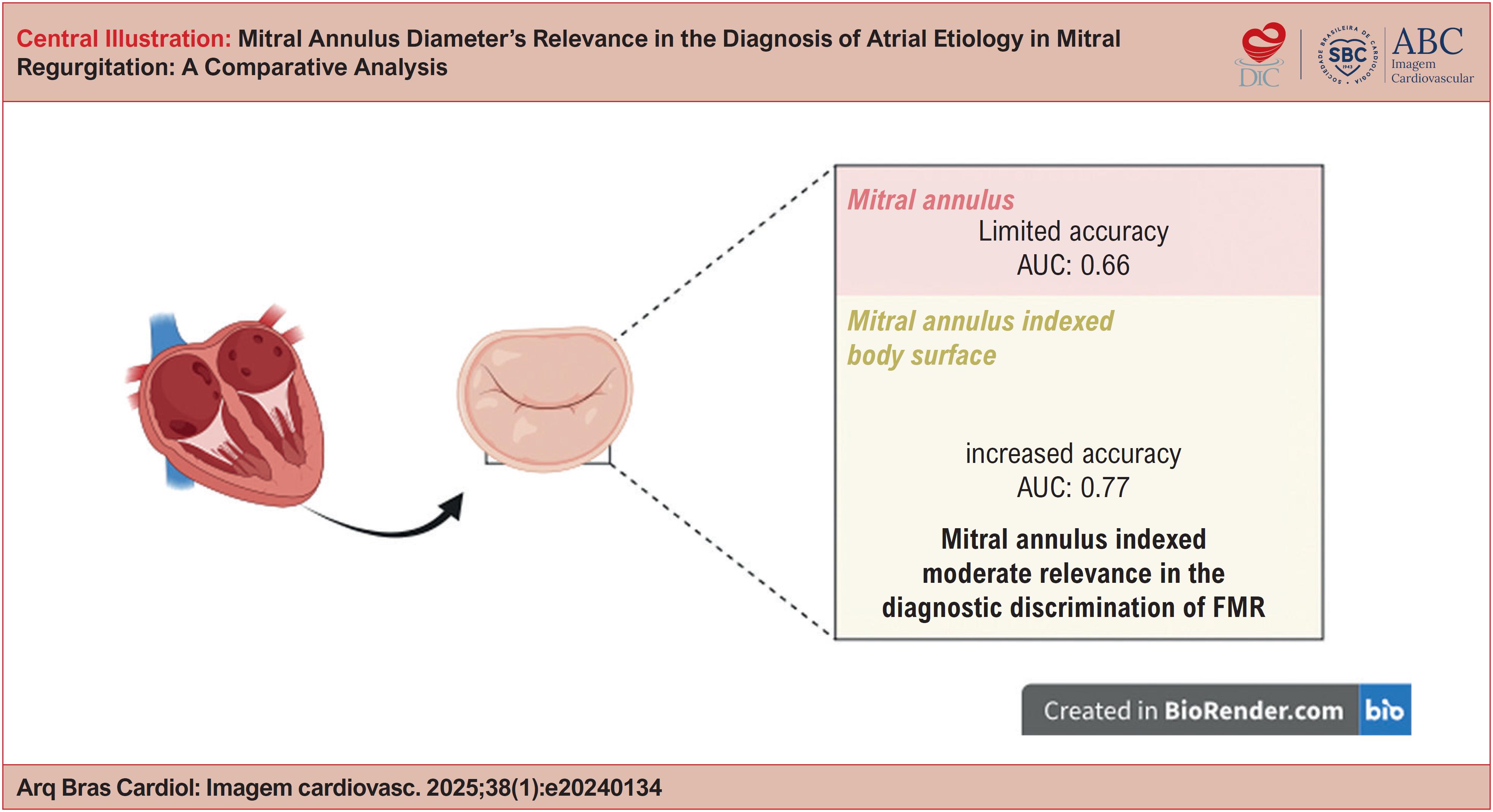
The diagnostic accuracy of the mitral annulus intercommissural diameter showed an area under the curve (AUC) of 0.659 (95% CI: 0.550 - 0.768), while the ROC curve analysis of the mitral diameter indexed through the body surface exhibited an AUC of 0.767 (95% CI: 0.675 - 0.859). The complementary analysis of the ROC curve identified an optimal cutoff value of 20.8750, at which the sensitivity and specificity were 67.9% ( Graph 1 ).
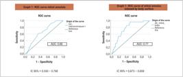
ROC Curves for evaluating atrial etiology. (A) Non-indexed mitral annulus: AUC = 0.66 (95% CI: 0.550–0.768), indicating moderate discriminatory ability. (B) Mitral annulus indexed by body surface area: AUC = 0.77 (95% CI: 0.675–0.859), suggesting higher diagnostic accuracy. The reference line represents the absence of discriminatory power (AUC = 0.5). The curves were adjusted based on the dimensions of the mitral annulus (intercommissural length or indexed value) and reflect the population heterogeneity, highlighting the superiority of the adjusted index in distinguishing atrial etiology. AUC: area under the curve; ROC: receiver operating characteristic.
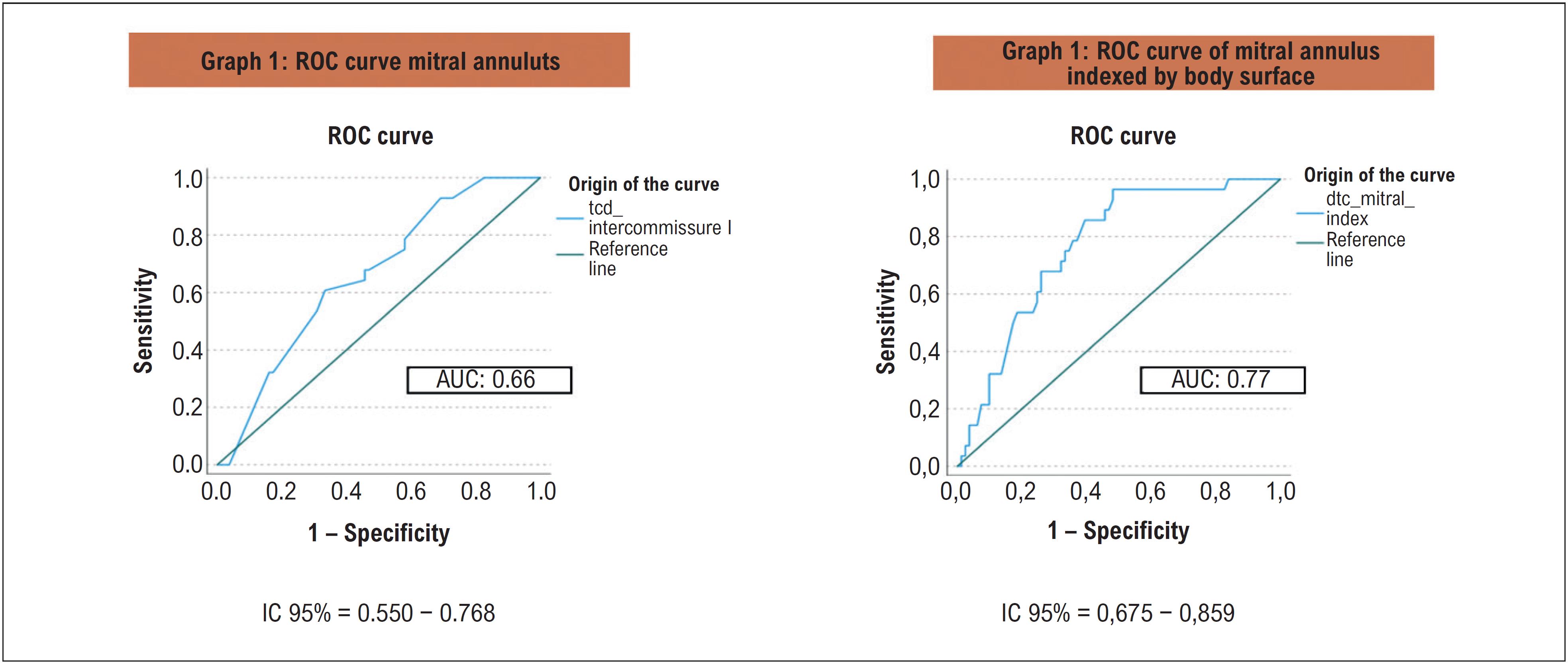
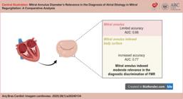
These findings highlight the relevance of the indexed intercommissural diameter as a potentially superior diagnostic metric to distinguish Atrial from Non-atrial etiologies of MR (Central Illustration).
Discussion
The prevalence of FMR in our study (25%) is consistent with the average number reported in previous observational studies, reflecting the relevance of this condition.2 , 7 , 17 Moreover, the analysis demonstrated that AF is a relevant marker for atrial etiology, with statistically significant differences between the Atrial and Non-atrial groups ( p < 0.001). AF prevalence reached 71.4% in the atrial MR group, underscoring its strong association with this etiology.18 - 20 These findings reinforce the importance of considering AF when identifying the atrial functional etiology in patients with MR.
In this study, a statistically significant difference was observed in the median ages between groups. Patients in the Atrial group had a higher median age (77 years; interquartile range: 73–84) compared to the Non-atrial group (67 years; interquartile range: 60–71; p < 0.001). This pattern is in agreement with data previously reported in the literature, which associates advanced age with a greater predisposition to the atrial etiology of MR.18 , 21 , 22 Aging is associated with structural changes such as Left Atrial dilation, which contribute to the development of FMR. These findings reinforce the relevance of age as a relevant pathophysiological factor in determining the etiology of MR.
In the Atrial etiology group, the gender distribution showed 42.9% males and 57.1% females, indicating a slight female predominance. In the Non-atrial group, the proportion of men was considerably higher, representing 67.9% of the participants (55 of 81 individuals). The difference in gender distribution between the groups was statistically significant ( p = 0.019), suggesting that gender may influence the etiology of MR. Although the exact mechanisms remain unclear, previous studies suggest anatomical and hormonal factors may contribute to atrial remodeling and the atrial etiology of MR development.3 , 5 However, these hypotheses still require further investigation.
In the Atrial group, LVEF showed a mean of 56.04 ± 7.53%, while in the Non-atrial group, the mean was significantly lower, reaching 44.42 ± 1.90% ( p < 0.001). The preservation of the ejection fraction in the atrial group reflects a striking characteristic of this group, since the reduction in LVEF, when present, occurs only in advanced stages of atrial FMR.21 , 23 - 25 FMR is recognized as an important prognostic indicator in patients with AF and HFpEF. This condition is associated with a higher risk of progressive systolic dysfunction and reduced LVEF, factors that contribute to clinical worsening and adverse outcomes.26 These findings reinforce the need for careful monitoring of left ventricular function in this clinical context.
The behavior of the mitral annulus and the mitral annulus indexed by body surface area was of particular interest in this study. Prior research on the mitral annulus as a diagnostic tool for FMR has yielded conflicting results, with most studies relying on absolute measurements obtained via transthoracic echocardiography.27 This study utilized transesophageal echocardiography due to its superior precision in assessing the mitral valve, annular complex, and valve morphology.27 - 29 Body surface area indexing was used to ensure greater reliability of measurements, especially in heterogeneous populations. This approach allows for a more standardized assessment adjusted to the individuality of patients, expanding its clinical applicability.20 These factors reinforce the importance of indexing in the context of FMR, where diagnostic accuracy is essential.
The intercommissural mitral annular diameter was compared between atrial and non-atrial MR groups, revealing significant differences in both absolute and indexed measurements. The mean absolute diameter was 35 mm in the Non-atrial group and 37.1 mm in the Atrial group ( p = 0.012), with a Cohen's d of 0.580, indicating a moderate effect size. When adjusted for body surface area, the difference became even more pronounced, with mean values of 19.35 mm/m² in the non-atrial group and 21.63 mm/m² in the atrial group ( p < 0.001, Cohen's d = 0.972), indicating a large effect size.
These findings confirm that indexing by body surface area enhances the detection of mitral annular changes, standardizing measurements to account for individual patient characteristics. This approach is particularly useful in heterogeneous populations and may improve clinical decision-making by refining the differentiation between atrial and non-atrial MR.20 , 29
The analysis of diagnostic accuracy using the ROC curve for the intercommissural diameter of the mitral annulus revealed an AUC of 0.659 (95% CI: 0.550–0.768), indicating only moderate accuracy in distinguishing between the atrial and non-atrial groups. On the other hand, indexing the mitral diameter by body surface area showed superior relevance, with an AUC of 0.767 (95% CI: 0.675–0.859), reflecting a more robust diagnostic discrimination capacity.
Additionally, the ROC curve analysis identified an optimal cutoff value of 20.8750 mm/m², with sensitivity and specificity of 67.9%. These findings reinforce the potential of indexed diameter as a superior tool when compared to the absolute value of the valve annulus in diagnostic algorithms, offering greater accuracy in differentiating between atrial and non-atrial etiologies of MR.
While indexing the mitral annular diameter increased diagnostic accuracy compared to absolute measurements, limitations remain. The C statistic and Youden index analysis suggest that, when used in isolation, the indexed annular diameter provides only modest diagnostic accuracy. This underscores the need for more comprehensive diagnostic models that integrate indexed diameter with additional clinical and echocardiographic variables to improve atrial MR identification.
Conclusion
The findings support the use of indexed diameter as a superior metric over absolute annular size in diagnostic algorithms, offering greater accuracy in differentiating MR etiologies. However, incorporating this measure into broader diagnostic frameworks may further enhance its clinical utility.
-
Sources of Funding
There were no external funding sources for this study.
-
Study Association
This article is part of the thesis of Doctoral submitted by Alexandre Costa Souza, from Instituto Dor de ensino e pesquisa (Idor).
-
Ethics Approval and Consent to Participate
This study was approved by the Ethics Committee of the Hospital São Rafael under the protocol number 5722007. All the procedures in this study were in accordance with the 1975 Helsinki Declaration, updated in 2013. Informed consent was obtained from all participants included in the study.
References
-
1 Deferm S, Bertrand PB, Verbrugge FH, Verhaert D, Rega F, Thomas JD, et al. Atrial Functional Mitral Regurgitation: JACC Review Topic of the Week. J Am Coll Cardiol. 2019;73(19):2465-76. doi: 10.1016/j.jacc.2019.02.061.
» https://doi.org/10.1016/j.jacc.2019.02.061 -
2 Pretto AS. Atrial Functional Mitral Regurgitation. Arq Bras Cardiol: Imagem Cardiovasc. 2024;37(1):e20230097. doi: 10.36660/abcimg.20230097i.
» https://doi.org/10.36660/abcimg.20230097i -
3 Doldi P, Stolz L, Orban M, Karam N, Praz F, Kalbacher D, et al. Transcatheter Mitral Valve Repair in Patients with Atrial Functional Mitral Regurgitation. JACC Cardiovasc Imaging. 2022;15(11):1843-51. doi: 10.1016/j.jcmg.2022.05.009.
» https://doi.org/10.1016/j.jcmg.2022.05.009 -
4 Mesi O, Gad MM, Crane AD, Ramchand J, Puri R, Layoun H, et al. Severe Atrial Functional Mitral Regurgitation: Clinical and Echocardiographic Characteristics, Management and Outcomes. JACC Cardiovasc Imaging. 2021;14(4):797-808. doi: 10.1016/j.jcmg.2021.02.008.
» https://doi.org/10.1016/j.jcmg.2021.02.008 -
5 Farhan S, Silbiger JJ, Halperin JL, Zhang L, Dukkipati SR, Vogel B, et al. Pathophysiology, Echocardiographic Diagnosis, and Treatment of Atrial Functional Mitral Regurgitation: JACC State-of-the-Art Review. J Am Coll Cardiol. 2022;80(24):2314-30. doi: 10.1016/j.jacc.2022.09.046.
» https://doi.org/10.1016/j.jacc.2022.09.046 -
6 Dziadzko V, Dziadzko M, Medina-Inojosa JR, Benfari G, Michelena HI, Crestanello JA, et al. Causes and Mechanisms of Isolated Mitral Regurgitation in the Community: Clinical Context and Outcome. Eur Heart J. 2019;40(27):2194-202. doi: 10.1093/eurheartj/ehz314.
» https://doi.org/10.1093/eurheartj/ehz314 -
7 Moonen A, Ng MKC, Playford D, Strange G, Scalia GM, Celermajer DS. Atrial Functional Mitral Regurgitation: Prevalence, Characteristics and Outcomes from the National Echo Database of Australia. Open Heart. 2023;10(1):e002180. doi: 10.1136/openhrt-2022-002180.
» https://doi.org/10.1136/openhrt-2022-002180 -
8 Muraru D, Guta AC, Ochoa-Jimenez RC, Bartos D, Aruta P, Mihaila S, et al. Functional Regurgitation of Atrioventricular Valves and Atrial Fibrillation: An Elusive Pathophysiological Link Deserving Further Attention. J Am Soc Echocardiogr. 2020;33(1):42-53. doi: 10.1016/j.echo.2019.08.016.
» https://doi.org/10.1016/j.echo.2019.08.016 -
9 Hoit BD. Atrial Functional Mitral Regurgitation: The Left Atrium Gets its Due Respect. J Am Coll Cardiol. 2011;58(14):1482-4. doi: 10.1016/j.jacc.2011.06.033.
» https://doi.org/10.1016/j.jacc.2011.06.033 -
10 Gertz ZM, Raina A, Saghy L, Zado ES, Callans DJ, Marchlinski FE, et al. Evidence of Atrial Functional Mitral Regurgitation Due to Atrial Fibrillation: Reversal with Arrhythmia Control. J Am Coll Cardiol. 2011;58(14):1474-81. doi: 10.1016/j.jacc.2011.06.032.
» https://doi.org/10.1016/j.jacc.2011.06.032 -
11 Silbiger JJ. Mechanistic Insights Into Atrial Functional Mitral Regurgitation: Far More Complicated than Just Left Atrial Remodeling. Echocardiography. 2019;36(1):164-9. doi: 10.1111/echo.14249.
» https://doi.org/10.1111/echo.14249 -
12 Kagiyama N, Mondillo S, Yoshida K, Mandoli GE, Cameli M. Subtypes of Atrial Functional Mitral Regurgitation: Imaging Insights Into Their Mechanisms and Therapeutic Implications. JACC Cardiovasc Imaging. 2020;13(3):820-35. doi: 10.1016/j.jcmg.2019.01.040.
» https://doi.org/10.1016/j.jcmg.2019.01.040 -
13 Machino-Ohtsuka T, Seo Y, Ishizu T, Sato K, Sugano A, Yamamoto M, et al. Novel Mechanistic Insights Into Atrial Functional Mitral Regurgitation - 3-Dimensional Echocardiographic Study. Circ J. 2016;80(10):2240-8. doi: 10.1253/circj.CJ-16-0435.
» https://doi.org/10.1253/circj.CJ-16-0435 -
14 Zoghbi WA, Adams D, Bonow RO, Enriquez-Sarano M, Foster E, Grayburn PA, et al. Recommendations for Noninvasive Evaluation of Native Valvular Regurgitation: A Report from the American Society of Echocardiography Developed in Collaboration with the Society for Cardiovascular Magnetic Resonance. J Am Soc Echocardiogr. 2017;30(4):303-71. doi: 10.1016/j.echo.2017.01.007.
» https://doi.org/10.1016/j.echo.2017.01.007 -
15 Grayburn PA, Thomas JD. Basic Principles of the Echocardiographic Evaluation of Mitral Regurgitation. JACC Cardiovasc Imaging. 2021;14(4):843-53. doi: 10.1016/j.jcmg.2020.06.049.
» https://doi.org/10.1016/j.jcmg.2020.06.049 -
16 Zoghbi WA, Levine RA, Flachskampf F, Grayburn P, Gillam L, Leipsic J, et al. Atrial Functional Mitral Regurgitation: A JACC: Cardiovascular Imaging Expert Panel Viewpoint. JACC Cardiovasc Imaging. 2022;15(11):1870-82. doi: 10.1016/j.jcmg.2022.08.016.
» https://doi.org/10.1016/j.jcmg.2022.08.016 -
17 Naser JA, Alexandrino FB, Harada T, Michelena HI, Borlaug BA, Eleid MF, et al. The Natural History of Atrial Functional Mitral Regurgitation. J Am Coll Cardiol. 2024;83(16):1495-507. doi: 10.1016/j.jacc.2024.02.026.
» https://doi.org/10.1016/j.jacc.2024.02.026 -
18 Kihara T, Gillinov AM, Takasaki K, Fukuda S, Song JM, Shiota M, et al. Mitral Regurgitation Associated with Mitral Annular Dilation in Patients with Lone Atrial Fibrillation: An Echocardiographic Study. Echocardiography. 2009;26(8):885-9. doi: 10.1111/j.1540-8175.2009.00904.x.
» https://doi.org/10.1111/j.1540-8175.2009.00904.x -
19 Cramariuc D, Alfraidi H, Nagata Y, Levine RA, van Kampen A, Andrews C, et al. Atrial Dysfunction in Significant Atrial Functional Mitral Regurgitation: Phenotypes and Prognostic Implications. Circ Cardiovasc Imaging. 2023;16(5):e015089. doi: 10.1161/CIRCIMAGING.122.015089.
» https://doi.org/10.1161/CIRCIMAGING.122.015089 -
20 Lang RM, Badano LP, Mor-Avi V, Afilalo J, Armstrong A, Ernande L, et al. Recommendations for Cardiac Chamber Quantification by Echocardiography in Adults: An Update from the American Society of Echocardiography and the European Association of Cardiovascular Imaging. J Am Soc Echocardiogr. 2015;28(1):1-39.e14. doi: 10.1016/j.echo.2014.10.003.
» https://doi.org/10.1016/j.echo.2014.10.003 -
21 Kim DH, Heo R, Handschumacher MD, Lee S, Choi YS, Kim KR, et al. Mitral Valve Adaptation to Isolated Annular Dilation: Insights Into the Mechanism of Atrial Functional Mitral Regurgitation. JACC Cardiovasc Imaging. 2019;12(4):665-77. doi: 10.1016/j.jcmg.2017.09.013.
» https://doi.org/10.1016/j.jcmg.2017.09.013 -
22 Naser JA, Michelena HI, Lin G, Scott CG, Lee E, Kennedy AM, et al. Incidence, Risk Factors, and Outcomes of Atrial Functional Mitral Regurgitation in Patients with Atrial Fibrillation or Sinus Rhythm. Eur Heart J Cardiovasc Imaging. 2023;24(11):1450-7. doi: 10.1093/ehjci/jead199.
» https://doi.org/10.1093/ehjci/jead199 -
23 Abe Y, Akamatsu K, Ito K, Matsumura Y, Shimeno K, Naruko T, et al. Prevalence and Prognostic Significance of Functional Mitral and Tricuspid Regurgitation Despite Preserved Left Ventricular Ejection Fraction in Atrial Fibrillation Patients. Circ J. 2018;82(5):1451-8. doi: 10.1253/circj.CJ-17-1334.
» https://doi.org/10.1253/circj.CJ-17-1334 -
24 Reddy YNV, Obokata M, Verbrugge FH, Lin G, Borlaug BA. Atrial Dysfunction in Patients with Heart Failure with Preserved Ejection Fraction and Atrial Fibrillation. J Am Coll Cardiol. 2020;76(9):1051-64. doi: 10.1016/j.jacc.2020.07.009.
» https://doi.org/10.1016/j.jacc.2020.07.009 -
25 Zakeri R, Chamberlain AM, Roger VL, Redfield MM. Temporal Relationship and Prognostic Significance of Atrial Fibrillation in Heart Failure Patients with Preserved Ejection Fraction: A Community-Based Study. Circulation. 2013;128(10):1085-93. doi: 10.1161/CIRCULATIONAHA.113.001475.
» https://doi.org/10.1161/CIRCULATIONAHA.113.001475 -
26 Tang Z, Fan YT, Wang Y, Jin CN, Kwok KW, Lee AP. Mitral Annular and Left Ventricular Dynamics in Atrial Functional Mitral Regurgitation: A Three-Dimensional and Speckle-Tracking Echocardiographic Study. J Am Soc Echocardiogr. 2019;32(4):503-13. doi: 10.1016/j.echo.2018.11.009.
» https://doi.org/10.1016/j.echo.2018.11.009 -
27 Akamatsu K, Abe Y, Matsumura Y, Shimeno K, Naruko T, Takahashi Y, et al. Etiology of Atrial Functional Mitral Regurgitation: Insights from Transthoracic Echocardiography in 159 Consecutive Patients with Atrial Fibrillation and Preserved Left Ventricular Ejection Fraction. Cardiology. 2020;145(8):511-21. doi: 10.1159/000508279.
» https://doi.org/10.1159/000508279 -
28 Kagiyama N, Toki M, Hara M, Fukuda S, Aritaka S, Miki T, et al. Efficacy and Accuracy of Novel Automated Mitral Valve Quantification: Three-Dimensional Transesophageal Echocardiographic Study. Echocardiography. 2016;33(5):756-63. doi: 10.1111/echo.13135.
» https://doi.org/10.1111/echo.13135 -
29 Ring L, Dutka DP, Boyd J, Parker K, Wendler O, Monaghan MJ, et al. The Normal Mitral Valve Annulus in Humans Defined Using 3-Dimensional Transesophageal Echocardiography. JACC Cardiovasc Imaging. 2018;11(3):510-2. doi: 10.1016/j.jcmg.2017.05.017.
» https://doi.org/10.1016/j.jcmg.2017.05.017
Edited by
-
Editor responsible for the review:
Marcelo Tavares

 AUC: area under the curve; MR: Mitral Regurgitation.