Diagnostic Performance Of Contrast-Enhanced Echocardiography In Differentiating Cardiac Masses: A Systematic Review And Meta-analysis
Abstract
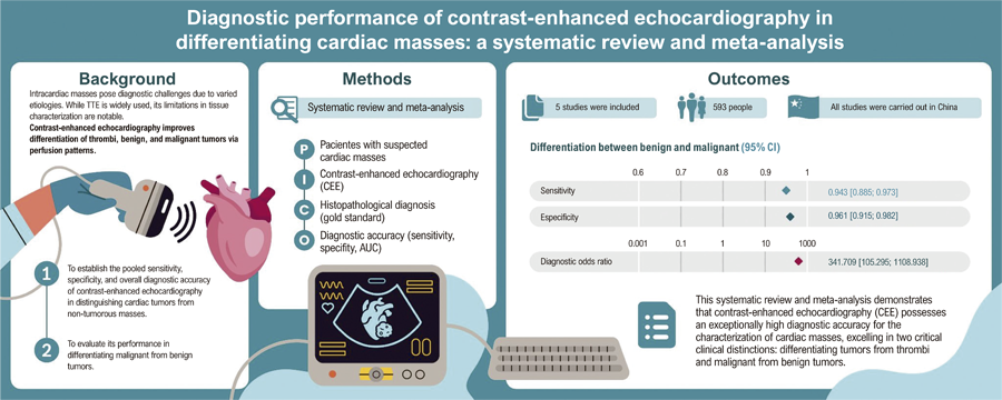
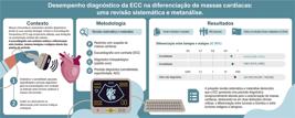
Background Conventional echocardiography often struggles to differentiate intracardiac masses, particularly in patients with poor acoustic windows. Contrast-enhanced Echocardiography (CEE) overcomes this limitation by visualizing perfusion patterns — distinguishing avascular thrombi from vascularized tumors. We aimed to synthesize existing evidence to evaluate the diagnostic accuracy of CEE.
Objectives To evaluate the diagnostic accuracy of CEE for differentiating cardiac masses in adults, using histopathology as reference and reporting AUC, sensitivity, specificity, PPV, and NPV.
Methods Systematic searches of PubMed, Web of Science, Cochrane Library, and EMBASE were performed on August 10, 2025. Studies meeting PICOTT criteria were included; extracted data included sensitivity, specificity, AUC, and 2×2 tables. Pooled estimates were obtained using standard bivariate and SROC models for diagnostic meta-analysis. Statistical significance set at P < 0.05.
Results Five prospective cohort studies (total n = 381 patients) were included. For tumor vs non-tumor, pooled sensitivity = 100% and specificity = 100% (95% CI 99.5–100%; I2 = 0%; heterogeneity P = 0.985), diagnostic odds ratio (DOR) = 3,890.65, AUC = 0.989. For malignant vs benign tumors, pooled sensitivity = 94.3% (95% CI 88.5–97.3%; I2 = 0%; P = 0.681), specificity = 96.1% (95% CI 91.5–98.2%; I2 = 0%; P = 0.970), DOR = 341.71, SROC AUC = 0.976.
Conclusions CEE showed very high diagnostic accuracy in the available prospective series. However, the small number of studies and limited sample sizes warrant cautious interpretation; larger prospective multicenter studies with standardized CEE protocols are needed to confirm these results.
Keywords:
Echocardiography; Contrast Media; Cardiac Neoplasms; Systematic Review; Meta-Analysis
Resumo
Fundamento Muitas vezes, a ecocardiografia convencional encontra dificuldade em diferenciar massas intracardíacas, especialmente em pacientes com janelas acústicas inadequadas. A Ecocardiografia com Contraste (ECC) supera essa limitação ao visualizar padrões de perfusão, distinguindo trombos avasculares de tumores vascularizados. Nosso objetivo foi sintetizar as evidências existentes para avaliar a precisão diagnóstica da ECC.
Objetivos Avaliar a precisão diagnóstica da ECC na diferenciação de massas cardíacas em adultos, utilizando a histopatologia como referência e relatando AUC, sensibilidade, especificidade, VPP e VPN.
Métodos Buscas sistemáticas foram realizadas nas bases de dados PubMed, Web of Science, Cochrane Library e EMBASE até 10 de agosto de 2025. Foram incluídos estudos que atendiam aos critérios PICOTT; os dados extraídos compreenderam sensibilidade, especificidade, AUC e tabelas 2×2. As estimativas agrupadas foram obtidas utilizando modelos bivariados padrão e modelos SROC para metanálise diagnóstica. A significância estatística foi definida como P < 0,05.
Resultados Foram incluídos cinco estudos de coorte prospectivos (total n = 381 pacientes). Para tumor versus não tumor, a sensibilidade agrupada foi de 100% e a especificidade foi de 100% (IC 95% 99,5–100%; I2 = 0%; heterogeneidade P = 0,985), odds ratio diagnóstico (DOR) = 3.890,65, AUC = 0,989. Para tumores malignos versus benignos, a sensibilidade agrupada foi de 94,3% (IC 95% 88,5–97,3%; I2 = 0%; P = 0,681), a especificidade foi de 96,1% (IC 95% 91,5–98,2%; I2 = 0%; P = 0,970), a DOR foi de 341,71 e a AUC da SROC foi de 0,976.
Conclusões A ECC demonstrou uma precisão diagnóstica muito alta nas séries prospectivas disponíveis. No entanto, o número reduzido de estudos e o tamanho limitado das amostras exigem uma interpretação cautelosa; estudos prospectivos multicêntricos de maior porte, com protocolos padronizados de ECC, são necessários para confirmar esses resultados.
Palavras-chave:
Ecocardiografia; Meios de Contraste; Neoplasias Cardíacas; Revisão Sistemática; Metanálise
Introduction
Intracardiac masses represent a diagnostic challenge due to their diverse etiologies, including thrombi, benign tumors (such as myxomas), and malignant lesions, all with significantly different prognoses and treatment strategies. Transthoracic echocardiography (TTE) remains the initial and most accessible imaging modality in clinical practice, offering real-time assessment of morphology and hemodynamic effects. However, its diagnostic yield is often limited in patients with poor acoustic windows or atypical mass locations, which may lead to underdetection or misclassification of masses.1,2 Transesophageal Echocardiography (TEE) imaging can improve visualization but still falls short in reliable tissue characterization, especially when compared with cardiac MRI or CT, which provide richer tissue contrast and spatial resolution but are more resource-intensive.1,3
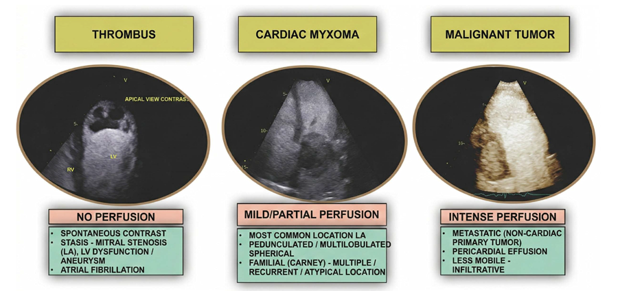
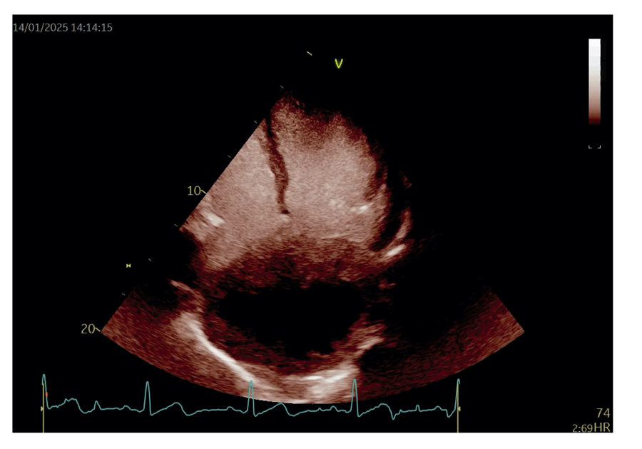
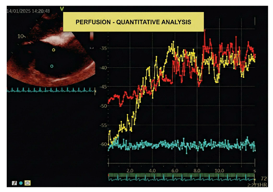
Contrast-enhanced Echocardiography (CEE) has emerged as a compelling adjunct to overcome these limitations. By enhancing perfusion imaging, CEE can differentiate avascular thrombi, mildly perfused benign tumors, and hypervascular malignant lesions based on distinct vascular patterns.4,5 For instance, the use of ultrasound-enhancing agents can vividly illustrate a mass’s perfusion characteristics (Figure 1). This technique can reveal details such as peripheral contrast uptake with a necrotic core in a cardiac paraganglioma (Figure 2) and enables quantitative analysis that differentiates perfused from non-perfused components (Figure 3).
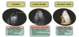
– Examples of the use of an ultrasound-enhancing agent for the evaluation of mass perfusion.
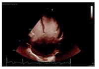
– Peripheral contrast uptake in a large mass inside the left atrium, without central uptake (necrosis) in a cardiac paraganglioma.
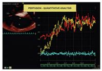
– Subcostal view. Cardiac paraganglioma. Quantitative analysis of the mass perfusion with an ultrasound-enhancing agent (contrast) - in yellow, the peripheral perfusion of the mass; in red, perfusion of the liver tissue for comparison; in blue, the absence of perfusion in the (necrotic) center of the mass.
Initial prospective data have shown that CEE correctly identifies cardiac mass types in 90%– 97% of cases, even with trainee observers, highlighting its potential for routine clinical use.4 Nonetheless, the current evidence base is characterized by small-scale studies, retrospective designs, and case reports, raising concern about generalizability and robustness.5
The primary objective of this meta-analysis is to establish the pooled sensitivity, specificity, and overall diagnostic accuracy of CEE in distinguishing cardiac tumors from non-tumorous masses. The secondary objective is to evaluate its performance in differentiating malignant from benign tumors. Ultimately, this study seeks to provide evidence to guide clinical decision-making and highlight priorities for future, large-scale prospective research.
Methods
Protocol and Registration
This systematic review and meta-analysis were developed strictly adhering to the recommendations of the Preferred Reporting Items for Systematic Reviews and Meta-Analyses6 (PRISMA 2020) statement, its extension for diagnostic test accuracy studies (PRISMA-DTA), and the Cochrane Handbook for Systematic Reviews of Diagnostic Test Accuracy.7 The study protocol was submitted to the International Prospective Register of Systematic Reviews8 (PROSPERO) under the registration number CRD420251142676.
Study Design
Diagnostic accuracy studies with a prospective or retrospective design were included. No time restrictions were applied, including articles from the earliest available date in the databases. Reviews, editorials, case reports, and case series with fewer than ten participants were excluded.
Eligibility Criteria
Studies were selected based on eligibility criteria defined by the PICOS framework. The eligible population (P) consisted of adult patients with suspected cardiac masses who underwent CEE. The results of the CEE were compared with the reference standard for a definitive diagnosis (Comparator), which was primarily based on histopathological analysis. However, diagnoses confirmed by other robust imaging modalities (e.g., Cardiac Magnetic Resonance) or by unequivocal therapeutic response (e.g., resolution of a thrombus after anticoagulation therapy) were also considered. The primary outcomes (O) of interest were diagnostic accuracy measures, including Area Under the Curve (AUC), sensitivity, specificity, Positive Predictive Value (PPV), and Negative Predictive Value (NPV).
Target Conditions
The target conditions for this review were the different subtypes of intracardiac masses. The primary condition to be identified was a cardiac tumor (benign or malignant), rather than an intracardiac thrombus.
Additionally, within the spectrum of tumors, a second target was to differentiate benign tumors (e.g., myxoma, fibroma) from malignant tumors (primary, such as sarcomas, or metastatic). The accuracy analyses were organized into subgroups to assess the test’s performance for each of these key clinical distinctions.
Index Test
The index test was defined as CEE, performed to characterize a previously identified or suspected cardiac mass. CEE was considered any echocardiogram that involved the intravenous administration of a microbubble contrast agent to assess the vascularity and perfusion of the mass. The index test result was not simply dichotomous (positive/negative), but rather a classification of the mass based on its perfusion patterns, with findings compared with the reference standard obtained at approximately the same time.
Information Sources and Search Strategy
A systematic and comprehensive search was conducted in the following electronic databases: PubMed, Embase, Cochrane Library, and Web of Science. The search was completed on August 10, 2025.
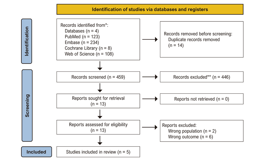
The initial search identified 473 articles (123 in PubMed, 234 in Embase, 8 in the Cochrane Library, and 108 in Web of Science) before removing duplicates. Additionally, the reference lists of included studies were manually searched to identify potentially eligible articles not captured in the initial search.
Study Selection and Data Extraction
The selection process was managed using the Rayyan software.9 Two independent reviewers (JP and AN) screened titles and abstracts, followed by a full-text assessment. Disagreements were resolved by consensus or through adjudication by a third reviewer. Data were extracted using a standardized form, which included study characteristics, population details, intervention specifics, and raw data for the 2x2 contingency table.
Risk of Bias Assessment
The methodological quality and risk of bias of each included study were independently assessed by two reviewers using the QUADAS-2 tool.10
Data Synthesis and Analysis
The accuracy data were synthesized through a meta-analysis using a bivariate random-effects model. From this model, summary estimates with 95% confidence intervals (CIs) for sensitivity and specificity were generated; the Summary Receiver Operating Characteristic (SROC) curve was constructed, and the diagnostic odds ratio was calculated. Heterogeneity was evaluated using I2 statistics, with P < 0.05 from Cochran’s Q test or I2 > 50% considered indicative of substantial heterogeneity. Statistical significance set at P < 0.05. Forest plots were used to illustrate individual and pooled effect sizes. Meta-analyses were performed in RStudio (RStudio 2025.09.0+387) for Windows using the “meta” and “mada” packages for data synthesis and visualization.
Results
Results of the Search
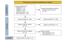
The initial search yielded 473 results. After removing duplicate records and ineligible studies, 13 remained and were fully reviewed against the inclusion criteria. Of these, 5 studies were included. The process is detailed in the PRISMA flow diagram (Figure 4).
The number of participants ranged from 32 to 236; all were adults. The studies varied in design, including prospective observational, cross-sectional, and retrospective approaches, and were conducted across both single and multicenter settings.
All studies employed Contrast-enhanced Echocardiography (CEE) using SonoVue (Bracco, Switzerland) as the contrast agent. The echocardiographic systems used included Philips iE33, in three studies11-13, and GE Vivid 7 Dimension, in one study14, with transducers and imaging protocols tailored to each study’s objectives. One study15 did not report the system used in echocardiography.
Quantitative parameters assessed across studies included mass area, peak intensity ratios (e.g., A1/A2, A1/A3), contrast enhancement intensity (A), replenishment rate (β or k), and perfusion ratios between cardiac masses and adjacent myocardium. Qualitative assessments encompassed echogenicity, contour, base morphology, mobility, perfusion characteristics, and presence of pericardial or pleural effusion.
The proportion of male participants ranged from 36.5% to 63.0%, and most studies focused on adult patients presenting with suspected cardiac masses after Transthoracic Echocardiography (TTE). One study13 included patients undergoing surgical treatment for cardiac masses, while another study15 targeted patients referred for myocardial contrast echocardiography.
Exclusion criteria were consistent across studies and were severe cardiac or systemic conditions (e.g., NYHA class IV heart failure, arrhythmias, hepatic or renal dysfunction), allergies to contrast agents or blood products, and neuropsychiatric disorders. Some studies also excluded patients lost to follow-up or managed conservatively. Other important characteristics of the studies included in this review are presented in Table 1.
We found no publication bias by visually analyzing the funnel plot (Figure S1), a linear regression for the asymmetry of the funnel plot was made by the Deek’s test, which was not statistically significant (Bias = -3.940, SE = 5.283, t = -0.75, p = 0.509), however because of the low number of included studies, the results should be regarded with care and are by themselves not enough to discard publication bias.
Methodological Quality of Included Studies
The methodological quality of the included studies was evaluated using the QUADAS-2 tool, which assesses four domains: patient selection, index test, reference standard, and flow & timing. Each domain was judged to be low risk, with some concerns, or high risk of bias. At the individual study level, two studies11,12 were rated as having an overall low risk of bias, while two14,15 raised some concerns, particularly regarding patient selection and flow & timing. One study13 was considered at high risk of bias due to inappropriate patient selection and concerns regarding the reference standard (Figure S2). In the domain-level analysis, patient selection and flow & timing were the areas with the highest frequency of concerns. At the same time, the index test and reference standard were generally well-conducted. Overall, the methodological quality of the included studies was considered acceptable, with most studies at low risk of bias, although relevant limitations were identified in specific domains (Figure S3).
Findings
To Differentiate Tumor from Thrombi
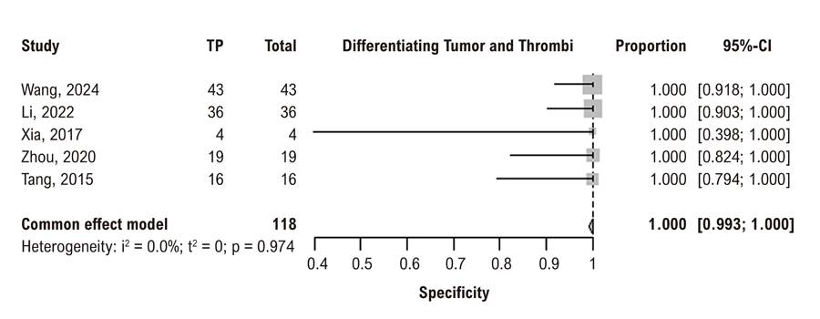
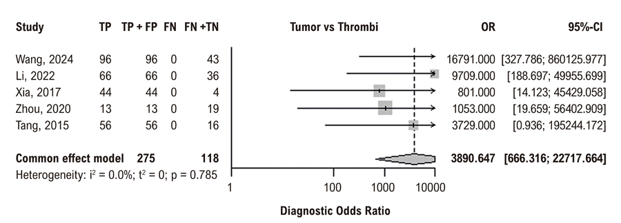
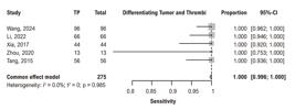
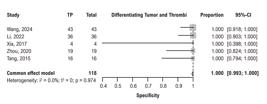
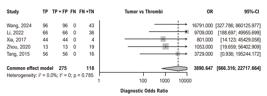
Every study reported high accuracy in differentiating tumors from thrombi using CEE, resulting in strong diagnostic performance parameters. The estimates for summary sensitivity (Figure 5), specificity (Figure 6), and Diagnostic Odds Ratio (Figure 7) confirm these findings. Due to the 100% accuracy, an SROC curve could not be plotted, but the AUC was 0.989.
To Differentiate Malignant Tumor from Benign
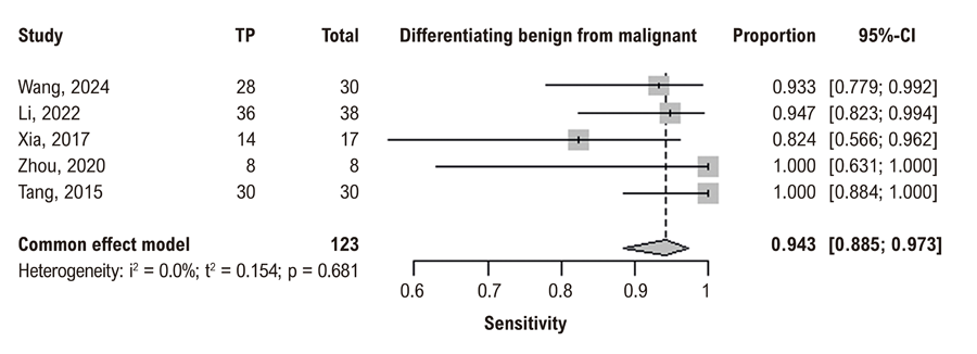
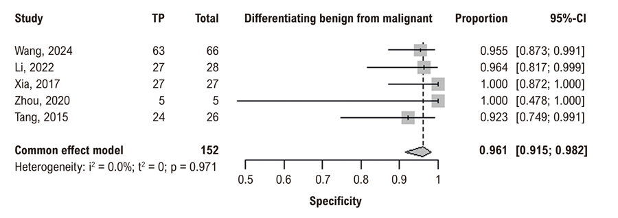
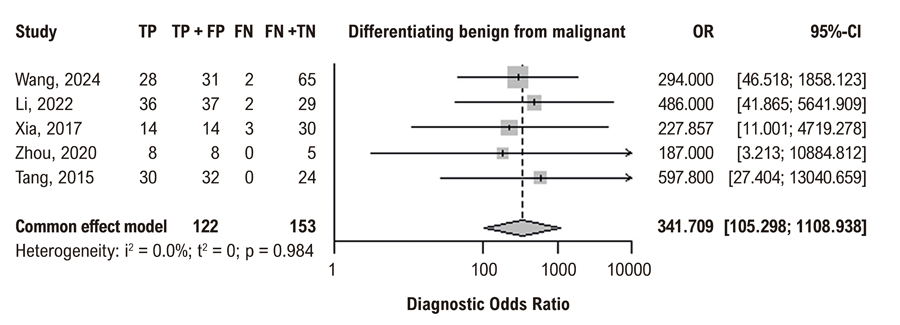
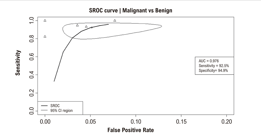
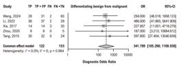
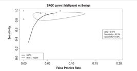
CEE had great results, with high estimates of summary sensitivity (Figure 8) and specificity (Figure 9). The summary Diagnostic Odds Ratio (Figure 10) further supports these findings. Additionally, the SROC curve was plotted (Figure 11).


Discussion
This systematic review and meta-analysis demonstrate that CEE has exceptionally high diagnostic accuracy for characterizing cardiac masses, excelling in two critical clinical distinctions: differentiating tumors from thrombi and malignant from benign tumors.
The first key finding of our analysis was the great pooled sensitivity and specificity (100%) of CEE for distinguishing cardiac tumors from thrombi. This result, while remarkable, is biologically plausible. Thrombi are inherently avascular structures, and the intravascular microbubbles used in CEE provide a stark contrast between the complete absence of perfusion within a thrombus and the variable but present vascularization of tumorous tissue, whether benign or malignant. This creates a binary, highly reliable diagnostic feature that is readily identifiable, even to less experienced operators, as suggested by some of the included studies. The near-perfect AUC of 0.989 resulted from model adjustments to prevent infinite values. Even so, these findings warrant cautious interpretation; the limited number of included studies (5) restricts statistical power and may mask potential small-study effects or reporting bias, despite the lack of observed heterogeneity.
The second finding concerns the distinction between benign and malignant tumors. Our pooled analysis yielded a sensitivity of 94.3% (95% CI 88.5% to 97.3%) and a specificity of 96.1% (95% CI 91.5% to 98.2%), with a summary AUC of 0.976. This indicates that CEE is not only excellent at identifying vascularization but also at interpreting its pattern — typically characterized by intense hypervascularity in malignant lesions compared with more moderate, slower perfusion in benign lesions. The high diagnostic odds ratio (DOR = 341.71) signifies a powerful test that can significantly increase or decrease the post-test probability of malignancy, directly informing critical management decisions regarding the urgency of intervention, biopsy planning, or surgical strategy.
Due to concerns of bias and heterogeneity with one study13 we conducted a post-hoc sensitivity analysis by redoing the meta-analysis and leaving the study out; however, it did not significantly alter the results (specificity of 0.952, sensitivity of 0.962, DOR of 374.767), which demonstrates the reliability of the results despite the concerns of bias.
It is the first meta-analysis, to our knowledge, to specifically synthesize the diagnostic performance of CEE for cardiac masses using a rigorous PRISMA-DTA methodology. Secondly, we employed robust statistical models (bivariate and SROC) specifically designed for diagnostic meta-analyses, which account for the potential correlation between sensitivity and specificity and provide more reliable pooled estimates. Thirdly, the included studies were all prospective cohorts, which strengthens the validity of the findings by minimizing selection and recall bias. Finally, the post hoc sensitivity analysis confirmed that the overall results were not unduly influenced by the study judged to be at high risk of bias, thereby enhancing the reliability of our conclusions.
Despite these robust findings, our results must be interpreted in light of several important limitations. The most significant limitation is the small number of included studies (n = 5) and the relatively modest total sample size (n = 381). This was due to most studies failing to report the necessary numbers to calculate the performance metrics. We also may have missed potential studies, as diagnostic accuracy studies are poorly tagged in electronic databases. Regarding publication bias, given the small number of studies, the linear regression Deek’s test did not yield the best results, but a visual analysis of the funnel plot showed no publication bias. While the statistical heterogeneity was negligible (I2 = 0%), the limited number of primary studies constrains the generalizability of our findings and the power to perform more extensive subgroup analyses (e.g., by tumor type, contrast agent generation, or by quantitative or qualitative analyses).
Furthermore, as highlighted by the QUADAS-2 assessment, certain methodological concerns were present in some studies, particularly regarding patient selection and the flow and timing between the index test and the reference standard. The great accuracy for thrombus differentiation, while compelling, should be viewed with cautious optimism until confirmed in larger, multi-center settings, as real-world performance can be influenced by image quality, interpreter expertise, and specific contrast protocols.
The clinical implication of our work is substantial. CEE emerges as a highly accurate, accessible, and cost-effective tool that can be integrated into the diagnostic pathway immediately after the initial detection of a mass on conventional echocardiography. It can confidently rule out thrombus, potentially avoiding the need for more expensive and less accessible cross-sectional imaging in many cases. For tumors, it provides a reliable non-invasive indicator of malignancy, helping to triage patients towards urgent intervention or more deliberate planning.
Conclusion
This meta-analysis provides compelling evidence that CEE is a powerful diagnostic tool with excellent accuracy for characterizing cardiac masses. It effectively differentiates tumors from thrombi and is highly proficient at distinguishing malignant from benign tumors. While limitations inherent in the available literature require cautious interpretation, CEE’s accessibility, safety, and demonstrated performance support its broader adoption in the standard diagnostic workflow for evaluating intracardiac masses.
Supplemental Materials
Supplemental Materials
References
-
1 L'Angiocola PD, Donati R. Cardiac Masses in Echocardiography: A Pragmatic Review. J Cardiovasc Echogr. 2020;30(1):5-14. doi: 10.4103/jcecho.jcecho_2_20.
» https://doi.org/10.4103/jcecho.jcecho_2_20 -
2 Kirkpatrick JN, Wong T, Bednarz JE, Spencer KT, Sugeng L, Ward RP, et al. Differential Diagnosis of Cardiac Masses Using Contrast Echocardiographic Perfusion Imaging. J Am Coll Cardiol. 2004;43(8):1412-9. doi: 10.1016/j.jacc.2003.09.065.
» https://doi.org/10.1016/j.jacc.2003.09.065 -
3 Uenishi EK, Caldas MA, Tsutsui JM, Abduch MC, Sbano JC, Kalil R Filho, et al. Evaluation of Cardiac Masses by Real-Time Perfusion Imaging Echocardiography. Cardiovasc Ultrasound. 2015;13:23. doi: 10.1186/s12947-015-0018-3.
» https://doi.org/10.1186/s12947-015-0018-3 -
4 Angeli F, Bodega F, Bergamaschi L, Armillotta M, Amicone S, Canton L, et al. Multimodality Imaging in the Diagnostic Work-Up of Patients with Cardiac Masses: JACC: CardioOncology State-of-the-Art Review. JACC CardioOncol. 2024;6(6):847-62. doi: 10.1016/j.jaccao.2024.09.006.
» https://doi.org/10.1016/j.jaccao.2024.09.006 -
5 Yang Z, Niu Y, Ma H, Gong W, Yu L, Liu L, et al. Contrast-Enhanced Echocardiographic Diagnosis of Benign and Malignant Cardiac Tumors and its Correlation with Pathology. Front Cardiovasc Med. 2023;10:1182334. doi: 10.3389/fcvm.2023.1182334.
» https://doi.org/10.3389/fcvm.2023.1182334 -
6 Page MJ, McKenzie JE, Bossuyt PM, Boutron I, Hoffmann TC, Mulrow CD, et al. The PRISMA 2020 Statement: An Updated Guideline for Reporting Systematic Reviews. BMJ. 2021;372:n71. doi: 10.1136/bmj.n71.
» https://doi.org/10.1136/bmj.n71 -
7 Deeks JJ, Bossuyt PM, Leeflang MMG, Takwoingi Y, editors. Cochrane Handbook for Systematic Reviews of Diagnostic Test Accuracy. Version 2.0 [Internet]. London: The Cochrane Collaboration; 2023 [cited 2025 Feb 08]. Available from: https://training.cochrane.org/handbook-dta
» https://training.cochrane.org/handbook-dta -
8 National Institute for Health and Care Research. PROSPERO: International Prospective Register of Systematic Reviews [Internet]. York: University of York; 2025 [cited 2026 Feb 08]. Available from: https://www.crd.york.ac.uk/prospero/
» https://www.crd.york.ac.uk/prospero/ -
9 Ouzzani M, Hammady H, Fedorowicz Z, Elmagarmid A. Rayyan-a Web and Mobile App for Systematic Reviews. Syst Rev. 2016;5(1):210. doi: 10.1186/s13643-016-0384-4.
» https://doi.org/10.1186/s13643-016-0384-4 -
10 Whiting PF, Rutjes AW, Westwood ME, Mallett S, Deeks JJ, Reitsma JB, et al. QUADAS-2: A Revised Tool for the Quality Assessment of Diagnostic Accuracy Studies. Ann Intern Med. 2011;155(8):529-36. doi: 10.7326/0003-4819-155-8-201110180-00009.
» https://doi.org/10.7326/0003-4819-155-8-201110180-00009 -
11 Wang Q, Wang B, Zhang X, Zhong X, Chang S, Yang J, et al. The Usefulness of Contrast Echocardiography in the Evaluation of Cardiac Masses: A Multicenter Study. BMC Cardiovasc Disord. 2024;24(1):43. doi: 10.1186/s12872-024-03708-2.
» https://doi.org/10.1186/s12872-024-03708-2 -
12 Li Y, Ren W, Wang X, Xiao Y, Feng Y, Shi P, et al. The Diagnostic Accuracy of Contrast Echocardiography in Patients with Suspected Cardiac Masses: A Preliminary Multicenter, Cross-Sectional Study. Front Cardiovasc Med. 2022;9:1011560. doi: 10.3389/fcvm.2022.1011560.
» https://doi.org/10.3389/fcvm.2022.1011560 -
13 Xia H, Gan L, Jiang Y, Tang Q, Zhang P, Tang X, et al. Use of Transesophageal Echocardiography and Contrast Echocardiography in the Evaluation of Cardiac Masses. Int J Cardiol. 2017;236:466-72. doi: 10.1016/j.ijcard.2017.01.073.
» https://doi.org/10.1016/j.ijcard.2017.01.073 -
14 Tang QY, Guo LD, Wang WX, Zhou W, Liu YN, Liu HY, et al. Usefulness of Contrast Perfusion Echocardiography for Differential Diagnosis of Cardiac Masses. Ultrasound Med Biol. 2015;41(9):2382-90. doi: 10.1016/j.ultrasmedbio.2015.05.010.
» https://doi.org/10.1016/j.ultrasmedbio.2015.05.010 - 15 Zhou Q, Xiong Y, Zhou Y. Feasibility of Myocardial Contrast Echocardiography Quantitative Analysis in Differentiating Cardiac Masses. JACC. 2020;75(11):1688.
-
Study Association:
This study is not associated with any thesis or dissertation work.
-
Ethics Approval and Consent to Participate:
This article does not contain any studies with human participants or animals performed by any of the authors.
-
Availability of Research Data:
The underlying content of the research text is contained within the manuscript.
-
Sources of Funding:
There were no external funding sources for this study.
Edited by
-
Editor responsible for the review:
Marcelo Tavares
The underlying content of the research text is contained within the manuscript.