Persistent Cephalgia Secondary to Carotid Web
Keywords
Headache; Internal Carotid Artery; Vascular Echo Doppler; Carotid Web
Palavras-chave
Cefaleia; Artéria Carótida Interna; Ecografia Vascular com Doppler; Membrana Carotidea
Keywords
Headache; Internal Carotid Artery; Vascular Echo Doppler; Carotid Web
Palavras-chave
Cefaleia; Artéria Carótida Interna; Ecografia Vascular com Doppler; Membrana Carotidea
Introduction
The Carotid Web (CW) consists of an intraluminal projection of the carotid artery’s intima, forming either as a membrane or tissue fold, and may cause arterial lumen stenosis, disrupt blood flow patterns, leading to vascular embolic events such as a Cerebrovascular Accident (CVA). 1 The Carotid Web can be asymptomatic or symptomatic, with symptoms most often presenting as persistent cephalgia (headache). CW is a congenital anomaly of embryological development at the carotid bifurcation level. Currently, due to technological advances, CW is more easily diagnosed through the following diagnostic methods: Color Doppler Echocardiography (CDE), Computed Tomography Angiography (CTA), Magnetic Resonance Angiography (MRA) and Angiography (AGP). CDE is a highly accurate exam, demonstrating high sensitivity (94%) and specificity (92%). However, its positive and negative predictive values can be influenced by the disease’s low prevalence, making the exam highly reliant on a qualified, experienced examiner. 4 , 5 , 6 , 7 , 8 , 9 , 10
Case Report
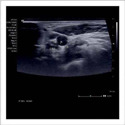
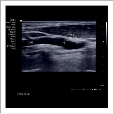
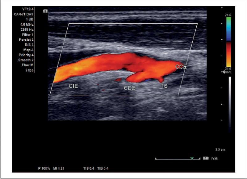
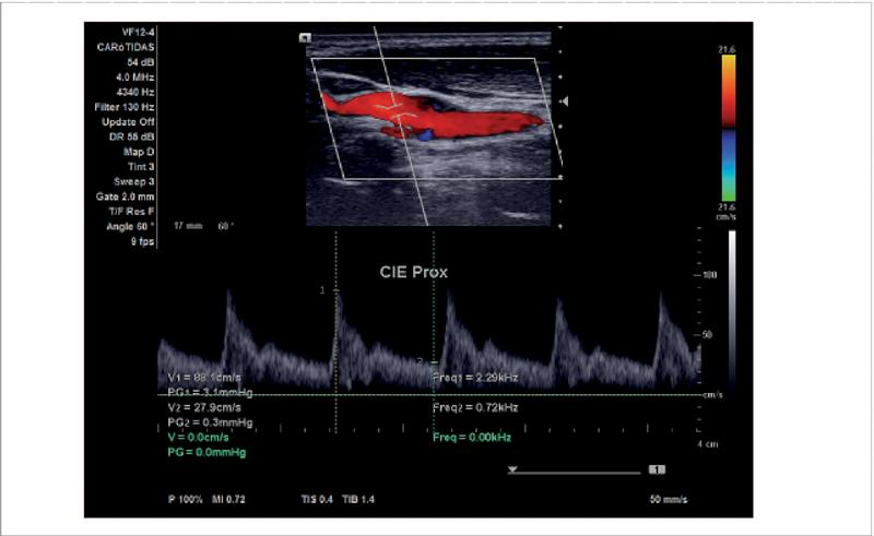
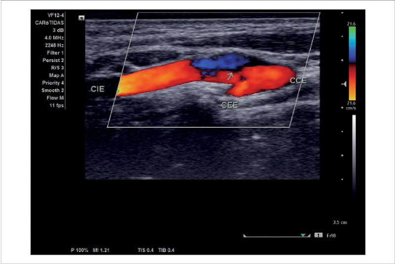
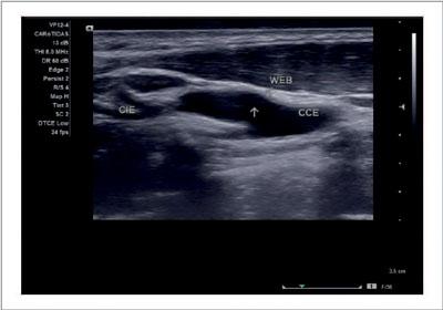
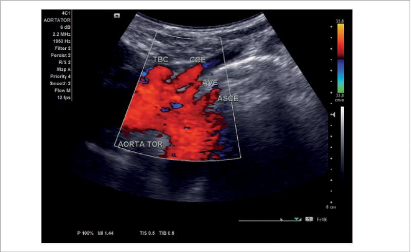
Patient P.F.A., a 33-year-old female, consulted a neurologiston December 6, 2023, with a primary complaint of intense, pulsating headaches, typically lasting long periods and predominantly affecting the left side of her head, suggesting possible vasospasm. Physical examination showed no apparent abnormalities, leading to a referral to a vascular surgeon for further investigation of possible vascularrelated headache. A Color Doppler exam of the bilateral carotid and vertebral arteries was performed, diagnosing a Carotid Web in the left carotid artery ( Figure 1 2 , 3 , 4 , 5 , 6 and 7 ). Imaging revealed afocal, triangular thickening of the intimalmedia layer with parietal irregularities and a small fibrotic area within the intimai layer, where a thin, mobile membrane was observed projecting into the lumen, intermittently contacting the vessel wall. The membrane’s proximal and distal ends were anchored in the internal carotid artery’s proximal segment ( Figure 2 and Video 2 ), which maintained a normal diameter but showed slight bulging and altered flow patterns consistent with the characteristics of a Carotid Membrane. ( Figure 3 and Video 2 ). Additionally, an anatomical variation related to the left vertebral artery, originating directly from the aortic arch was also observed in the CDE ( Figure 7 and 8 and Video 1 ).
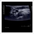
Cross-section in two-dimensional mode showing a thin membrane crossing the lumen with insertion into the wall of the left proximal internal carotid artery.
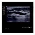
Longitudinal section in two-dimensional mode showing a triangular shape with a fibrosis point in the intimai layer where the thin membrane projecting into the lumen of the left proximal internal carotid artery is inserted. WEB: Carotid Web; CIE: Left Internal Carotid; CCE: Left Common Carotid.
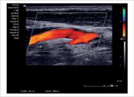
Longitudinal section with color Doppler showing a thin membrane inside the lumen of the left proximal internal carotid artery. CIE: Left Internal Carotid; CCE: Left Common Carotid; CEE: Left External Carotid; TS: Superior Thyroid.CEE: Left External Carotid; TS: Superior Thyroid.
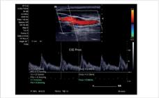
Longitudinal section with spectral pulsed Doppler showing the anterograde flow pattern and no significant increase in velocity in the left proximal internal carotid artery. CIE: Left Internal Carotid.
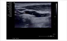
Longitudinal section with two-dimensional mode showing intimai thickening and slight bulging of the proximal left internal carotid artery wall. CIE: Left Internal Carotid; CCE: Left Common Carotid.
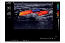
Longitudinal section with color Doppler showing the reverse flow division pattern through the presence of the membrane, and slight bulging of the proximal left internal carotid artery wall. CIE: Left Internal Carotid; CCE: Left Common Carotid; CEE: Left External Carotid.
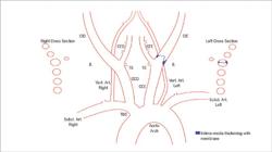
Cartographic drawing showing the site of the carotid web in the left carotid artery. CED: Right External Carotid; CID: Right Internal Carotid; CCD: Right Common Carotid; CIE: Left Internal Carotid; CCE: Left Common Carotid; CEE: Left External Carotid; TBC: Brachiocephalic Trunk.
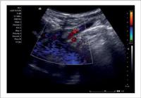
Longitudinal section with convex transducer and color Doppler demonstrating the anatomical variation of the left vertebral artery origin in the aortic arch, and the origins of the other supra-aortic trunk vessels. Em: https://abcimaging. org/supplementary-material/2025/3801/2024_0080_video_01.mp4
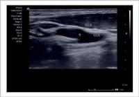
CDE showing the redundant membrane and flow alteration. Em: https://abcimaging.org/supplementary-material/2025/3801/2024_0080_video_02.mp4
The carotid system on the right side showed normal calibers and preserved flows.
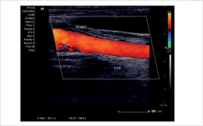
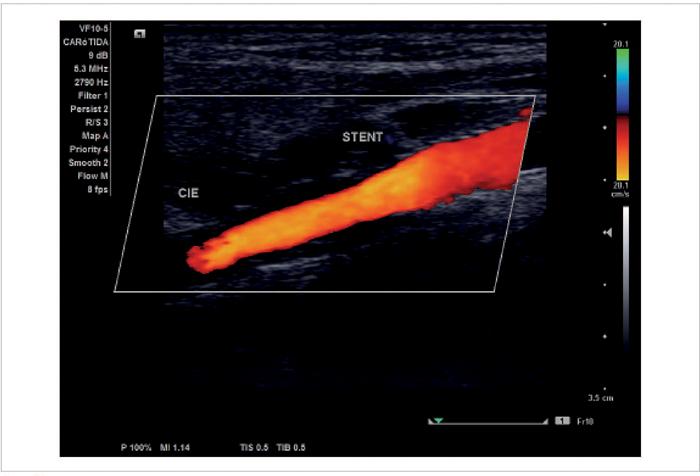
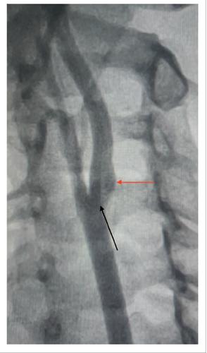
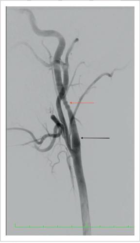
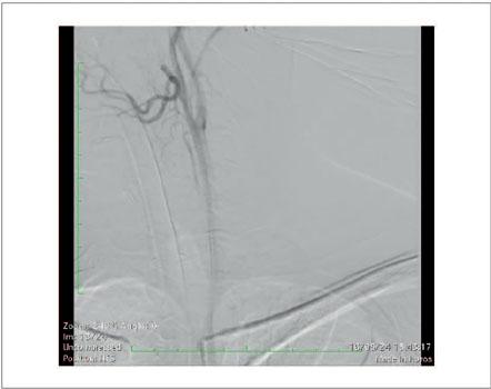
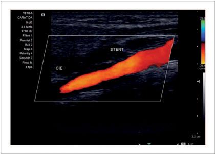
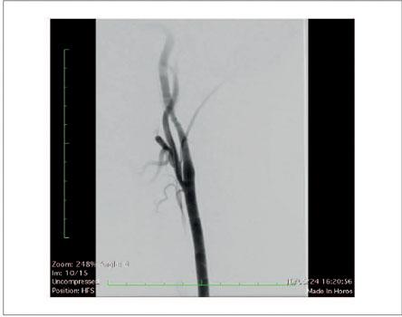
Angiography (AGP) ( Figure 12 and Video 3 ) confirmed the presence of the Carotid Web (CW), showing local flow pattern alterations without significant stenosis. Treatment options included clinical, open surgery and endovascular approaches with stent implantation, from which the patient decided on the endovascular therapeutic intervention with placement of a stent 3 in the left carotid, supported by the justification of minimizing the risks for cerebrovascular events, which was performed without angioplasty, having no complications or other relevant findings. At the end of the procedure, a control AGP was performed, still intraoperative ( Figure 13 and Video 5 ), where it was possible to note the absence of the aforementioned alterations, presenting only an episode of vasospasm. One month post-procedure, follow-up CDE confirmed that the stent was well-positioned at the site of the CW, with normal, low-resistance anterograde flow and no abnormalities or structural changes Figures ( 9 , 10 and 11 and Video 4 ). At follow-ups one and five months later, the patient reported no recurrence of the pulsating headache experienced pre-intervention.
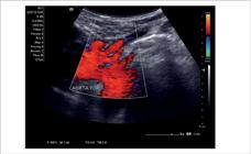
Longitudinal section with convex transducer and color Doppler demonstrating the anatomical variation of the left vertebral artery origin in the aortic arch, and the origins of the other supra-aortic trunk vessels. TBC: Brachiocephalic Trunk; AVE: Left Vertebral Artery; ASCE: Left Subclavian Artery; CEE: Left External Carotid.
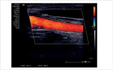
Longitudinal section with color Doppler showing a patent stent and normal anterograde flow within the left distal common carotid artery, without abnormalities. CCE: Left Common Carotid.
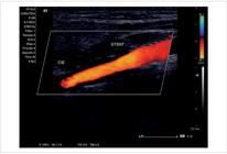
Longitudinal section with color Doppler showing a patent stent and normal anterograde flow supporting the carotid web within the left proximal internal carotid artery, without abnormalities. CIE: Left Internal Carotid.
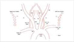
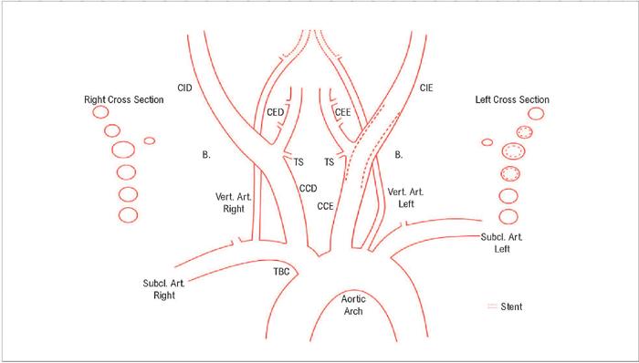
Cartographic drawing showing the stent site in the left carotid artery. CED: Right External Carotid; CID: Right Internal Carotid; CCD: Right Common Carotid; CIE: Left Internal Carotid; CEE: Left External Carotid; CCE: Left Common Carotid; TBC: Brachiocephalic Trunk.
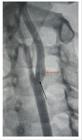
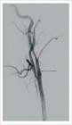
Trans-operative AGP of the carotid web stent (black arrow), with a slight final vasospasm (red arrow).
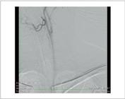
Trans-operative AGP showing the intra-carotid web (Black arrow) and rarefaction of contrast at the site of blood turbulence (Red arrow). Em: https://abcimaging.org/supplementary-material/2025/3801/2024_0080_video_03.mp4
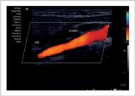
CDE showing normal flow in the stent. Em: https://abcimaging.org/supplementary-material/2025/3801/2024_0080_video_04.mp4
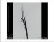
Trans-operative AGP showing well positioned Wallstent Carotid/Boston stent, with disappearance of the mobile membrane (Black arrow). Note the spasm induced by the presence of the stent - “rosary beads” aspect (Red arrow). Em: https://abcimaging.org/supplementary-material/2025/3801/2024_0080_video_05mp4
Literature Review
The Carotid Web has a 1 -7% 4 prevalence, representing up to 37% of cryptogenic ischemic CVAs, that is, of unidentified cause. It is responsible for thrombogenic hemodynamic changes distal to it, which can eventually cause embolization and consequent stroke. 4 Histopathologically, this variation (CW) represents a form of fibromuscular dysplasia characterized by intimai fibroelastic hyperplasia in the vascular endothelium, which forms a triangular intraluminal projection, possibly resulting in stenosis, aneurysm, or arterial dissection, and being most commonly found in the renal and carotid arteries. 6 In cases involving cervical arteries, typical symptoms include headache, vertigo, and pulsatile tinnitus, with presentation varying based on the CW pattern and degree of arterial narrowing. The carotid murmur may also be noted. 6 As described in other cases, distinctive changes have been observed in various blood flow imaging techniques, which may show a triangular isohypoechoic lesion that protrudes, with or without an accompanying membrane, appearing as a punctiform spicule, short or long trabeculae, or web-like structures seen in both axial and longitudinal ultrasound scans. At the same time, color-coded Doppler typically reveals areas of turbulence and reduced flow velocities behind the membrane. 5 Though often asymptomatic, this membrane can be redundant and mobile, causing hemodynamic disruptions that may exceed those associated with atherosclerotic disease. 4 The main differential diagnosis should consider segmental carotid dissection. Therapeutic follow-up should be tailored to each patient, as no standardized treatment approach is widely accepted in clinical practice. One potential therapeutic strategy includes the use of anticoagulant or antiplatelet medications, recommended for both symptomatic and asymptomatic patients when the goal is to lower the risk of cerebrovascular events, combined with healthier lifestyle changes. 6 In patients with a history of CVA, intervention in the affected carotid artery is recommended by the endovascular technique with stent 3 , or open surgery.
Remarks
The patient mentioned in this report did not experience thrombotic events, and her symptoms were limited to intense, pulsating, and persistent headaches. The diagnosis was established with Color Doppler Echocardiography (CDE), which detected structural changes, including a mobile and redundant membrane and alterations in the local flow pattern, later confirmed with Carotid Angiography.
Conclusion
CDE is a non-invasive and cost-effective method, comprising a valuable and accurate diagnostic tool for identifying the Carotid Web, and it plays an essential role in treatment planning, directly contributing to improved survival and quality of life for individuals with this condition.
-
Sources of Funding
There were no external funding sources for this study.
-
Study Association
This study is not associated with any thesis or dissertation work.
-
Ethics Approval and Consent to Participate
This article does not contain any studies with human participants or animals performed by any of the authors.
References
-
1 Natarajan S, Rahul S, Archana R, Sivarajan T. Carotid Web – An Unusual Cause for Ischemic Stroke in a Young Woman. IJVES CInI- Case reports, Innovations and Images. 2023;2(1):21-3. doi: 10.4103/IJCR.IJCR_8_24.
» https://doi.org/10.4103/IJCR.IJCR_8_24. -
2 Patel SD, Otite FO, Topiwala K, Saber H, Kaneko N, Sussman E, et al. Interventional Compared with Medical Management of Symptomatic Carotid Web: A Systematic Review. J Stroke Cerebrovasc Dis. 2022;31(10):106682. doi: 10.1016/j.jstrokecerebrovasdis.2022.106682.
» https://doi.org/10.1016/j.jstrokecerebrovasdis.2022.106682. -
3 Kamatani K, Yoshida S, Tashiro N, Hashiguchi Y, Takigawa K, Yasaka M, et al. The Case of Treatment for Carotid Web - Double-Layer Micromesh Stent Implantation in Our Hospital. Surg Neurol Int. 2023;14:339. doi: 10.25259/SNI_525_2023.
» https://doi.org/10.25259/SNI_525_2023. -
4 Park CC, El Sayed R, Risk BB, Haussen DC, Nogueira RG, Oshinski JN, et al. Carotid Webs Produce Greater Hemodynamic Disturbances than Atherosclerotic Disease: A DSA Time-density Curve Study. J Neurointerv Surg. 2022;14(7):729-33. doi: 10.1136/neurintsurg-2021-017588.
» https://doi.org/10.1136/neurintsurg-2021-017588. -
5 Fontaine L, Guidolin B, Viguier A, Gollion C, Barbieux M, Larrue V. Ultrasound Characteristics of Carotid Web. J Neuroimaging. 2022;32(5):894-901. doi: 10.1111/jon.13022.
» https://doi.org/10.1111/jon.13022. -
6 Touzé E, Southerland AM, Boulanger M, Labeyrie PE, Azizi M, Bouatia-Naji N, et al. Fibromuscular Dysplasia and Its Neurologic Manifestations: A Systematic Review. JAMA Neurol. 2019;76(2):217-26. doi: 10.1001/jamaneurol.2018.2848.
» https://doi.org/10.1001/jamaneurol.2018.2848. -
7 Ristow AVB, Massière B, Meirelles GV, Casella IB, Morales MM, Moreira RCR, et al. Brazilian Angiology and Vascular Surgery Society Guidelines for the Treatment of Extracranial Cerebrovascular Disease. J Vasc Bras. 2024;23:e20230094. doi: 10.1590/1677-5449.202300942.
» https://doi.org/10.1590/1677-5449.202300942. -
8 Ben Z, Wang J, Zhan J, Chen S. Ultrasonic Characteristics of Carotid Webs. Neuroradiology. 2022;64(1):95-98. doi: 10.1007/s00234-021-02757-0.
» https://doi.org/10.1007/s00234-021-02757-0. -
9 Vukasović R, Pintarić M, Lovrenčić-Huzjan A. Headache as a Symptom of Carotid Web. Archives of Psychiatry Research. 2023; 59:133-6. DOI:10.20471/may.2023.59.01.19.
» https://doi.org/10.20471/may.2023.59.01.19. - 10 Engelhorn AN, Engelhorn CA, Morais DM Filho, Barros FS, Coelho NA, editors. Guia Prático de Ultrassonografia Vascular. Rio de Janeiro: DiLivros; 2019.
Edited by
-
Editor responsible for the review: Simone Nascimento dos Santos