A Common Primary Cardiac Tumor With an Uncommon Presentation
Abstract
Background Cardiac tumors are rare, and most are secondary (metastatic) lesions. Primary tumors, though uncommon, are typically benign, with myxomas being the most frequent and occasionally displaying unique features. Diagnosis often requires multimodality imaging.
Case summary A 41-year-old man with a history of ischemic stroke and resection of an apical left ventricular myxoma presented for evaluation after a follow-up transthoracic echocardiogram (TTE) revealed a mobile, ovoid apical lesion with high embolic risk, despite being asymptomatic. Cardiac magnetic resonance (CMR) imaging suggested recurrence of the ventricular myxoma. Surgical resection was performed, and histopathology confirmed the diagnosis of cardiac myxoma (CM).
Discussion CMs are the most common primary benign cardiac tumors, typically located in the left atrium and attached to the interatrial septum. They are less frequently found in the ventricular cavities. About half of cases are asymptomatic, and recurrence is possible after resection. This case highlights a rare instance of late recurrence in an atypical location .
Heart Neoplasms; Myxoma; Magnetic Resonance Imaging; Echocardiography
Resumo
Fundamentos Tumores cardíacos são raros, e a maioria são lesões secundárias (metastáticas). Tumores primários, apesar de incomuns, são tipicamente benignos; os mixomas são os mais frequentes e ocasionalmente apresentam características únicas. O diagnóstico geralmente necessita de exames de imagem multimodais.
Resumo do caso Um homem de 41 anos com história de acidente vascular isquêmico e ressecção de um mixoma ventricular esquerdo apical apresentou-se para avaliação após um ecocardiograma transtorácico (ETT) de acompanhamento haver revelado uma lesão apical ovoide, móvel, com alto risco de embolia, apesar de assintomático. O exame de ressonância magnética cardíaca (RMC) sugeriu recorrência de mixoma ventricular. Realizou-se ressecção cirúrgica e o diagnóstico de mixoma cardíaco (MC) foi confirmado por histopatologia.
Discussão Os MCs são os tumores cardíacos benignos primários mais comuns, tipicamente localizados no átrio esquerdo e fixos ao septo interatrial. Eles são menos comuns nas cavidades ventriculares. Cerca de metade dos casos são assintomáticos, e a recorrência é possível após a ressecção. Este caso destaca um exemplo raro de recorrência tardia em um local atípico.
Neoplasias Cardíacas; Mixoma; Imageamento por Ressonância Magnética; Ecocardiografia
Introduction
Cardiac tumors are rare, and mostly secondary (metastatic), benign (90%) lesions.1 Among these, CMs are the most common, representing 50-60% of benign cases.2 , 3 Myxomas predominantly occur in the left atrium (75-85%), with fewer cases in the right atrium, ventricles, or valve structures.4 , 5 Once resected, tumor recurrence is possible, highlighting the need for proper postoperative follow-up. We report a case of a rapidly growing, late recurrent myxoma in an uncommon location.
Clinical case
A 41-year-old man with a history of ischemic stroke and resected left ventricular myxoma presented to the emergency room following a follow-up TTE that revealed a mobile, ovoid lesion in the left ventricular apex with high embolic risk, despite being asymptomatic. His stroke in 2014 had led to the diagnosis of a 20x15 mm pedunculated mass in the ventricular apex, which was resected via a transapical approach. Histology confirmed the lesion as a CM. The patient had no personal or family history of malignancy or primary cardiac tumors.
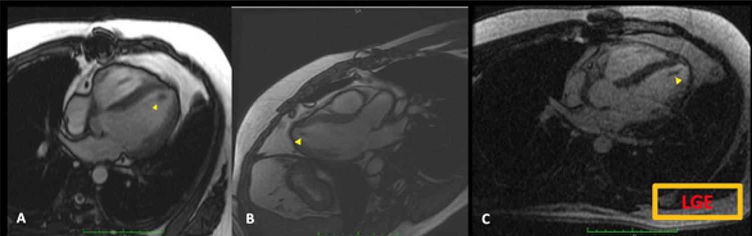
After the initial resection, serial TTEs were performed every two years to monitor for recurrence. In January 2023, a TTE revealed apical akinesia and a poorly characterized, linear, mobile structure. Due to the lack of ultrasound contrast, CMR was performed, showing apical scarring with akinesia, mild systolic dysfunction, and a 20x8 mm linear structure adherent to the apex, consistent with thrombus ( Figure.1 ). Warfarin therapy was initiated, but the patient did not achieve adequate anticoagulation (INR: 1.5).
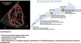
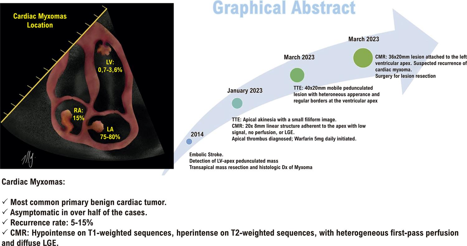
– Graphical Abstract. LV: left ventricle; RA: right atrium; LA: left atrium; CMR: cardiac magnetic resonance; LGE: late gadolinium enhancement; TTE: transthoracic echocardiogram.
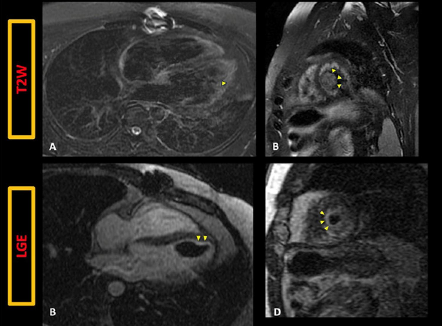
In March 2023, follow-up TTE revealed a large (40x20 mm), highly mobile, pedunculated lesion with a heterogeneous appearance ( Video.1 ). A repeat CMR confirmed a 36x20 mm lesion at the apex with heterogeneous T2 hyperintensity, mild gadolinium perfusion, and peripheral late enhancement, suggesting CM recurrence with possible thrombus ( Figures.2 - 3 and Video.2 ).
– TTE (A-B): Mobile, ovoid lesion in the left ventricular apex with a small pedicle, smooth edges, and heterogeneous content. http://abcimaging.org/supplementary-material/2025/3801/2024-0123_video_01.mp4
– CMR perfusion revealing mild, heterogeneous perfusion of the lesion. http://abcimaging.org/supplementary-material/2025/3801/2024-0123_video_02.mp4
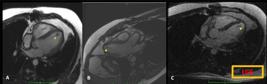
– CMR. A-B: Four-chamber and three-chamber cine showing a low-intensity lesion at the left ventricular apex; C: no late enhancement with gadolinium; LGE: late gadolinium enhancement.
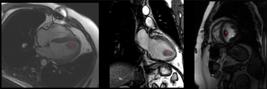
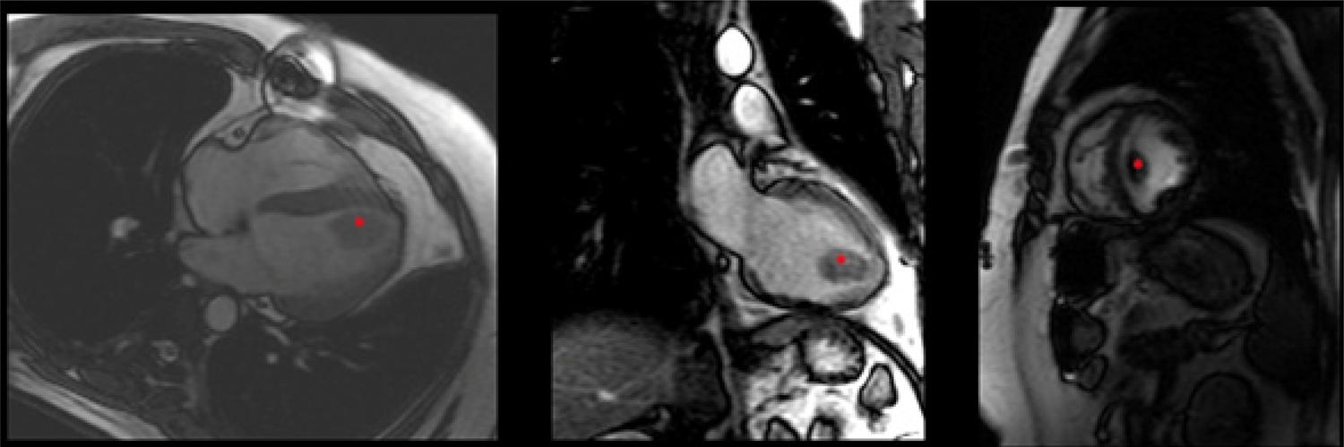
– CMR (four-chamber and two-chamber, short-axis images) showing a low-intensity lesion at the left ventricular apex
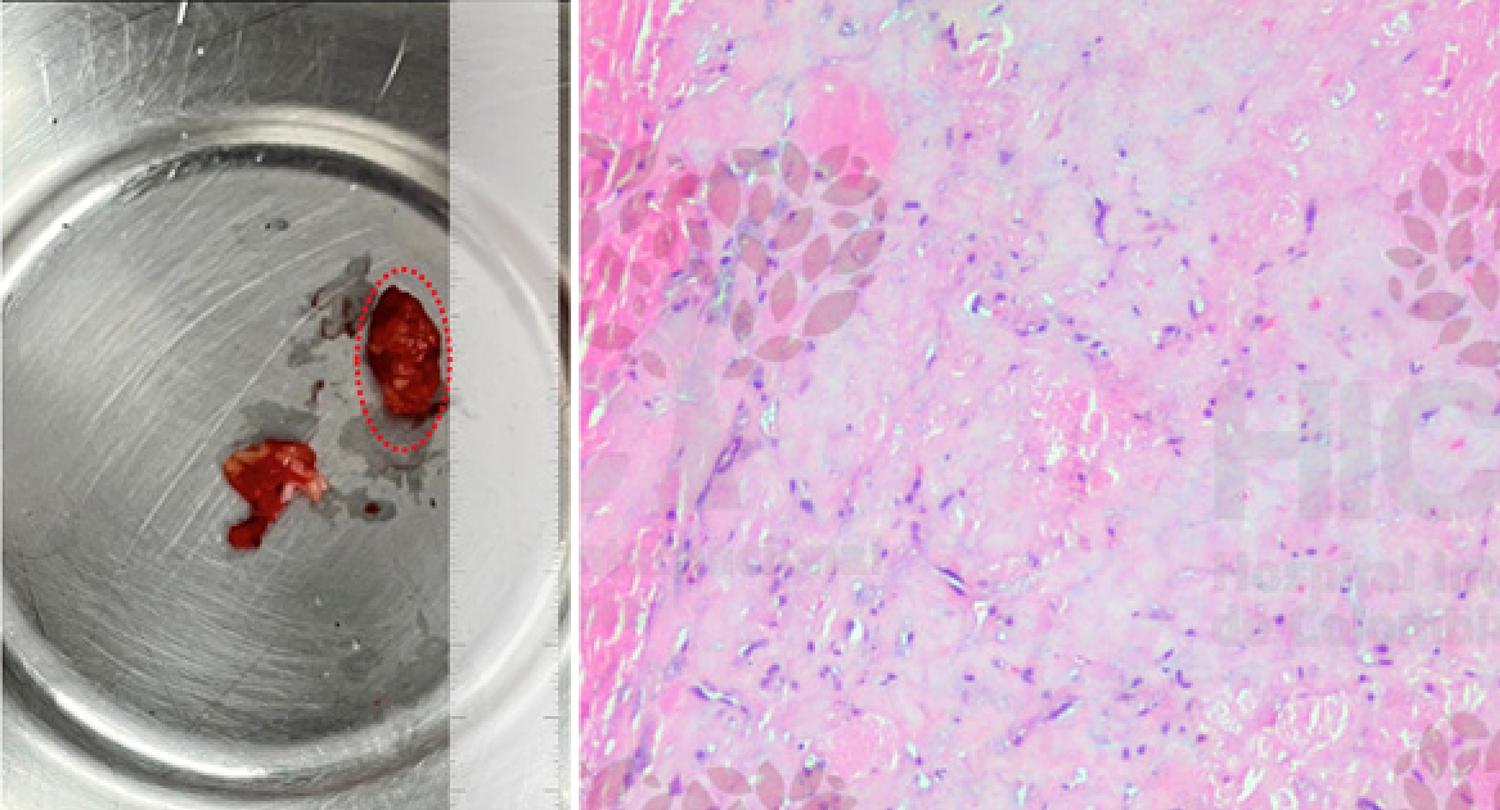
The patient underwent surgical resection of the lesion, which was identified as a fibrotic lesion with a thrombotic component. Histopathology confirmed the diagnosis of recurrent CM ( Figure.4 ). Postoperative recovery was uneventful, and the patient remains under regular follow-up without complications or further hospitalizations.

– CMR. A-B: T2W sequences showing heterogeneous hyperintensity at the apical lesion; C-D: LGE around the lesion’s periphery. LGE: late gadolinium enhancement; T2W: T2-weighted.
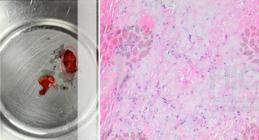
– Cardiac mass specimen. A: Ovoid lesion resected from the ventricular apex (dotted red line). B: Hematoxylin–eosin revealing myxomatous tissue with spindle-shaped cells (Myxoma Cells). Histology image includes a watermark from the pathology laboratory of Hospital Internacional de Colombia (HIC).
Discussion
Myxomas are the most frequent benign primary cardiac tumors, generally diagnosed in patients aged 40–70 years, with a female predominance. Left ventricular involvement is rare, accounting for 0.7–3.6% of cases.6 Up to half of patients are asymptomatic, while symptomatic presentations depend on the lesion’s location and its effect on adjacent structures. Obstructive symptoms (dyspnea, syncope, palpitations, lower extremity edema) occur in about two-thirds of symptomatic patients, especially with larger tumors, whereas the remaining third experience systemic or pulmonary embolism (depending on tumor location), particularly when lesions are small and friable. Rarely, systemic symptoms (anemia, fever, weight loss, fatigue, arthralgia, myalgia, Raynaud’s phenomenon) occur, linked to elevated cytokine (particularly IL-6) secretion by the tumor.7
Echocardiography is essential for diagnosing CM, offering a 90–96% sensitivity 8 During evaluation, key features such as the lesion’s location and impact should be noted. Myxomas exhibit two main morphologies: polypoid and papillary. Polypoid tumors are larger, with a smooth surface, small pedicle, and heterogeneous interior (including cystic or hemorrhagic areas), whereas papillary tumors are smaller, have a villous surface, and are more prone to embolic events.9 In this case, a polypoid CM was identified.
One of the primary differential diagnoses of CM is intracavitary thrombus, especially in the ventricles. In some cases, both may coexist. Additional imaging techniques, such as cardiac computed tomography and CMR, refine lesion characterization. On CMR, myxomas typically appear hypointense on T1-weighted sequences and hyperintense on T2-weighted sequences, reflecting their high extracellular water content.10 With gadolinium, they often show variable first-pass perfusion due to poor vascularity and display heterogeneous late enhancement with possible necrosis, as observed in our patient.
An important aspect with CMs is the recurrence rate, which range from 5% to 15% in retrospective studies and case series.1 , 11 Recurrences typically occur at the same location but may also appear elsewhere. Proposed causes include incomplete resection, metastatic spread, totipotent multicentricity, and familial inheritance.12 In a registry of 207 CMs followed for 9.5 ± 6.6 years, only multicentric myxomas were independently associated with recurrence.11
Another consideration is the recurrence-free interval after resection. In our case, the late recurrence was unexpected. Data from long-term registries indicate that most recurrences occur within the first five years, although there are isolated reports of recurrences 5–10 years after resection.11 , 13
Finally, the lesion’s rapid progression was striking, doubling in size (from 20×8mm to 40×20mm) over 2.5 months, indicating a growth rate of about 8mm/month. Some studies, based on echocardiography, report a growth rate of 4,9mm/month,14 while tomographic analyses suggest 1,2–2,2 mm/month.15 Notably, echocardiography often underestimates tumor size compared to surgical specimens. This case not only underscores the high growth potential of CM, but also its characteristics, including unusual location, recurrence, and complications.
Highlights
CMs are the most frequent benign tumors of the heart. The ventricular location of these is infrequent.
The main differential diagnosis for myxoma is the presence of thrombi. CMR is very useful for lesion characterization and differentiation. CMs have a significant recurrence rate as well as high growth rates.
Conclusion
CMs are the most common benign primary cardiac tumors. Their features can vary markedly in size, location, complications, recurrence, and growth. Although the most frequent patterns are well described, it is essential to recognize and understand the possibilities of less common presentations.
References
-
1 Tyebally S, Chen D, Bhattacharyya S, Mughrabi A, Hussain Z, Manisty C, et al. Cardiac Tumors: JACC CardioOncology State-of-the-Art Review. JACC CardioOncol. 2020;2(2):293-311. doi:10.1016/j.jaccao.2020.05.009.
» https://doi.org/10.1016/j.jaccao.2020.05.009 - 2 Paolisso P, Foà A, Bergamaschi L, Graziosi M, Rinaldi A, Magnani I, et al. Echocardiographic Markers in the Diagnosis of Cardiac Masses. J Am Soc Echocardiogr. 2023;36(5):464-73. doi:10.1016/j.echo.2022.12.022.
-
3 Bussani R, Castrichini M, Restivo L, Fabris E, Porcari A, Ferro F, et al. Cardiac Tumors: Diagnosis, Prognosis, and Treatment. Curr Cardiol Rep. 2020;22(12):169. doi: 10.1007/s11886-020-01420-z.
» https://doi.org/10.1007/s11886-020-01420-z -
4 Mando R, Barbat JJ, Vivacqua A. The Mysterious Mitral Mass: A Case of Valvular Myxoma. Case Rep Cardiol. 2018;1(1):3927948. doi:10.1155/2018/3927948.
» https://doi.org/10.1155/2018/3927948 -
5 Rahmanian PB, Castillo JG, Sanz J, Adams DH, Filsoufi F. Cardiac Myxoma: Preoperative Diagnosis Using a Multimodal Imaging Approach and Surgical Outcome in a Large Contemporary Series. Interact Cardiovasc Thorac Surg. 2007;6(4):479-83. doi:10.1510/icvts.2007.154096.
» https://doi.org/10.1510/icvts.2007.154096 -
6 Samanidis G, Khoury M, Balanika M, Perrea DN. Current Challenges in the Diagnosis and Treatment of Cardiac Myxoma. Kardiol Pol. 2020;78(4):269-77. doi:10.33963/KP.15254.
» https://doi.org/10.33963/KP.15254 -
7 Seino Y, Ikeda U, Shimada K. Increased Expression of Interleukin 6 mRNA in Cardiac Myxomas. Br Heart J. 1993;69(6):565-7. doi:10.1136/HRT.69.6.565.
» https://doi.org/10.1136/HRT.69.6.565 -
8 Koritnik P, Pavsic N, Bervar M, Prokselj K. Echocardiographic Characteristics of Cardiac Myxoma. Eur Heart J. 2021;42(Suppl 1). doi:10.1093/EURHEARTJ/EHAB724.0146.
» https://doi.org/10.1093/EURHEARTJ/EHAB724.0146 -
9 Mankad R, Herrmann J. Cardiac Tumors: Echo Assessment. Echo Res Pract. 2016;3(4):R65-R77. doi:10.1530/ERP-16-0035.
» https://doi.org/10.1530/ERP-16-0035 -
10 Aiello VD, Campos FPF. Cardiac Myxoma. Autops Case Rep. 2016;6(2):5-7. doi:10.4322/ACR.2016.030.
» https://doi.org/10.4322/ACR.2016.030 - 11 Wang Z, Chen S, Zhu M, Zhang W, Zhang H, Li H, et al. Risk Prediction for Emboli and Recurrence of Primary Cardiac Myxomas after Resection. J Cardiothorac Surg. 2016;11(1):22. doi:10.1186/S13019-016-0420-4/FIGURES/4.
- 12 Carvalho MS, Andrade MJ, Abecasis J, Gouveia R, Branco L, Neves JP, et al. Understanding Cardiac Myxoma Recurrence: A Case Report. Rev Port Cardiol. 2013;32(3):239-42. doi:10.1016/J.REPCE.2013.04.002.
-
13 Elbardissi AW, Dearani JA, Daly RC, Mullany CJ, Orszulak TA, Puga FJ, et al. Survival after Resection of Primary Cardiac Tumors: A 48-Year Experience. Circulation. 2008;118(14 Suppl):7-15. doi:10.1161/CIRCULATIONAHA.107.783126.
» https://doi.org/10.1161/CIRCULATIONAHA.107.783126 -
14 Gewehr DM, Neiverth A, Cavalcanti MS, Maestri TC, Haurani S, Kubrusly FB, et al. Fast Growth Rate of a Right Atrial Myxoma. Einstein. 2022;20:eRC6478. doi:10.31744/EINSTEIN_JOURNAL/2022RC6478.
» https://doi.org/10.31744/EINSTEIN_JOURNAL/2022RC6478 -
15 Hsi DH, Sosa A, Miller W, Oren T, Koulova A, Coady MA. A Giant Right Atrial Myxoma-The Growth Rate and Multi-Modality Imaging. Echocardiography. 2021;38(6):1057-60. doi:10.1111/echo.15049.
» https://doi.org/10.1111/echo.15049
-
Study Association:
This study is not associated with any thesis or dissertation work.
-
Ethics Approval and Consent to Participate:
This article does not contain any studies with human participants or animals performed by any of the authors.
-
Sources of Funding:
There were no external funding sources for this study.
Edited by
-
Editor responsible for the review:
Marcelo Tavares