 Comparative study between vasectomy, epididymectomy and cryosurgery for preparation of teaser goat (Capra hircus)
ABSTRACT
This study aimed to compare the efficiency of three sterilization techniques-cryosurgery of the tail of the epididymis, vasectomy, and epididymectomy-for obtaining teaser bucks. Thirty animals were divided into three groups (n=10). Pre-treatment seminal characteristics showed no significant differences among the groups. The average anesthetic recovery time was 94min 24s, with no significant variation between treatments. Cryosurgery was significantly faster (3min 30s) compared to vasectomy and epididymectomy (21min 08s and 22 min 07 s, respectively). After 90 days, the vasectomy and epididymectomy groups exhibited necrospermia or azoospermia in the first semen collection. In contrast, 6/10 animals in the cryosurgery group maintained viable sperm with increased motility, vigor, and concentration, while 4/10 showed necrospermia throughout the study. Ultrasonographic changes were observed in all groups, with cryosurgery causing significant alterations in epididymal size and texture. Histopathological findings in the vasectomy and epididymectomy groups included inflammatory infiltrates, fibrosis, lymphangiectasia, sperm granulomas, and necrosis. In the cryosurgery group, an enlarged epididymis, cystic areas, and sperm granulomas were observed. Although cryosurgery was less invasive, its effectiveness in achieving permanent azoospermia in goats was limited compared to vasectomy and epididymectomy. Further research is needed to refine and improve this technique.
Keywords:
surgery; andrological examination; ultrasound; histopathology
RESUMO
Este estudo teve como objetivo comparar a eficiência da criocirurgia da cauda do epidídimo, vasectomia e epididimectomia para obtenção de rufiões caprinos. Trinta animais foram divididos em três grupos (n=10). As características seminais pré-tratamento não apresentaram diferenças significativas. O tempo médio de recuperação anestésica foi de 94min e 24s, sem diferença entre os grupos. A criocirurgia foi significativamente mais rápida (3min e 30s) em comparação com a vasectomia e a epididimectomia (21min e 08s e 22min e 07s, respectivamente). Após 90 dias, os grupos de vasectomia e epididimectomia apresentaram necrospermia ou azoospermia já na primeira coleta. Em contraste, 6/10 animais do grupo criocirurgia mantiveram esperma viável, com aumento de motilidade, vigor e concentração espermática, enquanto 4/10 apresentaram necrospermia ao longo do estudo. Alterações ultrassonográficas foram observadas em todos os grupos, com a criocirurgia mostrando alterações significativas no tamanho e na textura do epidídimo. Achados histopatológicos revelaram infiltrados inflamatórios, fibrose, linfangiectasia, granulomas espermáticos e necrose nos grupos de vasectomia e epididimectomia. No grupo criocirurgia, foram observados aumento do epidídimo, áreas císticas e granulomas espermáticos. A criocirurgia foi menos invasiva, mas sua eficiência para obtenção de azoospermia permanente em caprinos foi limitada em comparação com vasectomia e epididimectomia. Novos estudos são necessários para refinar e aprimorar essa técnica.
Palavras-chave:
cirurgia; exame andrológico; ultrassonografia; histopatologia
INTRODUCTION
Reproductive efficiency in goat production relies on effective reproductive management (Simões et al., 2021). Among the reproductive practices employed, estrus detection using teaser bucks plays a crucial role in improving overall reproductive efficiency (Mellado and Hernandez, 1996), benefiting both natural mating and artificial insemination (Yotov et al., 2016).
For ruminant teaser formation, the most applied surgical techniques are vasectomy and epididymectomy (Morgan and Dawson, 2008). However, these procedures require skin incisions, ectomies, and post-surgical wound care. As a less invasive alternative for teaser buck preparation, cryosurgery techniques have emerged as a potential option (Dória et al., 2016).
Cryosurgery techniques are usually easier, less invasive, more cost-effective, and pose lower surgical risks than conventional methods (Souza et al., 2016; Ma et al., 2021). Therefore, cryosurgery applied to the tail of the epididymis may serve as an alternative for producing teaser bucks while reducing the incidence of postoperative complications.
A cryo-procedure targeting the tail of the epididymis could prevent sperm release in the ejaculate without compromising libido, as observed in rams by Dória et al. (2016). These authors demonstrated that an epididymal transfixation-based cryosurgery method induced azoospermia in rams without requiring skin incisions or ectomies, while also minimizing post-surgical wound care. This technique can be performed safely and efficiently on rural properties, reducing both intraoperative and postoperative complications and promoting a faster recovery. However, data on its application in goats remain limited. This cryo-procedure could offer an effective solution for reproductive management without raising major concerns regarding animal welfare. Thus, this study aimed to evaluate the efficiency of epididymal tail cryosurgery in goats for teaser formation, comparing its effectiveness to vasectomy and epididymectomy techniques.
MATERIAL AND METHODS
All experimental procedures were approved by the Universidade Federal Rural do Rio de Janeiro Animal Care and Committee (process number 003503/2019).
Thirty male Boer-Saanen cross goats, aged between 10 and 24 months and weighing between 19.5 and 48.5kg, were housed in collective stalls containing ten animals each. They were fed Pennisetum purpureum chopped grass, Cynodon dactylon hay, and a concentrated feed mix containing 14% crude protein on a dry matter basis. Water and commercial mineral salt were provided ad libitum. The study was conducted in Seropédica-RJ (latitude: -22o47´S, longitude: -43o40´W), where the climate is classified as Am according to the Koppen-Geiger system.
The animals were divided into three groups: vasectomy (VASC) and epididymectomy (EPID), both performed according to Morgan and Dawson (2008) and a non-surgical epididymal cryo-technique (CRYO).
An andrological examination was conducted at two time points: one week before the first surgical intervention and again 90 days later. Testicular biometrics were recorded during both assessments, including scrotal circumference (SC) using a tape measure, and scrotal width (SW), left (LLT) and right (LRT) testicular length, and left (TLT) and right (TRT) testicular thickness using a caliper (Chentouf et al., 2011). Additionally, SC and seminal quality were evaluated weekly between these two periods.
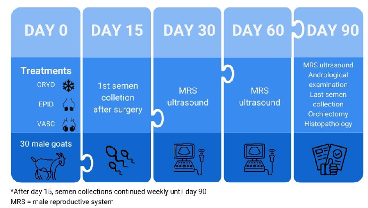
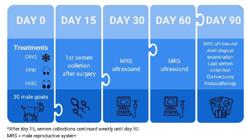
Semen collection was performed using an artificial vagina with a female in estrus as stimulus. To ensure conditioning, all animals were subjected to weekly artificial vagina semen collection sessions for 21 days. Immediately after collection, semen samples were evaluated macroscopically for color, appearance, and ejaculate volume (measured in a conical graduated tube). Microscopic analysis included mass motility (MMT), sperm motility (MOT), sperm vigor (VIG), and sperm concentration (Neubauer chamber). MOT and VIG were evaluated subjectively using semen aliquots diluted (1:200) in phosphate-buffered saline (PBS), incubated for 5min at 37°C, and examined under a phase-contrast microscope (×100). A schematic representation of the study, beginning on day 0 (the start of treatments), is shown in Fig. 1.
Three teaser preparation methods were performed: vasectomy (VASC) and epididymectomy (EPID), both based on the techniques described by Morgan and Dawson (2008), and a non-surgical epididymal cryo-technique (CRYO) following the methods of Dória et al. (2016). Thirty animals were evenly divided into three homogeneous groups (n=10) according to the teaser method. All procedures were performed by the same surgeon to ensure consistency.
For surgical teaser preparation, all animals were subjected to 24-h water and food fasting before the procedure. Sedation was induced using intramuscular administration of midazolam hydrochloride (0.1mg/kg) and 2% xylazine hydrochloride (0.05mg/kg). A local anesthetic block with 2% lidocaine (without vasoconstrictor) was applied at the incision site for the VASC and EPID groups and at the transfixation region for the CRYO group. Specifically, for vasectomy, the block was applied over the spermatic cord, while for epididymectomy, it was administered at the tail of the epididymis.
The vasectomy technique involved an incision between the testis and the inguinal ring, followed by an incision in the tunica vaginalis. The vas deferens was then isolated, ligated, and a segment excised. The epididymectomy procedure required an incision at the caudal pole of the testis, ligation of the tail and vas deferens, and subsequent resection. Both surgeries were completed with subcutaneous and skin sutures.
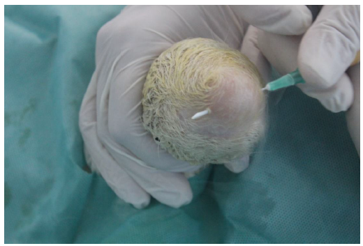
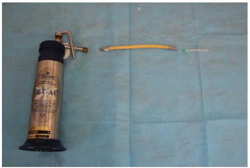
The CRYO technique involved lateromedial transfixation of the epididymal tail (Figure 2) using a 40×0.8mm hypodermic needle connected to a stainless-steel adapter, a 15cm rubber tube, and a cryosurgery device (Cry-Ac/Brymill®) containing liquid nitrogen (Figure 3). Two freeze/thaw cycles were applied: the first lasting 60 s and the second 45s.
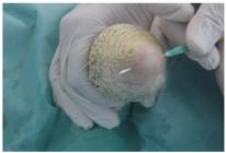
Transfixation of the epididymal tail in goats using a sterile disposable hypodermic needle. Detail for the needle tip frozen by liquid nitrogen (Font: Personal file).

Cryosurgery apparatus and circuit consisting of stainless-steel adapters, flexible rubber tube and disposable sterile hypodermic needle (Dória et al., 2016).
During the postoperative period, all animals received intramuscular oxytetracycline hydrochloride (0.1ml/kg) and Flunixin Meglumine (1.1mg/kg). Surgical times were recorded, including procedure time (PT) and total procedure time (TPT), which encompassed pre-anesthetic administration and anesthesia recovery. The duration of pre-operative, surgical, and post-operative phases was measured in minutes for all three groups.
Scrotal ultrasound examinations were performed by the same professional using a GE Logic Book XP® equipped with an 8.0 MHz linear transducer in B-mode. Longitudinal images of the testicular parenchyma were obtained from the caudal face of both testes. Ultrasound evaluations were performed at four time points: one week before surgery and at 30, 60, and 90 days post-procedure. For the ultrasound evaluation, Bucks were maintained in a quadrupedal position, and scrotal hair was left intact to minimize microtrauma (Dória et al., 2016).
Ninety days after the teaser procedures, all animals were subjected to a final andrological examination followed by testicular and epididymal collection via orchiectomy. Anesthesia was administered as previously described, with an additional local block at the scrotal incision site. Tissue samples from the testis, epididymal head and tail, and the transition between the testis and epididymal tail were fixed in 10% formaldehyde for histopathological examination.
Following fixation, microscope slides were prepared with sections of the epididymal head and tail and the testis-epididymis transition zone, stained with hematoxylin and eosin, and analyzed histopathologically. Lesions such as mononuclear inflammation, spermatic granulomas, necrosis, and lymphangiectasia were classified as mild, moderate, or severe (Fagundes et al., 2014; Guzelburc et al., 2019).
The experimental design followed a randomized block model, considering the effects of treatment, time, and experimental groups according to the model: Yijk=µ+Ti+Bj+Gk+(T×B)ij+ eijk where Y represents the observation, µ is the overall mean, T is the fixed effect of treatment, B is the effect of time, G is the random effect of the experimental groups, and e is the random error.
Normality was assessed using the Shapiro-Wilk test, and sphericity was verified with Bartlett’s test. Mixed model analysis was performed using the lme function in the nlme package. Means were compared utilizing Tukey’s test at a 5% significance level. The macroscopic aspects of semen were evaluated, and the frequency of each characteristic within treatments was recorded. Frequency comparisons were conducted using a proportion test at 5% significance. All statistical analyses were performed in R software.
RESULTS
In the testicular biometric evaluations, no significant differences (P>0.05) were observed between treatments for any variable (Table 1). In the pre-treatment seminal analysis, the animals exhibited normal ejaculations with a predominantly creamy appearance and coloration, with no significant differences (P>0.05).
The average time from premedication (MPA) to anesthetic recovery was 94:24 min and did not vary between groups (P>0.05). Additionally, there was no significant difference in the total procedure time (TPT) across the groups when considering anesthetic recovery. Regarding the surgical procedures, the average surgical time (PT) was 21:08 min for the vasectomy group and 22:07 min for the epididymectomy group. The CRYO technique, however, had a significantly shorter procedure time, totaling 3:30 min (P<0.05). The animals' responses to anesthesia and surgical intervention did not show notable differences in recovery times across the groups.
During the 90-day post-surgery evaluation period, the VASC and EPID groups presented zero values for mass motility, sperm motility, and sperm vigor, with low sperm concentrations (Table 2). Necrospermia or azoospermia was observed as early as the first collection (15 days post-treatment). In contrast, in the CRYO group, six out of ten animals (6/10) retained viable sperm, displaying higher values of mass motility, sperm motility, and sperm vigor throughout the evaluation period (P<0.05). Only four CRYO animals (4/10) exhibited necrospermia in the first week, and this condition persisted until the end of the experiment. Sperm concentration increased exclusively in the CRYO group, while the vasectomy and epididymectomy groups produced ejaculates with an aqueous and transparent appearance, in contrast to the creamy or milky appearance observed in six CRYO animals.
In the ultrasound evaluations, it was possible to observe notable differences in the testis and epididymis among the treatment groups. In vasectomized animals, four out of ten (4/10) showed enlargement of the epididymal head region, with cystic circular areas containing homogeneous, hypoechoic, or anechoic content inside and hyperechoic margins. In the VASC animals, two of those with a cystic lesion on the head also exhibited a similar lesion on the tail of the epididymis, along with increased thickness of the testicular mediastinum. Loss of definition, shape, and size of the epididymal head was observed in eight vasectomized animals (8/10).
Regarding the EPID group, all animals exhibited fibrosis in the resection region, while one animal showed testicular parenchyma degeneration, characterized by reduced organ size and increased tissue echogenicity.
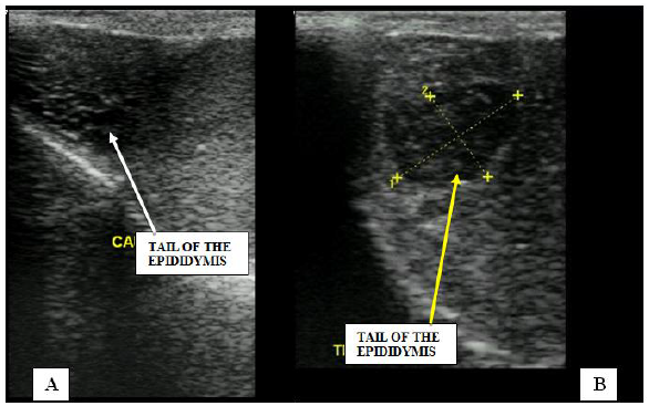
In the CRYO group, at 30 days, all animals showed alterations in the epididymal tail region, including an increase in shape and size, heterogeneous texture, and cystic circular areas with hyperechoic margins and content ranging from hypoechoic to anechoic. These lesions remained present until 90 days after the CRYO procedure (Figure 4).
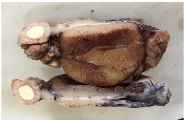
Ninety days after the surgical procedures, orchiectomy was performed in all animals, revealing a considerable amount of yellowish, cheese-like content accumulated between the tunics. Histological analysis characterized these findings as spermatic granulomas. In vasectomized animals, the epididymal head showed mononuclear inflammatory infiltrate (n=6), fibrosis (n=4), lymphangiectasia (n=3), hemorrhage (n=3), spermatic granulomas (n=4), and necrosis (n=1). In the tail region, inflammatory infiltrate and fibrosis were observed in four animals. Additionally, two animals in this group exhibited spermatic granulomas and hemorrhagic areas in the transition zone between the testicular parenchyma and the epididymis. In animals subjected to epididymectomy, fibrosis and adhesion were observed in the surgical region. CRYO animals showed an increased epididymal size, with cystic areas and yellowish content, diagnosed as spermatic granulomas (Figure 5). In the CRYO group, histopathological examination revealed lymphangiectasia and mild mononuclear inflammatory infiltrate in the epididymal head region in 5/10 animals. Additionally, all animals in this group showed lymphangiectasia and mild mononuclear inflammatory infiltrate in the epididymal tail region. Furthermore, spermatic granuloma formation was observed in eight bucks from the CRYO group, and fibrosis was noted in five animals. In the tail region, necrosis and mineralization were found in four animals. In the transition area between the testicular parenchyma and the epididymis, the most prevalent lesions were mononuclear inflammatory infiltrate (n=3), spermatic granulomas (n=3), and lymphangiectasia (n=3).
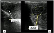
A- Longitudinal ultrasound image of the tail of the epididymis of an healthy animal (white arrow); B- Longitudinal ultrasound image of the tail of the epididymis of an animal submitted to cryosurgery. Circular cystic area, hyperechoic margin and contents inside (yellow arrow) (Font: Personal file).
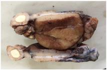
Region of the tail of the epididymis of an animal submitted to cryosurgery with the presence of a circular area containing yellowish material (sperm granuloma) (Font: Personal file).
In four animals subjected to epididymectomy, mononuclear inflammatory infiltrate was noted in both the epididymal head and the transition region between the testicular parenchyma and the surgical area, with spermatic granulomas and fibrosis observed in six animals that underwent the procedure.
DISCUSSION
The epididymal CRYO approach, designed to produce teaser bucks with permanent azoospermia, represents a significant advancement in reproductive management. To the best of our knowledge, this study is the first to evaluate this technique in goats, following the method previously established by Dória et al. (2016) in sheep. The importance of this technique lies in the detailed description of the needle insertion sites, positioning, and the freezing and thawing cycles applied to the epididymal tail. These factors are crucial for the effective application of the method across different species and highlight the need for specific adjustments in goats, contributing to advancements in caprine reproductive management.
Studies evaluating teaser methods in goats remain scarce. In the vasectomy and epididymectomy groups, all animals exhibited necrospermia or azoospermia in the first post-treatment collection. This outcome is expected, as both techniques involve the removal of a segment of the male reproductive system-whether the vas deferens in a vasectomy or the cauda epididymis in an epididymectomy-resulting in ejaculates devoid of viable sperm. The time required for semen to become completely sperm-free may vary, depending on the sperm reserve in the vas deferens. Tamadon et al. (2010) reported the absence of sperm motility and live sperm in the ejaculates of sheep subjected to epididymectomy three weeks post-surgery.
In the CRYO group, four out of ten animals exhibited necrospermia, while the remaining six continued to produce viable spermatozoa throughout the 90-day evaluation period. Although 40% of the CRYO-treated animals achieved azoospermia, which is a promising outcome, given that the primary goal of the technique is to ensure permanent infertility-the persistence of viable sperm in some animals suggests that the method has the potential to effectively induce azoospermia in a significant proportion of treated animals. This finding highlights the possibility of functional recovery in the epididymal ducts yet demonstrates that CRYO remains a viable alternative for producing teasers with a high success rate.
Dória et al. (2016) reported that in their study on sheep using CRYO, six out of nine animals exhibited azoospermia by the end of the 33-day observation period, with two achieving definitive azoospermia as early as postoperative day 18. However, their study only conducted seminal evaluations up to 33 days post-procedure. It is possible that some animals in their experiment may have later regained sperm production, as observed in the present study, where certain bucks initially displayed necrospermia but later resumed ejaculating viable sperm. Further research is needed to refine the CRYO technique, focusing on factors such as needle size, insertion points, animal weight, and freezing duration. Unlike vasectomy and epididymectomy-where no sperm was detected in ejaculates throughout the study owing to complete blockage of sperm passage-the CRYO method resulted in higher sperm concentrations in the ejaculate, with color and appearance resembling normal semen. Achieving consistent azoospermia is essential in teaser surgeries to ensure that treated males do not impregnate females.
As mentioned earlier, some animals in the CRYO group exhibited necrozoospermia. However, over the course of seminal evaluations, they began to release viable sperm in the ejaculate, with a progressive increase in concentration. This suggests that the damage caused by freezing was not sufficient to induce permanent necrosis of the epididymal ducts, indicating that the epididymis has the capacity for healing and functional recovery. Pinel et al. (2019) described the epididymis as having a notable regenerative capacity, likely associated with a resident stem cell population. Therefore, we speculate that a healing process promoted the recanalization of the epididymal duct, restoring caudal epididymal transit in goats subjected to CRYO. However, a modified approach using the cryo-technique on the epididymal tail with multiple transfixions and/or enhanced freezing cycles may be more effective in producing small ruminant teasers with permanent azoospermia.
In all groups, there was a significant increase in scrotal circumference over the weeks, likely owing to subclinical orchitis and epididymitis, which led to biometric changes in these organs. According to Flickinger et al. (1995), studies in rats show that obstruction of the vas deferens results in the accumulation of epididymal secretions with high sperm concentration in the caudal portion of the epididymis, increasing internal pressure and, consequently, overall size. Similar epididymal alterations have been reported in men and various species following vasectomy, leading to organ enlargement (O'Neil et al., 2007). In a study on sheep, Gouletsou et al. (2008) also observed transient epididymal enlargement post-vasectomy without significant ultrasound findings. Regarding postoperative complications, it is important to note that the incision site for the epididymectomy technique was over the epididymal tail, while the vasectomy incision was made over the spermatic cords. Literature suggests that incisions in the epididymal tail may be prone to infections due to direct contact with feces and the floor, increasing the risk of postoperative complications. In one animal subjected to this technique (1/10), infection was observed at the surgical incision site.
Dória et al. (2016) reported that sheep subjected to CRYO exhibited an increase in epididymal tail dimensions, alterations in shape, heterogeneous echotexture, and cystic cavities containing hypoechoic homogeneous content. These findings, consistent with the present study, were considered spermatoceles or spermatic granulomas. Additionally, these authors described spermatic granulomas in the epididymal head as anechoic or hyperechoic areas with well-defined margins, sometimes surrounded by hyperechoic capsules. Two vasectomized animals also showed increased testicular mediastinum thickness, aligning with observations by Gouletsou et al. (2008) and Cho et al. (2011), who reported spermatic granulomas in the epididymal tail of vasectomized sheep 1 week post-surgery.
The animals in the epididymectomy group exhibited areas of fibrosis in the tail resection region. A decrease in testicular size was also observed in one animal, with the parenchyma displaying heterogeneous echogenicity, hyperechoic areas with mineralization, and difficulty in visualizing the mediastinum. These findings were interpreted as testicular degeneration. The lesions observed are consistent with those reported by Ahmad and Noakes (1995), who also noted the presence of acoustic shadowing-an artifact caused by parenchymal mineralization.
The lesions identified in the CRYO group align with the findings of Dória et al. (2016) in sheep, who reported the presence of vacuoles occupying extensive areas of the epididymal caudal region, with sperm stasis inside. Additionally, zones of granulomatous inflammation surrounding these vacuoles were observed, characterized by the presence of macrophages, giant cells, fibroblastic reactions, semen extravasation into the interstitium, and hemorrhagic points.
CONCLUSION
Despite its simplicity, safety, short execution time, and low incidence of postoperative complications, the cryosurgery technique did not demonstrate superior efficiency compared to vasectomy and caudal epididymectomy in producing teasers with permanent azoospermia. Further studies exploring extended freezing times and additional penetration points should be considered.
ACKNOWLEDGEMENTS
This work was carried out with the support of the Coordination of Superior Level Staff Improvement - Brazil (CAPES) - Financing Code 001.
REFERENCES
- AHMAD, N.; NOAKES, D.E. Ultrasound imaging in determining the presence of testicular degeneration in two male goats. Br. Vet. J., v.151, p.101-110, 1995.
- CHENTOUF, M.; BISTER, J.L.; BOULANOUAR, B. Reproduction characteristics of North Moroccan indigenous goats. Small Ruminant Res., v.98, p.185-188, 2011.
- CHO, S.H.; MIN, S.K.; LEE, S.T. Associations of ultrasonographic features with scrotal pain after vasectomy. Korean J. Urol., v.52, p.782-786, 2011.
- DÓRIA, P.B.A.; RODRIGUES, A.L.R.; LEITE, C.R.et al. Esterilização cirúrgica de carneiros (Ovis aries) com o emprego da criocirurgia. Braz. J. Vet. Med., v.38, p.86-92, 2016.
- FAGUNDES, A.K.F.; OLIVEIRA, E.C.S. TENÓRIO, B.M. et al. Injection of a chemical castration agent, zinc gluconate, into the testes of cats results in the impairment of spermatogenesis: A potentially irreversible contraceptive approach for this species. Theriogenology, v.81, p.230-236, 2014.
- FLICKINGER, C.J.; HOWARS, S.S.; HERR, J.C. Effects of vasectomy on the epididymis. Microsc. Res. Techn.e, v.30, p.82-100, 1995.
- GOULETSOU, P.G.; GALATOS, G.; APOSTOLOS, D.et al. Clinical, ultrasonographic and pathological features following unilateral vasectomy in rams. Anim. Reprod. Sci., v.103, p.52-68, 2008.
- GUZELBURC, V.; BARAN, C.; YAFI, F.A.et al. Vasectomy with vessel sealing device: comparison of different diameters. Inte. J. Impotence Res., v.31, p.20-24, 2019.
- MA, J.; YU, X.L.; LV, JINBAO, L.V.et al. Cryotherapy mediates histopathological and microstructural changes during the treatment of skin and subcutaneous tumors in dogs. Cryobiology, v.98, p.164-171, 2021.
- MELLADO, M.; HERNANDES, J.R. Ability of androgenized goat wethers and does to induce oestrus in goats under extensive conditions during an oestrous and breeding season. Small Ruminant Res., v.23, p.37-42, 1996.
- MORGAN, G.L.; DAWSON, L.J. Development of teaser bulls under field conditions. Veterinary Clin. North Am. Food Anim. Pract., v.24, p.443-453, 2008.
- O’NEIL, D.A.; MCVICAR, C.M.; CCLURE, N.et al. Reduced sperm yield from testicular biopsies of vasectomized men is due to increased apoptosis. Fertil. Steril., v.87, p.834-841, 2007.
- PINEL, L.; MANDON, M.; CYR, D.G. Tissue regeneration and the epididymal stem cell. Andrology, v.7, p.618-630, 2019.
- SIMÕES, J.; ABECIA, J.A. CANNAS, A. et al. Review: managing sheep and goats for sustainable high yield production. Animal, v.15, p.100293, 2021.
- SOUZA, C.P.; LUCAS, R.; RAMADINHA. et al. Cryosurgery in association with itraconazole for the treatment of feline sporotrichosis. J. Feline Med. Surg., v.18, p.137-143, 2016.
- TAMADON, A.; NIKAHVAL, B.; SEPEHRIMANESH, M.et al. Epididymis ligation: a minimally invasive technique for preparation of teaser ram. Vet. Surg., v.39, p.121-127, 2010.
- YOTOV, S.A.; VELISLAVOVA, D.V.; DIMOVA, L.R. Pregnancy rate in Bulgarian White milk goats with natural and synchronized oestrus after artificial insemination by frozen semen during breeding season. Asian Pac. J. Reprod., v.5, p.144-147, 2016.