 Antimicrobial Action of a Biodegradable Thermoplastic Impregnated with Vancomycin for Use in 3D Printing Technology
Abstract
This study explores the potential of vancomycin-impregnated polylactic acid (V-PLA) as a novel biomaterial for orthopedic applications. V-PLA combines the biocompatibility of PLA with the antimicrobial properties of vancomycin, making it a promising candidate for managing orthopedic infections. We conducted a comprehensive assessment of V-PLA, including macroscopic characterization, biomechanical analysis, vancomycin release profiles, antimicrobial activity and antibiofilm effects on Staphylococcus aureus ATCCTM 25923. Filaments of V-PLA were manufactured by combining PLA pellets with vancomycin via extrusion and models produced by 3D printer. A biocompatibility test involved the insertion of PLA into a mouse calvaria model to evaluate the inflammatory response. Our results indicate that V-PLA exhibits a distinct macroscopic appearance and sustained vancomycin release over 28 days, surpassing minimal inhibitory concentrations for most Staphylococcus aureus strains. Moreover, V-PLA demonstrated the ability to prevent biofilm formation, a critical concern in orthopedic implant-related infections. While mechanical strength is identified as a limitation in certain applications, V-PLA's suitability varies depending on the clinical context. The V-PLA was biocompatible with a fibrous capsule similar to other prosthetic implants. This study sheds light on the potential of V-PLA for orthopedic spacers and implants, offering clinicians an innovative approach to infection management. Future research may explore its use in specific anatomical locations and clinical scenarios, advancing the field of orthopedic biomaterials.
Keywords:
Fused Deposition Modeling; impregnation; biomaterial; infection; antibiotics
HIGHLIGHTS
Polylactic acid (PLA) can be a polymer 3D printing for medical applications.
Vancomycin-impregnated PLA (V-PLA) is biocompatible and avoid biofilm formation.
The V-PLA presents a reduced tensile strength.
PLA with antimicrobial properties can be a future for medical devices.
INTRODUCTION
Given the limitations associated with traditional bone grafts, there is a growing trend in the adoption of synthetic bone substitutes for medical applications [1,2]. Biocompatibility, a fundamental property of biomaterials, whether of synthetic or natural origin, pertains to their ability to interact with the human body without inducing adverse reactions or harm [3]. This characteristic is indispensable for the successful utilization of biomaterials in medical contexts, particularly when they are intended for long-term implantation or tissue contact, as it ensures compatibility with the recipient's immune system and facilitates the healing process [4,5].
One such biomaterial that has garnered significant attention for clinical applications due to its mechanobiological attributes is polylactic acid (PLA), widely employed in additive manufacturing. PLA is a biodegradable and biocompatible thermoplastic derived from renewable sources such as corn starch and sugarcane [6]. Multiple investigations have substantiated the biocompatibility of PLA, reporting the absence of cytotoxic effects [7]. For instance, Chou and coauthors conducted experiments involving custom-printed PLA cages to address segmental femoral bone defects in rabbits, revealing favorable tissue growth outcomes. The authors concluded that the availability of cages with diverse geometric configurations could prove advantageous for the treatment of substantial segmental bone defects in human subjects [8].
Among antibiotics, vancomycin is an appealing candidate for incorporation into thermoplastics due to its excellent thermostability, long-term stability, and its lack of association with tissue damage [5,9-13]. However, incorporating vancomycin into PLA presents a challenge since vancomycin is hydrophilic, while PLA is hydrophobic [14]. In this scenario, surface modification of PLA becomes necessary to facilitate the impregnation of the antibiotic. One potential approach is to incorporate the drug into PLA through additive manufacturing processes involving high temperatures.
The potential to incorporate antibiotics into PLA extends its utility in developing implants or temporary spacers for managing orthopedic infections [6,15]. Moreover, PLA employed in 3D printing technology facilitates the creation of personalized spacers, a critical component in addressing substantial bone defects [16,17]. Considering the attributes of PLA and the antimicrobial properties of vancomycin, the primary objective of this study was to develop vancomycin-impregnated PLA and assess its antimicrobial properties, including its antibiofilm activity.
MATERIAL AND METHODS
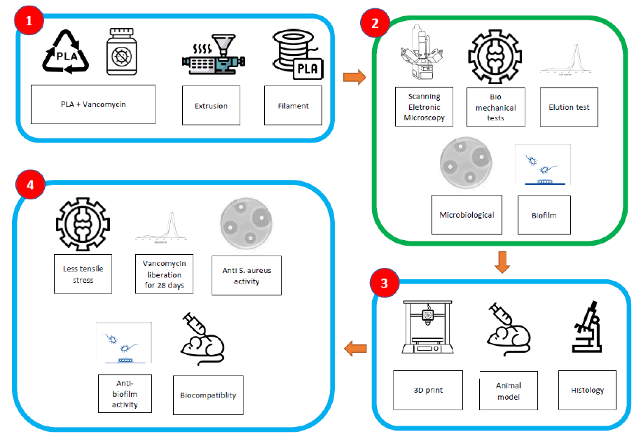
A schematic chart depicting the methods and results of the study with vancomycin-impregnated PLA is presented in the Figure 1.
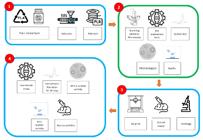
Schematic chart depicting the methods and results of the study with vancomycin-impregnated polylactic acid (PLA). In "1," we summarize the extrusion of the filament through the combination of PLA pellets with vancomycin using silicone oil. In 2, we describe the tests used for filament analysis, including scanning electron microscopy, biomechanical tests according to the ASTM (American Society for Testing and Materials) standard model, antibiotic elution tests, and microbiological tests. In 3, the process of 3D printing of models by FDM (fused deposition modeling) for the tests and their results in 4, which includes biocompatibility, antibiofilm and antimicrobial activity, as well as elution for at least 28 days and a reduction in the mechanical strength of PLA impregnated with vancomycin.
Reagents
Polylactic acid was obtained from Braskem (Sao Paulo, Brazil), vancomycin was obtained from Teuto (Anapolis, Brazil) and silicone oil obtained from Sigma-Aldrich (St Louis, MI). Mueller-Hinton agar and Tryptic soy broth were obtained from Sigma-Aldrich (St Louis, MI).
PLA impregnation
Filaments of vancomycin-impregnated PLA (V-PLA) were manufactured by combining PLA pellets with vancomycin via extrusion, following a protocol protected by a patent (INPI, Brazil, 102023018694-7). To provide a concise overview, PLA pellets were mixed with vancomycin at a ratio of 40 grams of PLA to every 1 gram of vancomycin. The PLA and vancomycin were blended in a conical tube of 50 mL under continuous agitation for 1 minute using silicone oil viscosity 5 cSt (25 °C) (Sigma-Aldrich, St Louis MI). Subsequently, the V-PLA material was subjected to a 30-minute drying process at 70°C and then extruded to produce filaments with a diameter of 1.75 mm.
Both control PLA (C-PLA), which lacked impregnation, and V-PLA samples were created using a commercial 3D printer (Creator Pro 3D printer, Flashforge Corporation, Zhejiang, China). Filaments with a 1.75 mm diameter were utilized for specimen preparation. The following parameters were employed for the 3D printing of the samples: nozzle movement speed (1800 mm/min), first layer speed (300 mm/min), nozzle temperature (220°C), bed temperature (60°C), layer height (0.1-0.3 mm), extrusion width (0.48 mm), and infill density (100%) for all specimens. During the fabrication process, the filaments were guided through a pinch roller feed mechanism to the heated nozzle. The filament was then melted and extruded through the nozzle, with the molten material deposited onto a preheated print bed. All biological and compression scaffolds were designed to have a relative density of 100% [18].
Scanning electronic microscopy
For qualitative SEM visualization, the specimens of each group was transferred into sterile glass Petri dishes. The models were metalized with gold particles in metalizing equipment with a Q150R ES rotary pump (Quorum Technologies, Lewes, UK), and later fixed in a metal base for observation under SEM, which was a PentaFET Precision (Oxford Instruments, Abingdon, UK) at 5.0 kV [15]. Observations were made with magnifications between 20 and 3.000×.
Vancomycin elution test
The elution test procedure entailed immersing a cylindrical V-PLA model, measuring 5 cm in height and 0.6 cm in diameter, in 10 mL of 0.9% physiological saline. Weekly measurements of vancomycin concentration were conducted by extracting 100 μL of the solution and analyzing it through high-performance liquid chromatography [19].
Biomechanical Assay
The biomechanical assessment of C-PLA and V-PLA involved conducting a tensile test to compare the impact of impregnation on the tensile strength properties of PLA. An EMIC DL 500 universal testing machine was employed to carry out these tests. Each group, comprising C-PLA and V-PLA, was represented by five samples. The dumbbell-shaped samples were designed in accordance with the ASTM D1708-13 standard. Prior to each test, the equipment was provided with sample thickness, width, and initial length values. Thickness and width measurements were essential for calculating the cross-sectional area and, consequently, the stress (as per equation 1). Thickness was measured at three points on the sample using a thickness gauge. The test involved the application of a pre-load of 0.1 N and a testing velocity of 5 mm/min [20].
Equation 1. Stress calculation.
where:
σ is the stress in megapascal [MPa];
F is the force in newton [N] and
A is the cross-sectional area in square millimeter [mm2].
Ethylene oxide toxic residues
The potential clinical application of V-PLA necessitates the consideration of a terminal sterilization procedure. Ethylene oxide was selected as the method to be tested, considering cost-effectiveness and the absence of water residue, thereby ensuring safety. Nevertheless, to ensure the safety of this sterilization process, an assessment of toxic residues of ethylene oxide was conducted. The determination of the concentration of toxic residues resulting from the use of ethylene oxide in sterilization was performed in accordance with the requirements specified by ISO10993-7 [21].
Antimicrobial activity
In each group, a total of five samples of C-PLA and V-PLA, each with a 6 mm diameter, were subjected to testing. Subsequently, these samples were placed on Mueller-Hinton agar plates containing S. aureus ATCCTM 25923, adjusted to a turbidity equivalent to 0.5 McFarland, corresponding to a concentration of 3 x 10⁸ cfu/mL (colony-forming units). The diameter of the inhibition zone surrounding the samples was measured after 24 hours of incubation at 37°C, serving as an indicator of their antibacterial activity [22, 23]. Vancomycin is thermostable at temperatures greater than 200 degrees Celsius. Nonetheless, we included a microbiological test comparing the V-PLA disc with PLA derived from pellets, commercially printed PLA, and a commercially available nitrocellulose diffusion disc (BD BBL™ Sensi-Disc™ Vancomycin VA-30ug) to assess comparative activity. Similarly, we included a V-PLA disc that was submerged in 10mL of ultrapure water for 28 days and then removed, air-dried, and subsequently placed on the same plate for comparison.
Antibiofilm activity
A 1:10 dilution was prepared in Tryptic soy broth (TSB) using a microbe 0.5 McFarland suspension of a S. aureus ATCCTM 25923. This dilution process was carried out until a bacterial concentration of 107 cfu/mL was achieved [24]. Next, 10 mL of TSB broth was added to sterile 24-well plates, completely covering the models (6 mm of diameter and 3 mm of height, n=4 for each group), and the plates were agitated at 120 rpm for 2 hours. This allowed the specimens to be exposed to the broth and facilitate bacterial adhesion [15, 25].
After the incubation period, the specimens were transferred to a new sterile 24-well plate containing 0.9% NaCl. This step was performed to remove any planktonic (free-floating) cells from the material. Following the removal of planktonic cells, the specimens were transferred to another sterile 24-well plate and submerged in 10 mL of TSB at 37°C for 24 hours without agitation. This allowed the cells adhering to the device surface to form a biofilm. After the biofilm formation, the specimens were submerged in 15 mL conical tubes filled with 10 mL of sterile 0.9% NaCl to remove any residual and unadhered/planktonic cells (step I). This washing step (step I) was repeated three times [26, 27].
Following the washing steps, the specimens were placed into 15 mL conical tubes filled with 10 mL of 0.9% NaCl for further processing. They were subjected to sonication for 5 minutes using an ultrasonic bath with a Soniclean 15 device (Sanders Medical, Santa Rita do Sapucaí, Brazil) operating at a frequency of approximately 40 kHz and a temperature of 35°C. [28]. After the sonication step (step II), the supernatant (100 μL) was collected and inoculated onto TSA agar plates for growth evaluation and cell count (cfu/mL). This allowed for the assessment of bacterial colonies and determination of the viable bacterial count in the samples.
Biocompatibility
The study was granted approval by the Ethical Committee of the PUCPR (Curitiba, Brazil). All procedures adhered to the regulations outlined in the Brazilian animal protection law. A total of twenty male C57BL/6N mice (FIOCRUZ, Curitiba, PR, Brazil) were included in the study, distributed across four study groups as follows: V-PLA (n=5), C-PLA (n=5), control (without graft) (n=5), and autograft (n=5).
The mice were anesthetized, and skin incisions were made at the top of the head, and the periosteum was detached. Then, bone defects 5 mm in diameter were made using a dental low-speed engine and a 5-mm diameter trephine bur (Implatex, Tokyo, Japan) [29]. The PLA model with 5mm of diameter was implanted into the defect site (groups C-PLA and V-PLA). In the group autograph, the bone removes from skull was macerated and reimplanted in the surgical site, and the group control, the skin was sutured without any implant. After the implants, the skin was tightly sutured. The mice were followed for 8 weeks before scheduled euthanasia. The surgical and euthanasia procedures were previously published, providing detailed information about the methods employed [30]. Following euthanasia, the skull and the surrounding soft tissue were immersed in 10% formalin for subsequent histological analysis.
Histological analysis
Histological analyses were conducted to evaluate inflammatory responses. Longitudinal cuts measuring 3.0 mm in length were made, and the tissue samples were fixed in neutral buffered formalin 10%. Subsequently, the samples were embedded in paraffin, and 4-µm thick sections were obtained.
To assess the morphological integrity of the grafts, hematoxylin, and eosin (H&E) staining was performed. An optical microscope BX51 (Olympus Tokyo, Japan) was used for visual examination of the stained sections. The prepared slides were scanned using an Axio Scan.Z1 slide scanner (Carl Zeiss Microscopy GmbH, Jena, Germany), and subsequent image processing and analysis were performed using Zen lite software (Carl Zeiss, Jena, Germany). This allowed for detailed examination and documentation of the histological features and changes observed in the bone samples.
Statistical analysis and data presentation
Continuous variables were expressed as medians, and the degree of dispersion was indicated by the 25th and 75th percentiles. For the comparison of medians, the Mann-Whitney test was conducted, and a significant difference between the medians was indicated when p < 0.05. There was no comparison of categorical data, and the other data were descriptive, not requiring statistical analysis. For the statistical analysis, the GraphPad Prism software, version 7.0, was used.
RESULTS
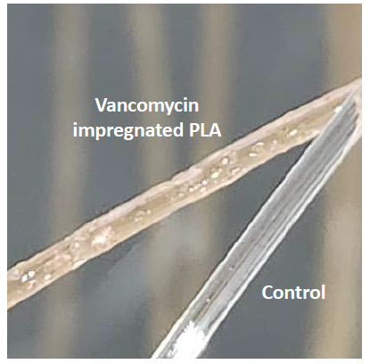
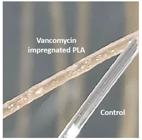
V-PLA displayed noticeable macroscopic distinctions compared to conventional PLA, featuring a more irregular surface and a yellowish color consistent with the presence of vancomycin (see Figure 2). The macroscopic characteristics of V-PLA were distinctly dissimilar from those of C-PLA filaments. It was observed that the V-PLA filament could be broken after a 90-degree bend, while the C-PLA filament would merely bend without breaking.
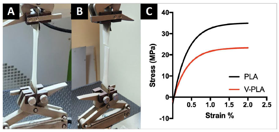
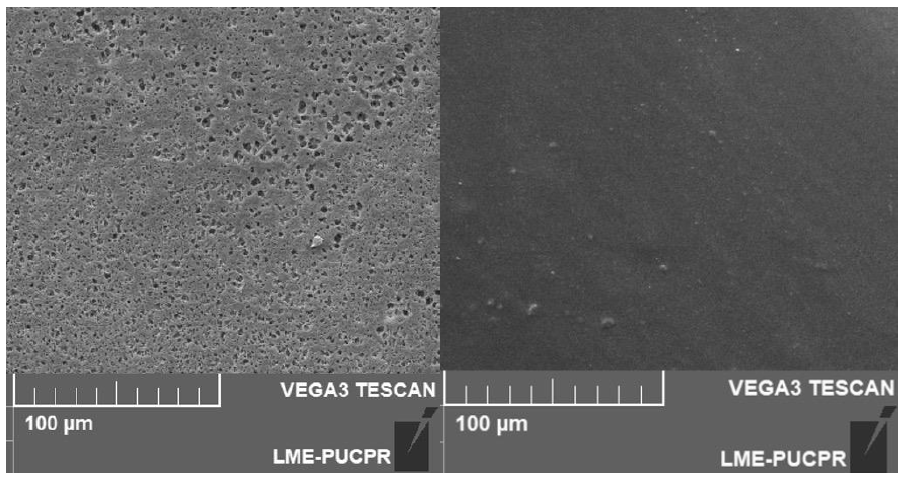
V-PLA exhibited an earlier rupture compared to C-PLA, signifying reduced resistance when exposed to mechanical stress (refer to Figure 3), achieving a median of 22 MPa of stress against 35 MPa of control group (n=5), a reduction of 37%. In the scanning electron microscopy (SEM) analysis, it was evident that the appearance of C-PLA remained consistent and uniform after the printing process. In contrast, the V-PLA group exhibited irregularities in the printed model, which consisted of 1 x 1 cm squares with a height of 3 mm. (Figure 4).
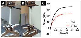
Biomechanical test of V-PLA and control to evaluate tensile. A - The test before rupture of test body; B - after rupture of test body; C - Curve of tensile stress of control (black) and V-PLA (red).
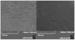
Scanning electronic microscopy of V-PLA (left) and PLA control (right). V-PLA presents modifications (roughness) on the surface.
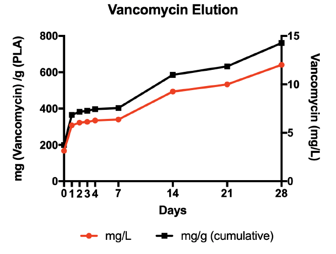
The vancomycin elution test indicated that vancomycin antibiotic concentrations consistently exceeded the minimum inhibitory concentrations for most vancomycin-sensitive S. aureus strains. Specifically, concentrations surpassed 2 mg/L from the initial day of measurement, with values starting at 3.15 mg/L and steadily rising to 12.02 mg/L by day 28 (see Figure 5 red line). When we analyzed the release of vancomycin per gram of PLA, the total concentration in 28 days reached 760 mg (Figure 5 black line). The tests for toxic residues of ethylene oxide were negative, indicating that sterilization using this method is a safe process.
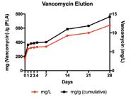
Vancomycin elution test of a V-PLA model for 28 days, showing the cumulative concentration of the drug in the water (red line) and cumulative vancomycin released by gram of PLA.
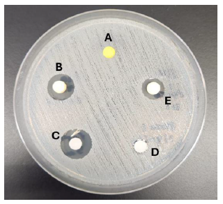
In addition to the elution test, a test of the impregnated disc was conducted, comparing it with a standard diffusion disc containing commercially available vancomycin (BD BBL) (Figure 6). Although the results are not superimposable, they demonstrate a similar halo of activity. Furthermore, the discs were kept for 28 days in ultrapure water, and a test on a plate was performed, showing a reduction in the drug but maintaining antimicrobial activity.
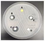
Plate test with S. aureus comparing the activity of a PLA disc impregnated with vancomycin (B), in relation to a nitrocellulose disc with 30ug vancomycin (C), a PLA disc produced from pellet but without impregnation (D), and a printed disc with commercially available PLA filament. The disc indicated as E is a model impregnated with vancomycin but left for 28 days in ultrapure water to evaluate the maintenance of activity.
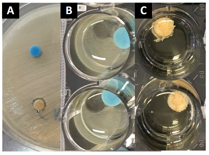
In the PLA disk susceptibility test, V-PLA exhibited the formation of a halo representing bacterial growth inhibition, a phenomenon not observed in non-impregnated PLA. It's worth noting that the halo was not extensive, indicating a low diffusion rate or potentially a lower concentration of vancomycin. Based on the elution graph, one plausible hypothesis is that the antibiotic release rate is relatively slow, which may explain the observed results (Figure 7 A).
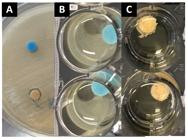
Microbiological tests of V-PLA (yellow and C-PLA (control in blue). A - disk diffusion test showing a small halo in the V-PLA; B - Control group of PLA with biofilm formation around the model and; C - V-PLA without biofilm formation in the well.
The biofilm test demonstrated an absence of biofilm formation in V-PLA, as evidenced by the absence of S. aureus counts following sonication. In contrast, the PLA group exhibited a median count of 3.4 x 10⁸ cfu/g, with an interquartile range (IQR) of 25-75% between 1.3 x 10⁸ and 6.2 x 10⁸ cfu/g. This indicates a substantial disparity in biofilm formation between V-PLA and C-PLA, with V-PLA effectively inhibiting biofilm development (p < 0.05). (Figure 7 B and C).
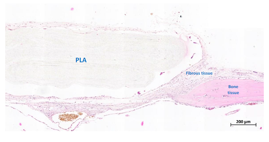
In the animal model, the primary goal was to assess the biocompatibility and the incorporation of PLA in contact with bone, as well as the potential for osteoconductivity. The introduction of both PLA and V-PLA led to the development of a fibrous capsule surrounded by fibroblasts, with no observable inflammatory reaction or osteointegration. In contrast, autografting elicited a robust inflammatory response and exhibited restricted bone neoformation (Figure 8). In light of these observations, it can be concluded that V-PLA demonstrates biocompatibility, but it is not associated with osteogenesis, osteoconductive, or osteoinductive.)
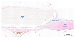
Histological characteristics of PLA implanted in mice. All images (A to E) present the relation of PLA with the tissue in different scales. There is a classic fibrous capsule around the model with fibroblasts and collagen fibers. In E is possible to visualize the PLA model and bone without osteointegration due to fibrous tissue around the PLA.
DISCUSSION
The successful inclusion of vancomycin into the thermoplastic has been demonstrated, enabling the preservation of its antimicrobial activity even after exposure to high temperatures, up to 200 degrees Celsius. It's important to note that vancomycin, like aminoglycosides, is a thermally stable antibiotic. In contrast, many other antibiotics are not as heat-resistant [31]. Indeed, it's worth noting that even thermally unstable antibiotics may retain a portion of their antimicrobial activity when exposed to elevated temperatures. The degree to which their activity is preserved can vary depending on the specific antibiotic and the temperature and duration of exposure. This preservation of activity after heat exposure may have implications for certain applications in which antibiotics need to be incorporated into materials or subjected to sterilization processes [32,33].
In the elution test, it is clear that the antibiotic is released at concentrations above the minimal inhibitory concentration (MIC), indicating at least bacteriostatic antimicrobial activity. To achieve bactericidal concentrations, it is likely that a higher antibiotic concentration would be necessary. Bactericidal concentrations typically exceed the MIC and are needed to effectively kill bacteria rather than just inhibit their growth. [17,34-36]. On the other hand, while the release of vancomycin may not reach the same levels as some other materials used in the field of orthopedics, such as orthopedic cement, it exhibits a prolonged release profile. This extended release ensures a long-term antimicrobial effect that persists for at least 28 days. This sustained antimicrobial activity can be valuable for preventing or managing infections in orthopedic applications over an extended period. [5,19,32,37]. It's worth noting that while a longer duration of antimicrobial activity could be investigated in your study, it's essential to consider the intended purpose of such materials [38,39]. These materials are typically viewed as temporary "bridges" used until the placement of a definitive prosthesis or implant [40]. In general, temporary materials in orthopedics should not exceed a duration of six months. Moreover, there is a global trend to further shorten this duration to help patients regain joint functionality as soon as possible [17]. Indeed, emphasizing materials with enhanced mechanical strength is paramount in preventing complications such as fractures and mechanical disorders in orthopedic applications. While V-PLA showed lower mechanical strength than C-PLA in the tensile test, this difference doesn't necessarily render the material impractical. The suitability of the material depends on the specific context and the intended clinical use.
The ideal mechanical test could vary based on the application, and in some cases, compression tests might be more relevant. Additionally, the appropriateness of a material can be highly location-specific within the human body. Factors such as the forces acting on the implant site, the surrounding tissues, and the intended function of the material all play a crucial role in determining the most suitable material for a particular orthopedic application.
Therefore, while mechanical strength is a vital consideration, the practicality of a material in clinical use is multifaceted and involves evaluating various factors beyond just tensile strength. Each application and location within the body may have unique requirements that need to be taken into account for an informed decision [41]. For the temporary replacement of a shoulder prosthesis, mechanical strength may have limited relevance, as is the case with filling bone defects in conjunction with plate and screw fixation. However, for applications like knee or hip spacers, conducting biomechanical tests that simulate the conditions at the implant site becomes imperative for assessing their viability [42]. One potential approach is to incorporate a robust material, such as steel, as a central support structure to provide mechanical strength, while applying a V-PLA coating to this material for the sole purpose of antibiotic release.
Alternatively, another option is to blend PLA with other biocompatible polymers to enhance its mechanical strength. This would involve combining PLA with other materials to create a composite that balances the desired mechanical properties with the need for controlled antibiotic release. [43]. The doping of PLA with antibiotics is a relatively recent modality following the spread of 3D printing technology. While there are some studies on the addition of antimicrobials, the literature is still not robust. The incorporation of gentamicin into PLA has been tested through a pressure-based surface modification method [44]. In another study, the extrusion method using gentamicin was tested, a technique similar to the one employed in our study. Although they did not mention the antibiotic concentration, it was surprising that there was no loss in strength, given that the antibiotic occupies a significant volume to maintain its long-term activity. It is also crucial that the antibiotic has the possibility of proper diffusion [45]. Another possibility for the addition of antibiotics to PLA involves their solubilization in a solvent that promotes the degradation of the polymer, followed by the subsequent removal of the solvent. However, none of these technologies are for use in 3D printing [46].
Considering its clinical use, we conducted tests for toxic residue of ethylene oxide, a form of terminal sterilization. The tests yielded negative results. We did not perform cytotoxicity tests as the literature is extensive in that regard, and vancomycin combined with other polymers has already been used safely for many decades [5,9,35,38,47]. Animal tests confirmed other findings in the literature regarding biocompatibility, where the implant did not demonstrate significant inflammatory reaction, only the formation of a fibrous capsule, which is common when performing an implant in animals [48]. It's important to note that PLA, when used on its own, does not exhibit the capacity for osseointegration, as confirmed by histological examinations. However, when combined with other materials, such as hydroxyapatite, it becomes feasible to confer properties conducive to bone integration. Nevertheless, it's essential to emphasize that our study's primary objective was not to achieve osseointegration. Instead, our main goal was to assess the feasibility of a temporary implant for the treatment or prevention of infections.
Since Staphylococcus species are the main pathogens associated with orthopedic infections, vancomycin was the drug of choice [17, 49, 50]. The qualitative disk diffusion test demonstrated its diffusion capacity and growth inhibition. In the biofilm model, it was clear that it had the ability to prevent biofilm formation, although it did not reach bactericidal concentrations [11,16,23,51,52]. This confers a very important property for its use as an orthopedic spacer.
Certainly, here are some limitations of this study. V-PLA exhibited lower mechanical strength compared to C-PLA. This could be a limitation if the material is intended for applications that require higher mechanical strength, such as load-bearing implants or sites subject to significant mechanical stresses. We have described visual macroscopic differences between V-PLA and C-PLA but does not provide quantitative analyses of these characteristics. The lack of precise measurements can be considered a limitation in characterizing the material. Vancomycin release was assessed for 28 days but we do not provide information on long-term release. The durability of antibiotic release beyond this period can be an important consideration for clinical applications. The study identified the formation of a fibrous capsule in response to the introduction of PLA but did not observe osteointegration. However, the assessment of osteointegration typically requires more in-depth analysis, including the observation of tissue interactions over a longer period.
CONCLUSION
While the study provided interesting results, it's essential to acknowledge that the conclusions are based on specific experiments and may not be directly generalizable to all clinical situations. The clinical applicability of V-PLA may depend on various factors, including anatomical location and specific use. V-PLA filament can be a future material to be used in the manufacture of several devices in medicine, avoiding or treating infections by susceptible microorganisms.
Acknowledgments
We thank Prof. Paulo Soares for technical support with scanning electronic microscopy
REFERENCES
- 1 Kirkpatrick JS, Cornell CN, Hoang BH, Hsu W, Watson JT, Watters WC, et al. Bone void fillers. J Am Acad Orthop Surg. 2010 Sep;18(9):576-9.
- 2 Evaniew N, Tan V, Parasu N, Jurriaans E, Finlay K, Deheshi B, et al. Use of a calcium sulfate-calcium phosphate synthetic bone graft composite in the surgical management of primary bone tumors. Orthopedics. 2013 Feb;36(2):e216-22.
- 3 Dantas LR, Ribeiro VST, Kraft L, Pinho RA, Suss PH, Vasconcellos FTF, et al. Collagen matrices are preserved following decellularization of a bovine bone scaffold. Cell Tissue Bank. 2022 Sep;23(3):531-40.
- 4 Winkler T, Sass FA, Duda GN, Schmidt-Bleek K. A review of biomaterials in bone defect healing, remaining shortcomings and future opportunities for bone tissue engineering: The unsolved challenge. Bone Joint Res. 2018 Mar;7(3):232-43.
- 5 Pedroni MA, Ribeiro VST, Cieslinski J, Lopes APA, Kraft L, Suss PH, et al. Different concentrations of vancomycin with gentamicin loaded PMMA to inhibit biofilm formation of Staphylococcus aureus and their implications. J Orthop Sci. 2022 Dec 14;14(14):S0949-2658(22)00336-0.
- 6 Ahmed W, Zhai Z, Gao C. Adaptive antibacterial biomaterial surfaces and their applications. Mater Today Bio. 2019 Mar;2:100017.
- 7 Kowalczyk P, Wojasinski M, Jaroszewicz J, Kopec K, Ciach T. Controlled formation of highly porous polylactic acid-calcium phosphate granules with defined structure. Biomater Adv. 2023 Jan;144:213195.
- 8 Chou YC, Lee D, Chang TM, Hsu YH, Yu YH, Chan EC, et al. Combination of a biodegradable three-dimensional (3D) - printed cage for mechanical support and nanofibrous membranes for sustainable release of antimicrobial agents for treating the femoral metaphyseal comminuted fracture. J Mech Behav Biomed Mater. 2017 Aug;72:209-18.
- 9 Telles JP, Morales R, Jr. Yamada CH, Marins TA, D'Amaro Juodinis V, Sztajnbok J, et al. Optimization of Antimicrobial Stewardship Programs Using Therapeutic Drug Monitoring and Pharmacokinetics-Pharmacodynamics Protocols: A Cost-Benefit Review. Ther Drug Monit. 2023 Apr 1;45(2):200-8.
- 10 Tuon FF, Yamada CH, Cieslinski J, Dos Santos Oliveira D, Ribeiro VST, Gasparetto J, et al. Cerebrospinal Fluid Penetration of Vancomycin During Continuous Infusion Therapy in Patients with Nosocomial Ventriculitis. Therapeutic Drug Monitoring. 2021;05.
- 11 Kraft L, Ribeiro VST, de Nazareno Wollmann LCF, Suss PH, Tuon FF. Determination of antibiotics and detergent residues in decellularized tissue-engineered heart valves using LC-MS/MS. Cell Tissue Bank. 2020 Dec;21(4):573-84.
- 12 Yamada CH, Telles JP, Oliveira DDS, Cieslinski J, Ribeiro VST, Gasparetto J, et al. Comparison of intermittent versus continuous-infusion vancomycin for treating severe patients in intensive care units. Braz J Infect Dis. 2020 Jul-Aug;24(4):356-9.
- 13 Tuon FF, Romero R, Gasparetto J, Cieslinski J. Vancomycin trough level and loading dose. Infect Drug Resist. 2018;11:2393-6.
- 14 Tuon FF, Rocha JL, Morales HM, Sakumoto MH, Miksza KF, Pecoit-Filho R. Modulation of inflammatory mediators during treatment of cellulitis with daptomycin or vancomycin/oxacillin. Int J Antimicrob Agents. 2015 Oct;46(4):476-8.
- 15 Da Rocha L, Ribeiro VST, de Andrade AP, Gonçalves GA, Kraft L, Cieslinski J, et al. Evaluation of Staphylococcus aureus and Candida albicans biofilms adherence to PEEK and titanium-alloy prosthetic spine devices. Eur J Orthop Surg Traumatol. 2022 Jul;32(5):981-9.
- 16 Dantas LR, Wollmann LC, Suss PH, Kraft L, Ribeiro VST, Tuon FF. Disinfection protocol for human musculoskeletal allografts in tissue banking using hydrogen peroxide 30. Cell Tissue Bank. 2021 Jun 6.
- 17 Telles JP, Cieslinski J, Tuon FF. Daptomycin to bone and joint infections and prosthesis joint infections: a systematic review. Braz J Infect Dis. 2019 May-Jun;23(3):191-6.
- 18 Alam F, Shukla VR, Varadarajan KM, Kumar S. Microarchitected 3D printed polylactic acid (PLA) nanocomposite scaffolds for biomedical applications. J Mech Behav Biomed Mater. 2020 Mar;103:103576.
- 19 Tseng TH, Chang CH, Chen CL, Chiang H, Hsieh HY, Wang JH, et al. A simple method to improve the antibiotic elution profiles from polymethylmethacrylate bone cement spacers by using rapid absorbable sutures. BMC Musculoskelet Disord. 2022 Oct 14;23(1):916.
- 20 Wollmann L, Suss P, Mendonca J, Luzia C, Schittini A, Rosa GWXD, et al. Characterization of Decellularized Human Pericardium for Tissue Engineering and Regenerative Medicine Applications. Arq Bras Cardiol. 2019 Jun 27;113(1):11-7.
- 21 Sladowski D, Grabska-Liberek I, Olkowska-Truchanowicz J, Lipski K, Gut G. An evaluation of sterilisation processes. Altern Lab Anim. 2008 Nov;36(5):585-90.
- 22 Cieslinski J, Ribeiro VST, Lima CK, Kraft L, Suss PH, Tuon FF. Sonication as a tool for disrupting biofilms and recovering microorganisms in bladder catheters. J Bras Nefrol. 2023 May 8.
- 23 Mendonca JR, Dantas LR, Tuon FF. Activity of multipurpose contact lens solutions against Staphylococcus aureus, Pseudomonas aeruginosa, Serratia marcescens and Candida albicans biofilms. Ophthalmic Physiol Opt. 2023 Sep;43(5):1092-9.
- 24 Franklin MJ, Chang C, Akiyama T, Bothner B. New Technologies for Studying Biofilms. Microbiol Spectr. 2015 Aug;3(4):10.1128.
- 25 Cieslinski J, Ribeiro VST, Kraft L, Suss PH, Rosa E, Morello LG, et al. Direct detection of microorganisms in sonicated orthopedic devices after in vitro biofilm production and different processing conditions. Eur J Orthop Surg Traumatol. 2021 Jan 4.
- 26 Kraft L, Ribeiro VST, Goncalves GA, Suss PH, Tuon FF. Comparison of amphotericin B lipid complex, deoxycholate amphotericin B, fluconazole, and anidulafungin activity against Candida albicans biofilm isolated from breakthrough candidemia. Enferm Infecc Microbiol Clin (Engl Ed). 2023 Jan 25.
- 27 Kraft L, Ribeiro VST, Petroski LP, Herai RH, Peronni KC, Figueiredo DLA, et al. Saprochaete clavata invasive infection: characterization, antifungal susceptibility, and biofilm evaluation of a rare yeast isolated in Brazil. Rev Inst Med Trop São Paulo. 2023;65:e12.
- 28 Trampuz A, Piper KE, Jacobson MJ, Hanssen AD, Unni KK, Osmon DR, et al. Sonication of removed hip and knee prostheses for diagnosis of infection. N Engl J Med. 2007 Aug 16;357(7):654-63.
- 29 Abe T, Sumi K, Kunimatsu R, Oki N, Tsuka Y, Nakajima K, et al. Dynamic imaging of the effect of mesenchymal stem cells on osteoclast precursor cell chemotaxis for bone defects in the mouse skull. J Dent Sci. 2018 Dec;13(4):354-9.
- 30 Wittmann C, Vanvelk N, Furst AE, Moriarty TF, Zeiter S. Development and Characterization of a Subcutaneous Implant-Related Infection Model in Mice to Test Novel Antimicrobial Treatment Strategies. Biomedicines. 2022 Dec 24;11(1):40.
- 31 Koo KH, Yang JW, Cho SH, Song HR, Park HB, Ha YC, et al. Impregnation of vancomycin, gentamicin, and cefotaxime in a cement spacer for two-stage cementless reconstruction in infected total hip arthroplasty. J Arthroplasty. 2001 Oct;16(7):882-92.
- 32 Singh KA, Jaunky C, Shah H. An unusual presentation, novel treatment with Meropenem PMMA beads and complications of Klebsiella osteomyelitis in a healthy adult- A case report. J Clin Orthop Trauma. 2022 Jan;24:101719.
- 33 Galvez-Lopez R, Pena-Monje A, Antelo-Lorenzo R, Guardia-Olmedo J, Moliz J, Hernández-Quero J, et al. Elution kinetics, antimicrobial activity, and mechanical properties of 11 different antibiotic loaded acrylic bone cement. Diagn Microbiol Infect Dis. 2014 Jan;78(1):70-4.
- 34 Britt NS, Patel N, Horvat RT, Steed ME. Vancomycin 24-Hour Area under the Curve/Minimum Bactericidal Concentration Ratio as a Novel Predictor of Mortality in Methicillin-Resistant Staphylococcus aureus Bacteremia. Antimicrob Agents Chemother. 2016 May;60(5):3070-5.
- 35 Schwerdt C, Rohner E, Bohle S, Jacob B, Matziolis G. The bactericidal effect of vancomycin is not altered by tranexamic acid, adrenalin, dexamethasone, or lidocaine in vitro. Sci Rep. 2021 May 24;11(1):10739.
- 36 Chuang YC, Wang JT, Lin HY, Chang SC. Daptomycin versus linezolid for treatment of vancomycin-resistant enterococcal bacteremia: systematic review and meta-analysis. BMC Infect Dis. 2014 Dec 13;14:687.
- 37 Beenken KE, Bradney L, Bellamy W, Skinner RA, McLaren SG, Gruenwald MJ, et al. Use of xylitol to enhance the therapeutic efficacy of polymethylmethacrylate-based antibiotic therapy in treatment of chronic osteomyelitis. Antimicrob Agents Chemother. 2012 Nov;56(11):5839-44.
- 38 Shen SC, Letchmanan K, Chow PS, Tan RBH. Antibiotic elution and mechanical property of TiO(2) nanotubes functionalized PMMA-based bone cements. J Mech Behav Biomed Mater.2019 Mar;91:91-8.
- 39 Cyphert EL, Kanagasegar N, Zhang N, Learn GD, von Recum HA. PMMA Bone Cement Composite Functions as an Adjuvant Chemotherapeutic Platform for Localized and Multi-Window Release during Bone Reconstruction. Macromol Biosci. 2022 May;22(5):e2100415.
- 40 Hollyer I, Ivanov D, Kappagoda S, Lowenberg DW, Goodman SB, Amanatullah DF. Selecting a high-dose antibiotic-laden cement knee spacer. J Orthop Res. 2023 Jul;41(7):1383-96.
- 41 Ashraf M, Zahra SG, Farooq M, Choudhary N, Ashraf N. PMMA Cranioplasty Making by Using Open-Source CAD Software, PLA Printers, and Silicone Rubber Molds: Technical Note with Two Illustrative Cases. Asian J Neurosurg. 2023 Jun;18(2):420-2.
- 42 Zhang X, Wang Y, Zhang L, Yu K, Ding Z, Zhang Y, et al. Biomechanical Properties of Bionic Collum Femoris Preserving Hip Prosthesis: A Finite Element Analysis. Orthop Surg. 2023 Apr;15(4):1126-35.
- 43 Ramot Y, Haim-Zada M, Domb AJ, Nyska A. Biocompatibility and safety of PLA and its copolymers. Adv Drug Deliv Rev. 2016 Dec 15;107:153-62.
- 44 Malhotra A, Krishna A, Garg SK, Gupta S, Jindal R, Jain G. Short term results with the use of PLA antibiotic coated nail in open tibia fractures: A prospective study. J Orthop. 2022 Aug 28;34:183-8.
- 45 Tappa K, Jammalamadaka U, Weisman JA, Ballard DH, Wolford DD, Pascual-Garrido C, et al. 3D Printing Custom Bioactive and Absorbable Surgical Screws, Pins, and Bone Plates for Localized Drug Delivery. J Funct Biomater. 2019 Apr 1;10(2):17.
- 46 Meyer JD, Falk RF, Kelly RM, Shively JE, Withrow SJ, Dernell WS, et al. Preparation and in vitro characterization of gentamycin-impregnated biodegradable beads suitable for treatment of osteomyelitis. J Pharm Sci. 1998 Sep;87(9):1149-54.
- 47 Peng Z, Lin X, Kuang X, Teng Z, Lu S. The application of topical vancomycin powder for the prevention of surgical site infections in primary total hip and knee arthroplasty: A meta-analysis. Orthop Traumatol Surg Res. 2021 Jun;107(4):102741.
- 48 Singhvi MS, Zinjarde SS, Gokhale DV. Polylactic acid: synthesis and biomedical applications. J Appl Microbiol. 2019 Dec;127(6):1612-26.
- 49 Gasparetto J, Pitta R, Cordova K, et al. Acute Kidney Injury in Patients Using Amikacin in Intensive Care Unit-A Paired Case-Control Study With Meropenem. Am J Ther. 2020 Jul/Aug;27(4):e403-e5.
- 50 Tuon FF, Rigatto MH, Lopes CK, Kamei LK, Rocha JL, Zavascki AP. Risk factors for acute kidney injury in patients treated with polymyxin B or colistin methanesulfonate sodium. Int J Antimicrob Agents. 2014 Apr;43(4):349-52.
- 51 Tuon FF, Suss PH, Telles JP, Dantas LR, Borges NH, Ribeiro VST. Antimicrobial Treatment of Staphylococcus aureus Biofilms. Antibiotics (Basel). 2023 Jan 4;12(1).
- 52 Chaiben V, Yamada CH, Telles JP, de Andrade AP, Arend LNVS, Ribeiro VST, et al. A carbapenem-resistant Acinetobacter baumannii outbreak associated with a polymyxin shortage during the COVID pandemic: an in vitro and biofilm analysis of synergy between meropenem, gentamicin and sulbactam. J Antimicrob Chemother. 2022 May 29;77(6):1676-84.