
 Histological evaluation of bone graft healing in maxillary sinus floor augmentation at two healing time points: a randomized clinical trial
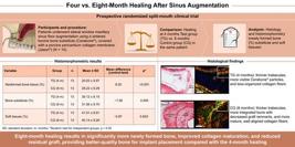
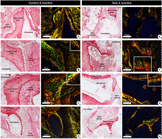
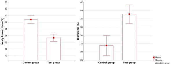
Este estudo tem como objetivo comparar os efeitos de dois períodos distintos de cicatrização (4 e 8 meses) na formação óssea após cirurgias de elevação do assoalho do seio maxilar usando enxertos ósseos bovinos sinterizados por meio de análises histológicas e histomorfométricas. Usando um delineamento de boca dividida, quatorze participantes com edentulismo bilateral na maxila posterior foram incluídos e randomizados em dois grupos com 4 meses (Grupo Teste - GT) e 8 meses (Grupo Controle - GC) de períodos de cicatrização. Após o período de cicatrização, amostras ósseas foram coletadas e submetidas à análise histomorfométrica para quantificar as porcentagens de osso neoformado, biomaterial residual e tecidos moles. A comparação entre os grupos foi realizada usando o teste t de Student pareado (p < 0,05). Vinte e oito enxertos de seio maxilar foram realizados e 54 implantes dentários foram instalados. A análise histológica revelou tecido ósseo trabecular neoformado em contato próximo com a superfície do biomaterial, com maior espessura observada no GC. As fibras de colágeno no GC apresentaram maior birrefringência, indicando aumento de espessura e organização em comparação ao GT. A análise histomorfométrica demonstrou diferença significativa na porcentagem de formação óssea nova entre o GC (28,22 ± 6,29%) e o GT (20,02 ± 5,97%; p < 0,001), bem como na porcentagem de biomaterial residual (GT: 39,12 ± 8,15%; GC: 31,65 ± 8,70%; p = 0,005). A proporção de tecidos moles permaneceu estável entre os grupos. O período de cicatrização de 8 meses aumentou significativamente a formação e a maturação do tecido ósseo, sugerindo que esse intervalo otimiza a qualidade do osso enxertado ao utilizar substituto ósseo bovino sinterizado.
Autoria
person Ricardo Pasquini Filho
schoolDepartment of Implant Dentistry at the Latin American Dental Research and Teaching Institute College(ILAPEO). CuritibaPR/ Brazil. Latin American Dental Research and Teaching Institute CollegeBrazilCuritiba, PR, BrazilDepartment of Implant Dentistry at the Latin American Dental Research and Teaching Institute College(ILAPEO). CuritibaPR/ Brazil.
person Leandro Eduardo Klüppel
schoolDepartment of Implant Dentistry at the Latin American Dental Research and Teaching Institute College(ILAPEO). CuritibaPR/ Brazil. Latin American Dental Research and Teaching Institute CollegeBrazilCuritiba, PR, BrazilDepartment of Implant Dentistry at the Latin American Dental Research and Teaching Institute College(ILAPEO). CuritibaPR/ Brazil.
person Rubens Moreno de Freitas
schoolDepartment of Implant Dentistry at the Latin American Dental Research and Teaching Institute College(ILAPEO). CuritibaPR/ Brazil. Latin American Dental Research and Teaching Institute CollegeBrazilCuritiba, PR, BrazilDepartment of Implant Dentistry at the Latin American Dental Research and Teaching Institute College(ILAPEO). CuritibaPR/ Brazil.
person Luíz Antônio Violin
schoolDepartment of Implant Dentistry at the Latin American Dental Research and Teaching Institute College(ILAPEO). CuritibaPR/ Brazil. Latin American Dental Research and Teaching Institute CollegeBrazilCuritiba, PR, BrazilDepartment of Implant Dentistry at the Latin American Dental Research and Teaching Institute College(ILAPEO). CuritibaPR/ Brazil. school Department of Biochemistry and Tissue Biology at the State University of Campinas- Institute of Biology (Unicamp-IB). Campinas Brazil State University of CampinasBrazilCampinas, Brazil Department of Biochemistry and Tissue Biology at the State University of Campinas- Institute of Biology (Unicamp-IB). Campinas Brazil
person Silvio Roberto Consonni
school Department of Biochemistry and Tissue Biology at the State University of Campinas- Institute of Biology (Unicamp-IB). Campinas Brazil State University of CampinasBrazilCampinas, Brazil Department of Biochemistry and Tissue Biology at the State University of Campinas- Institute of Biology (Unicamp-IB). Campinas Brazil
person Elcio Marcantonio Junior
schoolDepartment of Implant Dentistry at the Latin American Dental Research and Teaching Institute College(ILAPEO). CuritibaPR/ Brazil. Latin American Dental Research and Teaching Institute CollegeBrazilCuritiba, PR, BrazilDepartment of Implant Dentistry at the Latin American Dental Research and Teaching Institute College(ILAPEO). CuritibaPR/ Brazil. schoolDepartment of Periodontology, UNESP - University Estadual Paulista, Araraquara Dental School, Araraquara, São Paulo, BrazilUniversity Estadual PaulistaBrazilAraraquara, São Paulo, BrazilDepartment of Periodontology, UNESP - University Estadual Paulista, Araraquara Dental School, Araraquara, São Paulo, Brazil
person Tatiana Miranda Deliberador
schoolDepartment of Implant Dentistry at the Latin American Dental Research and Teaching Institute College(ILAPEO). CuritibaPR/ Brazil. Latin American Dental Research and Teaching Institute CollegeBrazilCuritiba, PR, BrazilDepartment of Implant Dentistry at the Latin American Dental Research and Teaching Institute College(ILAPEO). CuritibaPR/ Brazil.
Correspondence: Ricardo Pasquini Filho, Department of Implantology, ILAPEO College. Rua Jacarezinho 656, Curitiba, Paraná, Brazil, Zip code: 80810-130. E-mail address:
pasquinirpf@terra.com.br
pasquinirpf@terra.com.br
Responsible Editor
Manoel Damião de Souza-Neto
SCIMAGO INSTITUTIONS RANKINGS
Department of Implant Dentistry at the Latin American Dental Research and Teaching Institute College(ILAPEO). CuritibaPR/ Brazil. Latin American Dental Research and Teaching Institute CollegeBrazilCuritiba, PR, BrazilDepartment of Implant Dentistry at the Latin American Dental Research and Teaching Institute College(ILAPEO). CuritibaPR/ Brazil.
Department of Biochemistry and Tissue Biology at the State University of Campinas- Institute of Biology (Unicamp-IB). Campinas Brazil State University of CampinasBrazilCampinas, Brazil Department of Biochemistry and Tissue Biology at the State University of Campinas- Institute of Biology (Unicamp-IB). Campinas Brazil
Department of Periodontology, UNESP - University Estadual Paulista, Araraquara Dental School, Araraquara, São Paulo, BrazilUniversity Estadual PaulistaBrazilAraraquara, São Paulo, BrazilDepartment of Periodontology, UNESP - University Estadual Paulista, Araraquara Dental School, Araraquara, São Paulo, Brazil
Figuras | Tabelas
 Thumbnail
 Thumbnail
 Thumbnail
 Thumbnail
imageopen_in_new
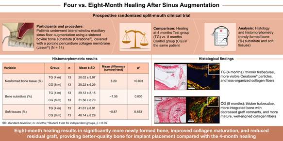
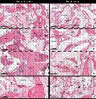
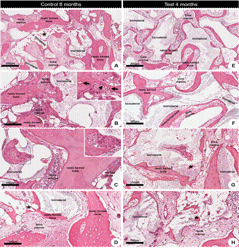
imageFigure 1 Representative photomicrographs of longitudinal sections of biopsies after 4 (A-D) and 8 (E-F) months of grafting. (A and E) At lower magnification, the trabeculae areas of neoformed bone tissue are around bone substitute areas or filled with bone marrow. It is possible to observe a difference in the thickness of these trabecular bones between the biopsies taken after 4 and 8 months of grafting. (B-D; F-H) At higher magnification, there is a flocculated bone substitute with an osteocyte nucleus (black arrow) within thin trabeculae of neoformed bone tissue. In a cross-sectional plane, these trabeculae permeate the interior of the region where the bone substitute is located (asterisk). In the bone marrow interface with the bone trabecula, there are bone lining cells, osteoblasts (black arrowhead), and osteoclasts (white arrowhead). Qualitatively, in the biopsies taken 8 months after grafting, the trabeculae of neoformed bone tissue are thicker than those taken 4 months after grafting. Furthermore, in both study groups, no gaps were found in the graft area without an osteocyte nucleus. Hematoxylin and Eosin (H&E) staining. Scale bars A and E = 200 µm; B-D; F-H = 100 µm. open_in_new
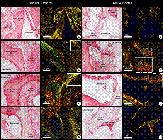
imageFigure 2 Representative photomicrographs of longitudinal sections of biopsies after 4 (A-D) and 8 (E-F) months of grafting under conventional light (left panel) and polarized light (right panel). (A and E) At lower magnification, the areas of trabeculae of neoformed bone tissue surround bone substitute areas or are filled with bone marrow. (B-D; F-H) At higher magnification, the organization of collagen fibers in the neoformed bone tissue in biopsies taken after 4 and 8 months of grafting. A progressive increase in collagen fiber thickness is observed as it transitions from orange and red to yellow and green, which is associated with mature collagen. The dark areas indicate a lack of collagen organization. Sirius Red and Hematoxylin staining. Scale bar A and E = 100 µm; B-D, F-H = 50 µm. open_in_new
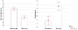
imageFigure 3 Histomorphometric analysis of the percentage of neoformed bone tissue and residual bone substitute between the groups. open_in_new
table_chartTable 1
Histomorphometric results after 4 and 8 months of healing for the CG and TG.
| Variable | Group | n | Mean ± sd | Median (min - max) | Mean Diff (control-test) | p* |
|---|---|---|---|---|---|---|
| Neoformed bone tissue | TG (4m) | 13 | 20.02 ± 5.97 | 19.79 (11.66 - 36.00) | 8.20 | <0.001 |
| (%) | CG (8m) | 13 | 28.22 ± 6.29 | 25.25 (20.35 - 41.12) | ||
| Bone substitute (%) | TG (4m) | 13 | 39.12 ± 8.15 | 41.40 (26.63 - 54.27) | -7.56 | 0.005 |
| CG (8m) | 13 | 31.56 ± 8.70 | 33.87 (6.47 - 40.21) | |||
| Soft tissues (%) | TG (4m) | 13 | 41.01 ± 6.91 | 40.89 (31.05 - 51.36) | -0.87 | 0.653 |
| CG (8m) | 13 | 40.14 ± 8.29 | 40.22 (22.86 - 59.57) |
-
SD: standard deviation; m: months; %: percentage; CG: Control Group; TG: Test Group, Min: minimum; Max: maximum, Diff: difference
Como citar
location_on
Fundação Odontológica de Ribeirão Preto
Av. do Café, S/N, 14040-904 Ribeirão Preto SP Brasil, Tel.: (55 16) 3602-3982, Fax: (55 16) 3633-0999 -
Ribeirão Preto -
SP -
Brazil
E-mail: bdj@forp.usp.br
E-mail: bdj@forp.usp.br
rss_feed
Acompañe los números de esta revista en su lector de RSS
Versão para download de PDF
Artículos relacionados
Lista de links para artigos relacionados. Os links abrem em nova aba.
Versões e tradução automática
Escolha a versão original do texto ou utilize um serviço de tradução automática.
Versão original do texto
Como citar
Escolha um formato para exportar ou selecione um estilo de citação. O conteúdo abaixo pode ser atualizado após a seleção.