
 Kennedy class III and IV dental arches: Trueness analysis of digitization methods and 3D-printing step
Este estudo tem como objetivo avaliar a veracidade de arcadas dentárias Classe III e IV de Kennedy digitalizadas por diferentes métodos e impressas tridimensionalmente (3D) usando tecnologia de estereolitografia em cenário in vitro. Modelos de referência (maxilares classes Kennedy III e IV) foram produzidos por desenho e manufatura assistidos por computador e medidos linearmente nas dimensões oclusocervicais, interarcos, e no espaço edêntulo. Scanner intraoral (IOS), scanner extraoral (EOS) e tomografia computadorizada de feixe cônico (CBCT) digitalizaram os modelos de referência. Cada arquivo digital foi impresso em 3D utilizando tecnologia de estereolitografia, totalizando sessenta moldes experimentais (n=10 por grupo). As mesmas medidas retiradas dos modelos de referência foram realizadas nos modelos experimentais. Two-way ANOVA e pós-teste de Bonferroni foram utilizados para veracidade (distorção entre os modelos experimental e de referência). A distorção foi significativamente maior para a classe IV quando comparada com a classe III e aumentou após a etapa de impressão 3D. Entre os métodos de digitalização, IOS e EOS tiveram desempenho semelhante e os modelos da CBCT apresentaram maior distorção, atingindo -1.0 e -1.4 mm nos espaços edêntulos de modelos digitais e impressos em 3D, respectivamente. Foi possível concluir que a veracidade dos arcos classes III e IV de Kennedy foram diferentes de acordo com os processos de digitalização com maior distorção nos espaços edêntulos, quando o modelo foi digitalizado por CBCT e convertido para um modelo 3D em comparação com IOS e EOS; e na condição de arcada dentária classe IV de Kennedy.
Autoria
person Lucas Moreira Mendonça
school Department of Dental Materials and Prosthodontics, School of Dentistry of Ribeirão Preto, University of São Paulo, Ribeirão Preto, SP, BrazilUniversity of São PauloBrazilRibeirão Preto, SP, Brazil Department of Dental Materials and Prosthodontics, School of Dentistry of Ribeirão Preto, University of São Paulo, Ribeirão Preto, SP, Brazil
person Marianna Soares Nogueira Borges
school Department of Dental Materials and Prosthodontics, School of Dentistry of Ribeirão Preto, University of São Paulo, Ribeirão Preto, SP, BrazilUniversity of São PauloBrazilRibeirão Preto, SP, Brazil Department of Dental Materials and Prosthodontics, School of Dentistry of Ribeirão Preto, University of São Paulo, Ribeirão Preto, SP, Brazil
person Ayodele Alves Amorim
school Department of Dental Materials and Prosthodontics, School of Dentistry of Ribeirão Preto, University of São Paulo, Ribeirão Preto, SP, BrazilUniversity of São PauloBrazilRibeirão Preto, SP, Brazil Department of Dental Materials and Prosthodontics, School of Dentistry of Ribeirão Preto, University of São Paulo, Ribeirão Preto, SP, Brazil
person Bruna Neves de Freitas
school Department of Dental Materials and Prosthodontics, School of Dentistry of Ribeirão Preto, University of São Paulo, Ribeirão Preto, SP, BrazilUniversity of São PauloBrazilRibeirão Preto, SP, Brazil Department of Dental Materials and Prosthodontics, School of Dentistry of Ribeirão Preto, University of São Paulo, Ribeirão Preto, SP, Brazilschool Department of Dentistry and Oral Health, Aarhus University, Aarhus, DenmarkAarhus UniversityDenmarkAarhus, Denmark Department of Dentistry and Oral Health, Aarhus University, Aarhus, Denmark
person Camila Tirapelli
school Department of Dental Materials and Prosthodontics, School of Dentistry of Ribeirão Preto, University of São Paulo, Ribeirão Preto, SP, BrazilUniversity of São PauloBrazilRibeirão Preto, SP, Brazil Department of Dental Materials and Prosthodontics, School of Dentistry of Ribeirão Preto, University of São Paulo, Ribeirão Preto, SP, Brazil
Correspondence: Bruna Neves de Freitas Department of Dentistry and Oral Health, Aarhus University, Vennelyst Boulevard 9, 8000, Aarhus, Denmark. Email:
brunanf@dent.au.dk
brunanf@dent.au.dk
SCIMAGO INSTITUTIONS RANKINGS
Department of Dental Materials and Prosthodontics, School of Dentistry of Ribeirão Preto, University of São Paulo, Ribeirão Preto, SP, BrazilUniversity of São PauloBrazilRibeirão Preto, SP, Brazil Department of Dental Materials and Prosthodontics, School of Dentistry of Ribeirão Preto, University of São Paulo, Ribeirão Preto, SP, Brazil
Figuras
 Thumbnail
 Thumbnail
 Thumbnail
 Thumbnail
 Thumbnail
imageFigure 1 Flowchart of study design. IOS: intraoral scanning. EOS: Extraoral scanning. CBCT: cone beam computed tomography. open_in_new

imageFigure 2 Visual demonstration of dental arch casts and dimensions taken from reference cast and experimental groups: a and b) Occlusal view of Kennedy class III and IV dental arches, respectively, showing interarch (distance between tip of the canines cusps and between the tip of the mesiobuccal cusps of second molars) and edentulous space dimensions (distance between reference points located at the mesiodistal boundaries of the edentulous spaces); c and d) Lateral view of Kennedy class III and IV dental arches, respectively, showing occlusocervical dimensions (distance between reference points located at cervical third of the buccal surface of the canines and second molars to their respective cusps tips). Note the reference measurement points at the tips of each arrow. open_in_new

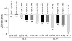
imageFigure 3 Trueness considering the distortion from occlusocervical (OC) dimensions in Kennedy class III and IV. OC-C: occlusocervical dimension from canine; OC-2M: occlusocervical dimension from second molar. Numerical values indicate the distortion (mm) and confidence interval. The same color bars are compared: different capital letters indicate statistical difference between casts (digital vs digital and 3D-printed vs 3D-printed) for the same arch classification; different lower-case letters indicate difference between casts (digital vs digital and 3D-printed vs 3D-printed) for different arch classifications. open_in_new

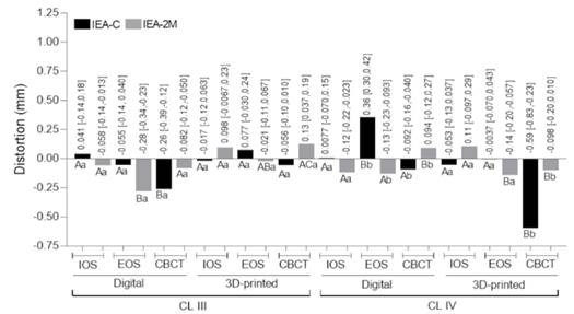
imageFigure 4 Trueness considering the distortion from interarch (IEA) dimensions in Kennedy class III and IV. IEA-C: interarch dimension between canines; IEA-2M: interarch dimension between second molars. Numerical values indicate the distortion (mm) and confidence interval. The same color bars are compared: different capital letters indicate statistical difference between casts (digital vs digital and 3D-printed vs 3D-printed) for the same arch classification; different lower-case letters indicate difference between casts (digital vs digital and 3D-printed vs 3D-printed) for different arch classifications. open_in_new
imageFigure 5 Trueness considering the distortion from edentulous space in the Kennedy class III and IV. Numerical values indicate the distortion (mm) and confidence interval. The same color bars were compared: different capital letters indicate statistical difference between casts (digital vs digital and 3D-printed vs 3D-printed) for the same arch classification; different lower-case letters indicate difference between casts (digital vs digital and 3D-printed vs 3D-printed) for different arch classifications open_in_new

Como citar
location_on
Fundação Odontológica de Ribeirão Preto
Av. do Café, S/N, 14040-904 Ribeirão Preto SP Brasil, Tel.: (55 16) 3602-3982, Fax: (55 16) 3633-0999 -
Ribeirão Preto -
SP -
Brazil
E-mail: bdj@forp.usp.br
E-mail: bdj@forp.usp.br
rss_feed
Acompañe los números de esta revista en su lector de RSS
Versão para download de PDF
Artículos relacionados
Lista de links para artigos relacionados. Os links abrem em nova aba.
Versões e tradução automática
Escolha a versão original do texto ou utilize um serviço de tradução automática.
Versão original do texto
Como citar
Escolha um formato para exportar ou selecione um estilo de citação. O conteúdo abaixo pode ser atualizado após a seleção.