 Dexamethasone-induced suppression of steroidogenic proteins and altered tyrosine phosphorylation patterns compromise testicular function in adult rats
Abstract
Dexamethasone (DEX), a synthetic glucocorticoid widely prescribed for chronic inflammatory conditions, autoimmune disorders, and acute pain management, has been implicated in male reproductive dysfunction. Although many studies have documented DEX-induced oxidative stress, apoptosis, and testosterone reduction in rodent models, the molecular mechanisms underlying steroidogenic disruption and tyrosine phosphorylation remain incompletely understood. Aim of this study was to investigate the effects of chronic DEX administration on key steroidogenic proteins and tyrosine phosphorylated (TyrPho) protein profiles in rat testis. Adult rats received either DEX (1.5 mg/kg body weight, intraperitoneally) or vehicle control daily for 21 consecutive days. Results showed that DEX treatment significantly reduced testicular weight, seminiferous tubule diameter, and epithelial height compared to those of control. Tubular atrophy and interstitial tissue degeneration were observed in DEX-treated testes. Western blot analysis demonstrated significant downregulation of steroidogenic acute regulatory protein (StAR) and cytochrome P450 family 11 subfamily A member 1 (CYP11A1), while heat shock protein 70 (HSP70) expression was not different. Immunofluorescence confirmed the reduced StAR localization in Leydig cells of DEX group. TyrPho protein profiling revealed differential responses, with DEX significantly increasing a 70 kDa protein band while decreasing a 25 kDa protein band; other Tyrpho proteins (50, 42, and 38 kDa, respectively) not affected. It was concluded that chronic DEX exposure compromised the testicular architecture and suppressed critical steroidogenic machinery, providing mechanistic insights into glucocorticoid-induced hypogonadism and reproductive toxicity in male mammals.
Keywords:
dexamethasone; steroidogenesis; StAR; CYP11A1; tyrosine phosphorylation; testicular dysfunction; glucocorticoid toxicity
Resumo
A dexametasona (DEX), um glicocorticoide sintético amplamente prescrito para condições inflamatórias crônicas, doenças autoimunes e tratamento da dor aguda, tem sido associada à disfunção reprodutiva masculina. Embora muitos estudos tenham documentado o estresse oxidativo, a apoptose e a redução da testosterona induzidos por DEX em modelos de roedores, os mecanismos moleculares subjacentes à disrupção esteroidogênica e à fosforilação de tirosina permanecem incompletamente compreendidos. O objetivo deste estudo foi investigar os efeitos da administração crônica de DEX sobre as principais proteínas esteroidogênicas e perfis de proteínas fosforiladas em tirosina (TyrPho) em testículos de ratos. Ratos adultos receberam DEX (1,5 mg/kg de peso corporal, por via intraperitoneal) ou veículo controle diariamente por 21 dias consecutivos. Os resultados mostraram que o tratamento com DEX reduziu significativamente o peso testicular, o diâmetro dos túbulos seminíferos e a altura do epitélio em comparação com o grupo controle. Atrofia tubular e degeneração do tecido intersticial foram observadas nos testículos tratados com DEX. A análise por Western blot demonstrou uma regulação negativa significativa da proteína reguladora aguda da esteroidogênese (StAR) e do membro 1 da subfamília A da família 11 do citocromo P450 (CYP11A1), enquanto a expressão da proteína de choque térmico 70 (HSP70) não apresentou diferença. A imunofluorescência confirmou a localização reduzida da StAR nas células de Leydig do grupo DEX. O perfil de proteínas TyrPho revelou respostas diferenciais, com a DEX aumentando significativamente uma banda de proteína de 70 kDa, enquanto diminuía uma banda de proteína de 25 kDa; outras proteínas TyrPho (50, 42 e 38 kDa, respectivamente) não foram afetadas. Concluiu-se que a exposição crônica à DEX comprometeu a arquitetura testicular e suprimiu a maquinaria esteroidogênica crítica, fornecendo informações mecanicistas sobre o hipogonadismo induzido por glicocorticoides e a toxicidade reprodutiva em mamíferos machos.
Palavras-chave:
dexametasona; esteroidogênese; StAR; CYP11A1; fosforilação de tirosina; disfunção testicular; toxicidade por glicocorticoide
1. Introduction
In vertebrate animals including human, the chronic stress (CS) can increase the cortisol levels, primarily via activation of the hypothalamic-pituitary-adrenal axis (Wang et al., 2025; Sigwalt et al., 2011). For male reproduction, it has been shown that elevated cortisol enhances reactive oxygen species (ROS) generation, resulting in increased lipid peroxidation as evidenced by higher malondialdehyde (MDA) levels in the testis (Choowong-In et al., 2021; Sadeghzadeh et al., 2019; Arun et al., 2018). Consequently, such oxidative stress leads to the activation of the intrinsic apoptotic pathway like expressions of caspase 9 and 3 proteins, and subsequently promotes apoptosis in seminiferous germ and interstitial cells (Choowong-In et al., 2021; Lapyuneyong et al., 2022). Indeed, those apoptotic events in the Leydig cells contribute to reduced testosterone synthesis, as revealed by the decreased expressions of steroidogenic acute regulatory (StAR) and cytochrome P450 family 11 subfamily a member 1 or CYP11A1 proteins (Choowong-In et al., 2021; Lapyuneyong et al., 2022; Arun et al., 2018). Previously, the increased apoptosis was demonstrated in both Leydig and spermatogenic cells, associated with sperm quality impairments in CS animal models (Choowong-In et al., 2021; Shen et al., 2020; Sadeghzadeh et al., 2019).
Dexamethasone (DEX), a synthetic glucocorticoid analog, is commonly used in clinical settings to treat chronic inflammations, such as chronic obstructive pulmonary disease, acute pain, and autoimmune disorders (Huabbangyang et al., 2023; Black and Grodzinsky, 2019; Vizzardelli et al., 2006; Rhen and Cidlowski, 2005). Recent studies have employed DEX to induce chronic stress in animal models, demonstrating its impact on male reproductive organs (Chaimontri et al., 2025; Chawalchitiporn et al., 2025; Innoi et al., 2025). Specifically, DEX administration has been shown to decrease testosterone levels, leading to reductions in seminal vesicle weights and seminal fluid (Innoi et al., 2025). Moreover, DEX treatment in rodent models has resulted in increased testicular MDA levels and upregulation of the caspase 9 and 3 protein expressions, leading to decreased testosterone levels and sperm concentration as previously demonstrated (Chaimontri et al., 2025; Chawalchitiporn et al., 2025; Jeje et al., 2020; Sigwalt et al., 2011). However, the effects of DEX on the expression of key role proteins involved in testosterone biosynthesis, such as StAR, CYP11A1, HSP70, and testicular TyrPho proteins, have not been fully revealed. This recent study; therefore, aimed to investigate the effect of DEX administration on such critical protein expressions, providing insights into the additional molecular mechanisms underlying stress-induced disruptions in testicular function.
2. Materials and Methods
2.1. Drug
Dexamethasone (DEX) was obtained from a veterinary pharmacy in Khon Kaen province, Thailand. This drug product, containing a concentration of 5 mg DEX/ml sodium phosphate solution, was manufactured by Trading Co., Ltd. (18/1 Sukhaphiban 2, Prawet, Bangkok, Thailand).
2.2. Animals, experimental design, and animal ethics
Adult male Sprague Dawley rats (270-300 g) were purchased from the Laboratory Animal Unit, Faculty of Medicine, Khon Kaen University, Thailand. Animals were housed in the cleaned plastic cages under standard laboratory conditions (25 ± 3°C; 12 h light/dark cycle; sound < 85 dB; humidity 30-60%; light intensity 330-400 Lux) and fed ad libitum with commercial pellets and distilled water. Rats were grouped into control and DEX groups (n = 4 per group). Control rats received sodium phosphate by intraperitoneal (i.p.) injection, while the DEX animals were injected with DEX (1.5 mg/kg BW, i.p.) once daily for 21 consecutive days (Laaziz et al., 2022). At the end of the experiment, rats were anesthetized with thiopental sodium (80 mg/kg BW, i.p.) and euthanized by cardiac puncture and cervical dislocation. This animal experiment has been approved by the Animal Ethics Committee of the Northeast Laboratory Animal Center, Khon Kaen University, Thailand (No. IACUC-KKU 14/66).
2.3. Testis collection
After euthanasia, the rat testes were collected and weighed to determine the absolute and relative weights. Then, the left testis was stored at -20°C before protein extraction, while the right one was fixed in 10% formalin fixative for routine histological processing.
2.4. Histological processing and histomorphometric analyses
Fixed testis was bisected, processed through graded alcohols, cleared in xylene, and infiltrated with liquid paraffin under 58°C before embedding. Then, each paraffin tissue block was sectioned at 6 μm thickness by using a semi-automatic microtome (ERM3100 HESTION, Australia). The sections were deparaffinized and stained with hematoxylin and eosin Y (Sigma-Aldrich, USA). For histomorphometric analysis, 50 circular cross-sections of seminiferous tubules were randomly selected and observed under light microscope before capturing with an Axio Cam ICc 5 digital camera (ZEISS). Epithelial height and diameter of seminiferous tubules were measured by using ImageJ software. Data are expressed as mean ± S.D.
2.5. Western blot analysis
The testis (0.3 g) was homogenized in 1X RIPA buffer (Cell Signaling Technology, USA) containing protease inhibitor cocktail (Sigma-Aldrich, USA). Then, testicular lysates were sonicated on ice (20 Hz, 60 pulses; Cole-Parmer), and centrifuged at 14,000 rpm for 15 minutes to separate supernatant total protein from the pellet. Total protein concentration (µg/µl) was determined by using the Nanodrop spectrophotometer (ND-1000, NanoDrop, v3.5, Thermo Fisher Scientific, USA), measured at an absorbance of 280 nm.
Subsequently, total testicular protein samples (150 µg/lane) were separated on 10% SDS-PAGE and transferred onto nitrocellulose membranes (110 V, 2 h). The nonspecific protein bindings on membranes were blocked with 5% BSA diluted in 0.1% TBST (pH 7.4) for 1 h. Then, membranes were individually incubated with primary antibody against HSP70, StAR (1:3,000; Merk Millipore Co., USA), CYP11A1, or TyrPho (1:3,000; Santa Cruz Biotechnology, USA) at 4°C overnight. After washing with 0.1% TBST, they were incubated with HRP-conjugated secondary antibody (1:3,000; Santa Cruz Biotechnology, USA). All targeted protein bands were detected by using ECL substrate (GE Healthcare Life Science), visualized, and captured under the Image Quant 600 system (GE Healthcare, USA). To detect the internal control protein, previous membranes were subsequently stripped by using stripping buffer (pH 2.2) to reprobe with β-actin antibody (1:3,000; Santa Cruz Biotechnology, USA) overnight at 4°C. For TyrPho protein determination, EGF (Millipore Co., USA) and BSA (EMD Millipore Corp., USA) were used as the positive and negative controls, respectively. The intensities of TyrPho protein expression were quantified and normalized to the corresponding β-actin band by using ImageJ software version 1.54k (https://imagej.en.download.it/). The data are expressed as relative intensity (mean ± S.D.).
2.6. Immunofluorescence
The paraffinized testicular section on a glass slide was melted in a hot air oven (60° C) before deparaffinization with xylene and rehydrating with descending serial ethanol alcohol and distilled water. After that, the epitopes on testicular tissue were retrieved by incubation with 1X citrate buffer (pH 6) in a microwave at 560 watts. Then, the sections were permeabilized with 0.2% Triton X-100 diluted in PBS (pH 7.4) for 30 min in the moisture chamber before incubating with 3% BSA diluted in PBS (pH 7.4) for 30 min to block the nonspecific binding proteins. Subsequently, the primary antibody against StAR (1:300 in PBS; Millipore, USA) was applied and incubated in a moist chamber at 4°C overnight. After washing, sections were incubated with secondary antibody conjugated with Alexa Fluor 488 (1:300 in PBS; Millipore, USA) for 2 h at room temperature (dark condition). After that, nuclei on the tissue section were counterstained with Hoechst 33342 (1:4,000 in PBS; Abcam, Cambridge, UK) for 50 min. Then, sections were dehydrated and mounted before visualizing the positive antigen-antibody complex of StAR expression under a fluorescence microscope (Nikon Eclipse 80i; Melville, NY, USA) by using FITC filter (green fluorescence) and DAPI filter (blue fluorescence), respectively.
2.7. Statistical analysis
All data are expressed as mean ± S.D. The normal distribution test of data was performed by using IBM SPSS software version 19 (installed from KKU software center, Khon Kaen University, Thailand) before further statistical analysis. To compare the difference between control and DEX groups, normalized data were analyzed using the independent samples t-test and P value that was less than 0.05 was considered as a statistically significant difference.
3. Results
3.1. Effects of DEX testicular sizes and weights
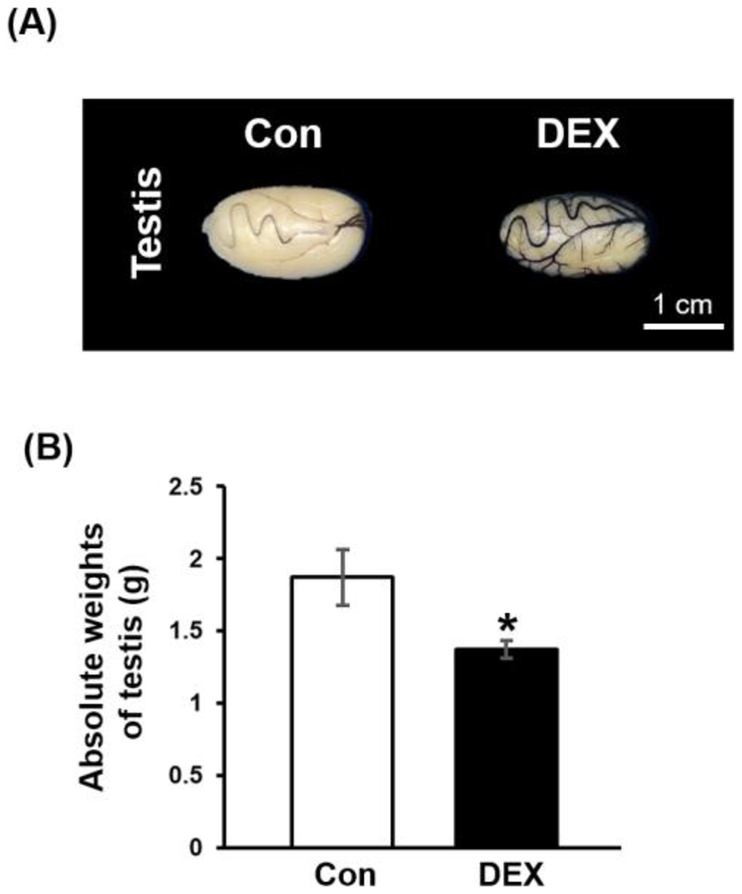
Figure 1 shows that DEX could significantly reduce the size and weight of the rat testis as compared to the control (Figure 1A and B). It also significantly decreased testicular absolute weight (Figure 1B).
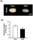
Representative photograph of testes (A) and its absolute weights (B) compared between control (Con) and dexamethasone (DEX) groups. *Significant difference (p< 0.05).
3.2. Effects of DEX on the seminiferous morphometrics
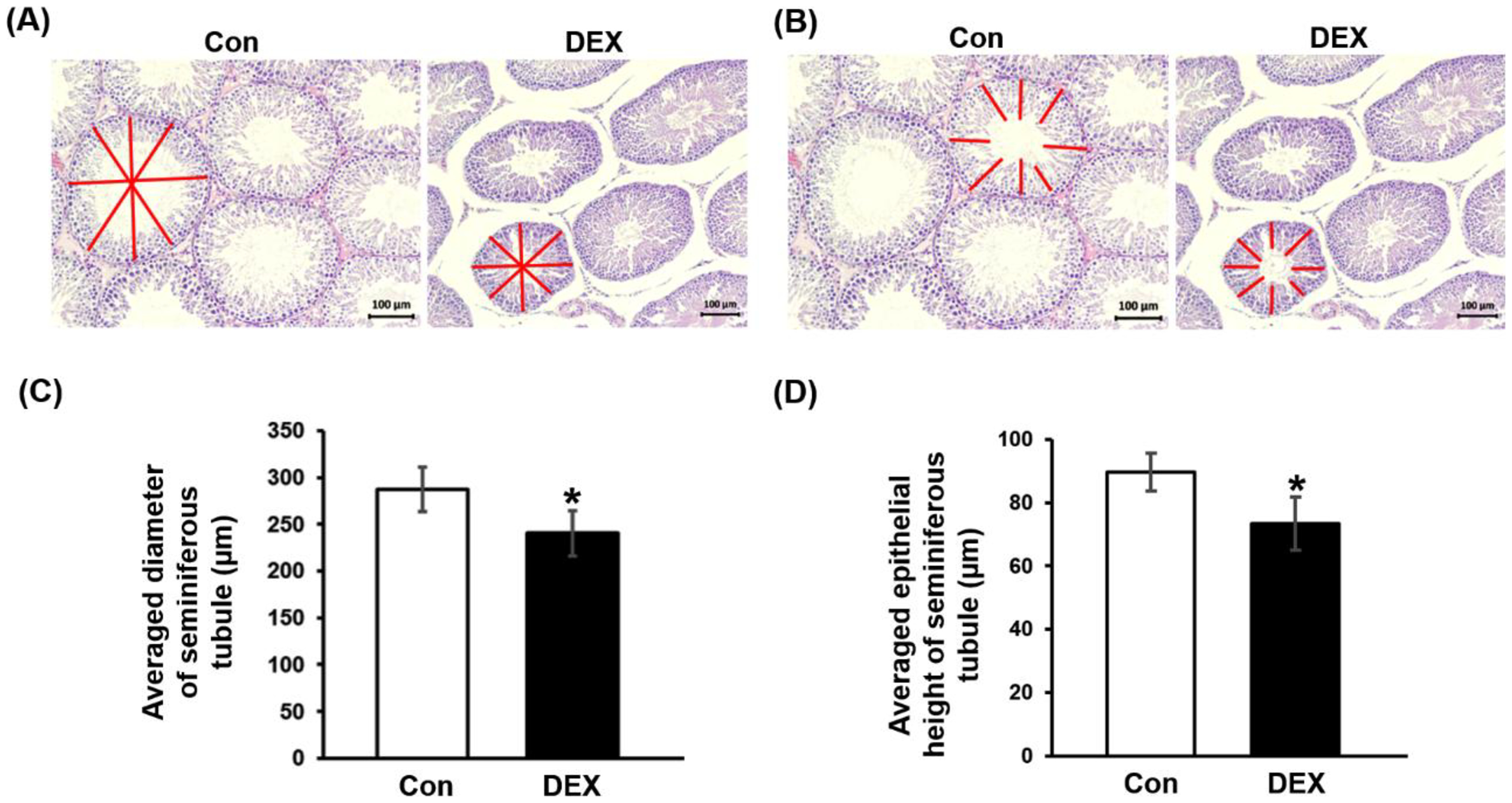
In Figure 2, the diameter and epithelial heights of seminiferous tubules induced with DEX were significantly decreased when compared to those of the control.
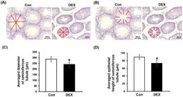
Showing seminiferous diameter (A) and epithelial height (B) measurements. Representative averaged diameter (C) and epithelial height (D) graphs of seminiferous tubules. *Significant difference (p< 0.05).
3.3. DEX damaged the testicular histology
The testicular histology of control and DEX groups is shown in Figure 3. The number of atrophic seminiferous tubules was increased in DEX group as compared to that of control (Figures 3A and C). Additionally, the interstitial tissue degeneration was mostly found in DEX group (Figure 3D).

Histology of H&E testicular sections compared between control (A and B) and DEX (C and D) groups revealed with both low and high magnifications.
3.4. Effect of DEX on expressions of testicular proteins
The western blot results show that HSP70 expression in the DEX testis did not differ from that of control group (Figure 4A and B). However, DEX significantly decreased the testicular StAR and CYP11A1 expressions compared to those of control group (Figures 4A and B).
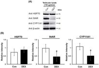
The expressions of heat shock protein 70 (HSP70), steroidogenic acute regulatory protein (StAR), and cytochrome P450 family 11 subfamily a member 1 (CYP11A1) proteins (A) and their relative intensities (B) in testicular lysates compared between Con and DEX groups. *Significant difference (p< 0.05).
3.5. The alterations of TyrPho protein expressions
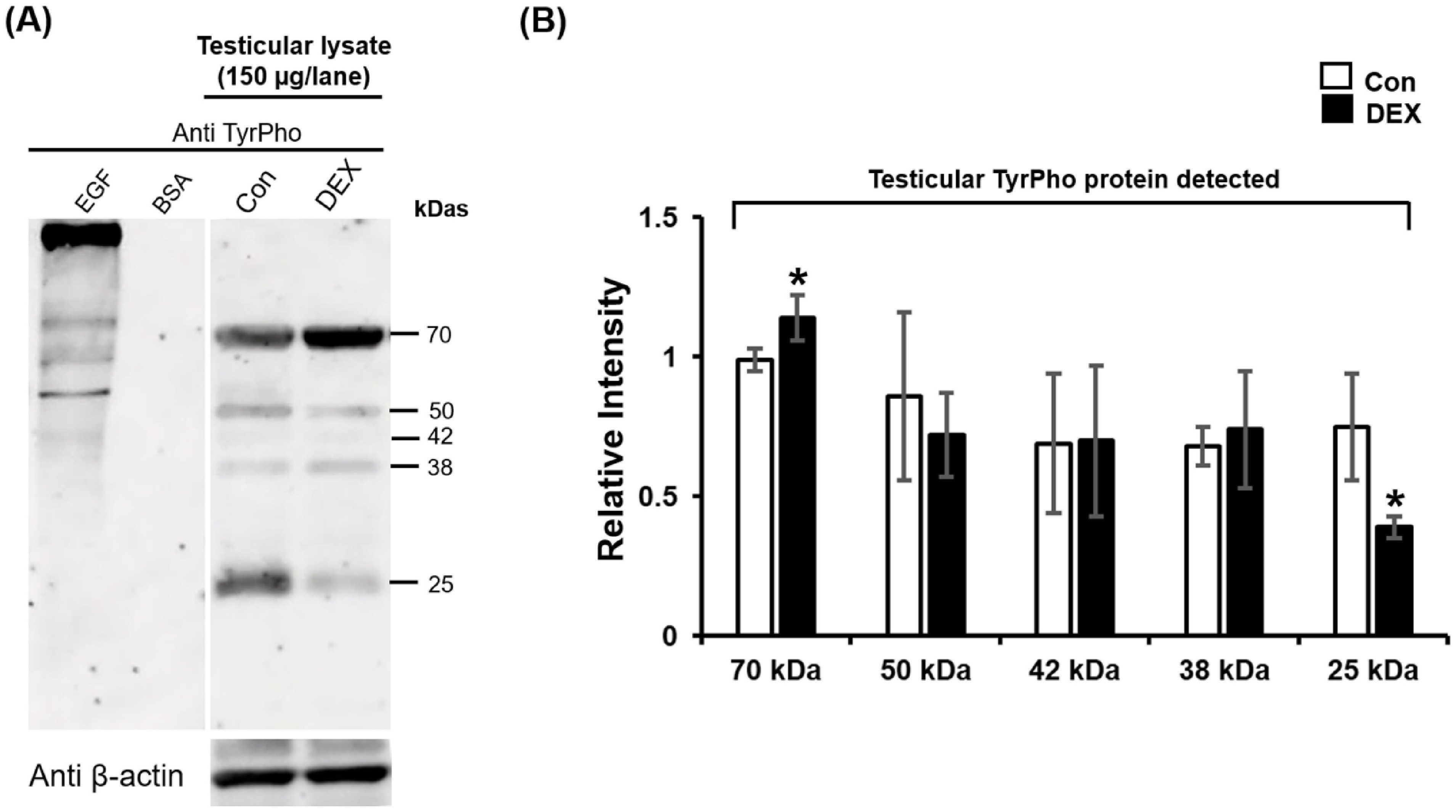
It was found that profiles of the tyrosine phosphorylation (TyrPho) protein expression in testis reveal 5 TyrPho protein bands including 70, 50, 42, 38, and 25 kDas, respectively in both groups (Figure 5A). In contrast to control, TyrPho protein expression of a 70 kDa was significantly increased, while that of 25 kDa when was decreased in DEX testis (Figures 5A and B). However, intensities of testicular TyrPho proteins at 50, 42, and 38 kDas were not significantly different compared between groups (Figure 5B).
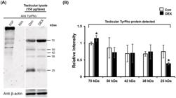
Expressions of tyrosine phosphorylated (TyrPho) proteins in testicular lysates (A) and their intensities (B) compared between groups. EGF (epidermal growth factor) used as a positive control. BSA (bovine serum albumin) used as a negative control. *Significant difference (p< 0.05).
3.6. Effect of DEX on StAR expression in Leydig cells
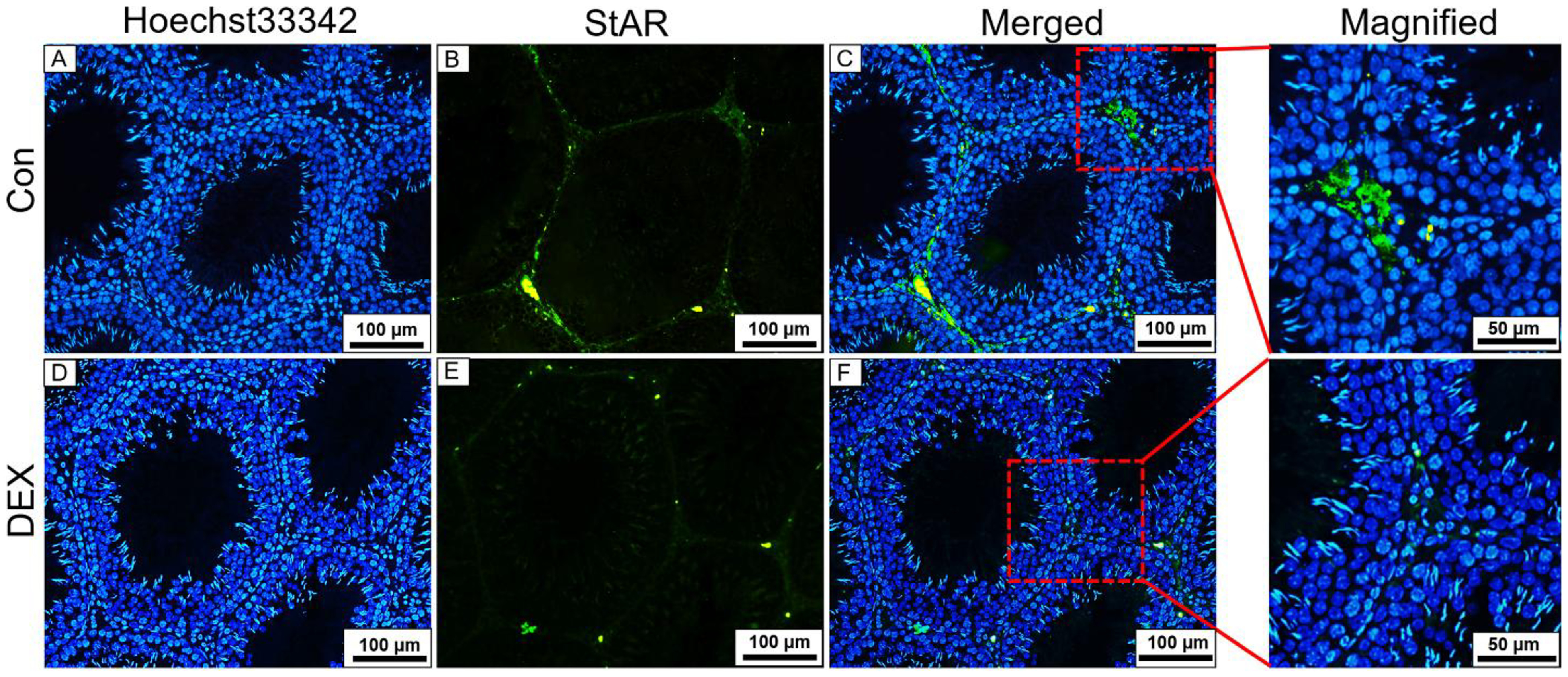
For immunofluorescent staining, the StAR protein expression of DEX interstitial tissue (Figures 6E and F) was decreased in the cytoplasm of Leydig cells when compared with control group (Figures 6B and C).
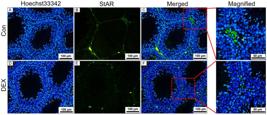
Representative immunofluorescent micrographs showing expression of steroidogenic acute regulatory (StAR) protein in interstitial tissue compared between control (A, B, and C) and DEX (D, E, and F) groups. Hoechst 33342 staining emitted as blue signals at cell nuclei. Positive StAR (green signals) protein expressed and localized in cytoplasm.
4. Discussion
The present study has demonstrated that chronic DEX administration (1.5 mg/kg BW for 21 consecutive days) significantly reduced testicular weight and disrupted seminiferous tubule architecture in adult rats. These findings were partially different from previous reports showing no significant testicular weight reduction in DEX-treated mice (Chaimontri et al., 2025; Mehranjani et al., 2022; Sadeghzadeh et al., 2019) and rats (Aziz et al., 2024; Jeje et al., 2020). However, the recent results are similar to other studies demonstrating DEX-induced reductions in testicular size and weight (Zangabad et al., 2023; Hasona, 2018; Khosravanian et al., 2015). The observed decreases in seminiferous tubule diameter and epithelial height, accompanied by tubular atrophy and interstitial tissue degeneration, were consistent with DEX-induced oxidative stress, as evidenced by elevated malondialdehyde levels in testicular tissue as previously reported (Aziz et al., 2024; Nassan et al., 2021). These morphological alterations suggested that DEX might compromise the testicular architecture through oxidative damage-mediated cellular degeneration.
Under physiological conditions, testosterone biosynthesis in Leydig cells is regulated by luteinizing hormone-mediated signaling pathways involving downstream phosphorylation cascades that can control the expression and activities of key steroidogenic proteins such as StAR and CYP11A1 (Mattos et al., 2023; Narayan et al., 2019; Ayaz and Howlett, 2015). In this study, DEX treatment significantly suppressed testicular StAR and CYP11A1 protein expressions, while HSP70 levels remained unchanged, though a trend toward increased expression was investigated. Immunofluorescence analysis could confirm the reduced StAR expression in the interstitial compartment of DEX-treated testes, which was corroborated with the Western blot results. The downregulation of StAR and CYP11A1 strongly suggests the impaired testosterone synthesis, which was supported by a recent study demonstrating that 21-day DEX administration decreases serum testosterone levels (Innoi et al., 2025).
Protein phosphorylation, particularly tyrosine phosphorylation (TyrPho), is known to play a critical role in regulating steroidogenic gene expression and testosterone biosynthesis (Mattos et al., 2023). Our analysis revealed the differential expression patterns of testicular TyrPho proteins in response to DEX treatment. Particularly, DEX significantly decreased the expression of a 25 kDa TyrPho protein, which may correspond to mouse epididymal luminal fluid protein (called MEP-9). This protein has been localized in the cytoplasm of elongated spermatids and the caput epididymis, where it functions to disrupt adhesion between elongated spermatids and Sertoli cells, also facilitating spermiation process (Vierula et al., 1992). The reduction in this protein may indicate the disrupted spermatid release mechanisms in the adluminal seminiferous tubules. Furthermore, a previous study showed that a heat shock protein 25 (HSP25) prevented protein misfolding during cell division (Ehrnsperger et al., 1997). In 1999, Wakayama and Iseki demonstrated that the HSP25 used to prevent the misfolding of proteins has been localized in primary spermatocytes, particularly in leptotene, zygotene, and early pachytene phases (Wakayama and Iseki, 1999). In this study, the 25 kDa TyrPho protein might be HSP25 and the decrease of its expression in the DEX group was associated with decreasing of epithelial height of the seminiferous tubule by delaying spermatogenesis (Gispert et al., 2013; Linn et al., 2021). In addition, the 25 kDa TyrPho protein might be proteasome subunit alpha 8 (PSMA8), which has been localized at the cytoplasm of spermatocytes in the pachytene phase (Zhang et al., 2019). Moreover, the decreasing of PSMA8 expression might indicate that the meiotic 1 division (M phase) in spermatocytes was delayed, resulting in decreased epithelial height of the seminiferous tubule (Zhang et al., 2019). In our study, the DEX treatment significantly decreased the expression of 25 kDa TyrPho protein, leading to spermatogenesis disruption and reduction of the tubular epithelial height. However, further investigation is necessary to confirm the actual protein of the 25 kDa TyrPho protein.
In contrast, DEX treatment significantly increased the expression of a 70 kDa TyrPho protein, which may represent the zeta chain of T cell receptor-associated protein kinase 70 (ZAP-70). Bartis et al. (2006) have reported that DEX treatment could upregulate the ZAP-70 expression in T-cell leukemia cells. Possibly, the elevation of this 70 kDa TyrPho protein in this recent study may indicate DEX-induced testicular inflammation or altered immune cell signaling within the testicular microenvironment. The expressions of TyrPho proteins at 50, 42, and 38 kDas were not significantly different between groups, suggesting these proteins may not be directly involved in testosterone biosynthesis or were not responsive to DEX-induced stress in rat model.
Taken together, these findings indicated that chronic DEX administration could disrupt the testicular function through some mechanisms: (1) structural damage to seminiferous tubules and interstitial tissue, (2) suppression of key steroidogenic proteins (StAR and CYP11A1) essential for testosterone synthesis, and (3) altered TyrPho patterns that may affect spermatogenesis and inflammatory responses. These molecular alterations have provided the mechanistic insights into DEX-induced hypogonadism and testicular dysfunction.
5. Conclusion
The present study demonstrated that chronic DEX administration induced the significant testicular damage in rats, characterized by disrupted seminiferous tubule architecture, and suppressed the expressions of steroidogenic proteins like StAR and CYP11A1. Additionally, DEX altered the testicular TyrPho profile, particularly decreasing 25 kDa TyrPho protein expression. These molecular changes impaired the androgen production and suggested that DEX-induced testicular dysfunction may involve both steroidogenic suppression and altered phosphorylation-dependent signaling pathways.
Acknowledgements
Natthapol Lapyuneyong was supported by Invitation Research (Grant Number IN67072), Faculty of Medicine, Khon Kaen University, Thailand.
Data Availability Statement
Research data is only available upon request.
References
-
ARUN, S., BURAWAT, J., YANNASITHINON, S., SUKHORUM, W., LIMPONGSA, A. and IAMSAARD, S., 2018. Phyllanthus Emblica leaf extract ameliorates testicular damage in rats with chronic stress. Journal of Zhejiang University. Science. B, vol. 19, no. 12, pp. 948-959. https://doi.org/10.1631/jzus.B1800362 PMid:30507078.
» https://doi.org/10.1631/jzus.B1800362 -
AYAZ, O. and HOWLETT, S.E., 2015. Testosterone modulates cardiac contraction and calcium homeostasis: cellular and molecular mechanisms. Biology of Sex Differences, vol. 6, no. 1, pp. 9. https://doi.org/10.1186/s13293-015-0027-9 PMid:25922656.
» https://doi.org/10.1186/s13293-015-0027-9 -
AZIZ, R.L.A., WAHAB, A.A., RAZIK, A.R.H.A., KAMEL, S., FARGHALI, A.A., SALEH, R., MAHMOUD, R., IBRAHIM, M.A., NABIL, T.M. and ELA, F.I.A.E., 2024. Physiological roles of propolis and red ginseng nanoplatforms in alleviating dexamethasone-induced male reproductive challenges in a rat model. Molecular Biology Reports, vol. 51, no. 1, pp. 72. https://doi.org/10.1007/s11033-023-08991-4 PMid:38175282.
» https://doi.org/10.1007/s11033-023-08991-4 -
BARTIS, D., BOLDIZSAR, F., SZABO, M., PALINKAS, L., NEMETH, P. and BERKI, T., 2006. Dexamethasone induces rapid tyrosine-phosphorylation of ZAP-70 in Jurkat cells. he. The Journal of Steroid Biochemistry and Molecular Biology, vol. 98, no. 2-3, pp. 147-154. https://doi.org/10.1016/j.jsbmb.2005.01.032 PMid:16406604.
» https://doi.org/10.1016/j.jsbmb.2005.01.032 -
BLACK, R. and GRODZINSKY, A.J., 2019. Dexamethasone: chondroprotective corticosteroid or catabolic killer? European Cells & Materials, vol. 38, pp. 246-263. https://doi.org/10.22203/eCM.v038a17 PMid:31755076.
» https://doi.org/10.22203/eCM.v038a17 -
CHAIMONTRI, C., ARUN, S., KAMOLLERD, T., LAPYUNEYONG, N., TAOTO, C., INNOI, S., CHAWALCHITIPORN, T., SAWATPANICH, T., UABUNDIT, N. and IAMSAARD, S., 2025. Low sperm quality and increased testicular caspases in chronic stress mice induced by dexamethasone. Brazilian Journal of Biology = Revista Brasileira de Biologia, vol. 85, pp. e289850. https://doi.org/10.1590/1519-6984.289850 PMid:40465972.
» https://doi.org/10.1590/1519-6984.289850 -
CHAWALCHITIPORN, T., INNOI, S., CHAIMONTRI, C., TAOTO, C., KAMOLLERD, T., KERDSANG, P., KOEDBUA, N., THUKHAMMEE, W., POODENDAEN, C., DUANGCHIT, S., IAMSAARD, S. and CHAIYAMOON, A., 2025. Histopathology and changes of pro-caspase 3 and androgen receptor expressions with increased MDA in epididymis of a depressive-like behavior rat model. International Journal of Morphology, vol. 43, no. 4, pp. 1321-1328. https://doi.org/10.4067/s0717-95022025000401321
» https://doi.org/10.4067/s0717-95022025000401321 -
CHOOWONG-IN, P., SATTAYASAI, J., POODENDAEN, C. and IAMSAARD, S., 2021. Decreased expression of AKAP4 and TyrPho proteins in testis, epididymis, and spermatozoa with low sexual performance of mice induced by modified CUMS. Andrologia, vol. 53, no. 3, pp. e13977. https://doi.org/10.1111/and.13977 PMid:33486757.
» https://doi.org/10.1111/and.13977 -
EHRNSPERGER, M., GRABER, S., GAESTEL, M. and BUCHNER, J., 1997. Binding of non-native protein to Hsp25 during heat shock creates a reservoir of folding intermediates for reactivation. The EMBO Journal, vol. 16, no. 2, pp. 221-229. https://doi.org/10.1093/emboj/16.2.221 PMid:9029143.
» https://doi.org/10.1093/emboj/16.2.221 -
GISPERT, S., PARGANLIJA, D., KLINKENBERG, M., DROSE, S., WITTIG, I., MITTELBRONN, M., GRZMIL, P., KOOB, S., HAMANN, A., WALTER, M., BUCHEL, F., ADLER, T., HRABE DE ANGELIS, M., BUSCH, D.H., ZELL, A., REICHERT, A.S., BRANDT, U., OSIEWACZ, H.D., JENDRACH, M. and AUBURGER, G., 2013. Loss of mitochondrial peptidase Clpp leads to infertility, hearing loss plus growth retardation via accumulation of CLPX, mtDNA and inflammatory factors. Human Molecular Genetics, vol. 22, no. 24, pp. 4871-4887. https://doi.org/10.1093/hmg/ddt338 PMid:23851121.
» https://doi.org/10.1093/hmg/ddt338 -
HASONA, N.A., 2018. Grape seed extract attenuates dexamethasone-induced testicular and thyroid dysfunction in male albino rats. Andrologia, vol. 50, no. 5, pp. e13002. https://doi.org/10.1111/and.13002
» https://doi.org/10.1111/and.13002 -
HUABBANGYANG, T., SILAKOON, A., SANGKETCHON, C., SUKHUNTEE, J., KUMKONG, J., SRITHANAYUCHET, T., CHAMNANPOL, P. and MEECHAI, T., 2023. Effects of pre-hospital dexamethasone administration on outcomes of patients with COPD and asthma exacerbation; a cross-sectional study. Archives of Academic Emergency Medicine, vol. 11, no. 1, pp. e56. https://doi.org/10.22037/aaem.v11i1.2037 PMid:37671276.
» https://doi.org/10.22037/aaem.v11i1.2037 -
INNOI, S., CHAWALCHITIPORN, T., SAWATPANICH, T., TANGSRISAKDA, N., CHAIMONTRI, C., TAOTO, C., KAMOLLERD, T., PATJORN, Y., WONGSOMWONG, A., CHAIYAMART, A., THUKHAMMEE, W., POODENDAEN, C., DUANGCHIT, S. and IAMSAARD, S., 2025. Dexamethasone-induced chronic stress decreases biochemical fluid components and seminal vesicle secreting protein 4 expression via epithelial cell apoptosis. Andrologia, vol. 1, no. 1, pp. 7168333. https://doi.org/10.1155/and/7168333
» https://doi.org/10.1155/and/7168333 -
JEJE, S.O., ADEGBITE, L.O., AKINDELE, O.O., KUNLE-ALABI, O.T. and RAJI, Y., 2020. Allium cepa Linn juice protect against alterations in reproductive functions induced by maternal dexamethasone treatment during lactation in male offspring of Wistar rats. Heliyon, vol. 6, no. 5, pp. e03872. https://doi.org/10.1016/j.heliyon.2020.e03872 PMid:32395653.
» https://doi.org/10.1016/j.heliyon.2020.e03872 -
KHOSRAVANIAN, H., RAZI, M., FAROKHI, F. and KHOSRAVANIAN, N., 2015. Simultaneous administration of dexamethasone and vitamin E reversed experimental varicocele-induced impact in testicular tissue in rats; Correlation with Hsp70-2 chaperone expression. International Braz J Urol, vol. 41, no. 4, pp. 773-790. https://doi.org/10.1590/S1677-5538.IBJU.2013.0148
» https://doi.org/10.1590/S1677-5538.IBJU.2013.0148 -
LAAZIZ, A., MOSTAFI, H.E., ELHESSNI, A., TOUIL, T., DOUMAR, H. and MESFIOUI, A., 2022. Chronic clomipramine treatment reverses depressogenic-like effects of a chronic treatment with dexamethasone in rats. IBRO Neuroscience Reports, vol. 13, pp. 147-155. https://doi.org/10.1016/j.ibneur.2022.07.007 PMid:36035970.
» https://doi.org/10.1016/j.ibneur.2022.07.007 -
LAPYUNEYONG, N., TANGSRISAKDA, N., CHOOWONG-IN, P., CHAISIWAMONGKOL, K., UABUNDIT, N., SAWATPANICH, T., ARUN, S., WU, A.T. and IAMSAARD, S., 2022. Seed extract of Thai Mucuna pruriens reduced male reproductive damage in rats induced by chronic stress. Pharmaceutical Biology, vol. 60, no. 1, pp. 374-383. https://doi.org/10.1080/13880209.2022.2034896 PMid:35180038.
» https://doi.org/10.1080/13880209.2022.2034896 -
LINN, E., GHANEM, L., BHAKTA, H., GREER, C. and AVELLA, M., 2021. Genes regulating spermatogenesis and sperm function associated with rare disorders. Frontiers in Cell and Developmental Biology, vol. 9, pp. 634536. https://doi.org/10.3389/fcell.2021.634536 PMid:33665191.
» https://doi.org/10.3389/fcell.2021.634536 -
MATTOS, K.D., PIERRE, K.J. and TREMBLAY, J.T., 2023. Hormones and signaling pathways involved in the stimulation of leydig cell steroidogenesis. Endocrines, vol. 4, no. 3, pp. 573-594. https://doi.org/10.3390/endocrines4030041
» https://doi.org/10.3390/endocrines4030041 -
MEHRANJANI, M.S., AZIZI, M. and SADEGHZADEH, F., 2022. The effect of melatonin on testis histological changes and spermatogenesis indexes in mice following treatment with dexamethasone. Drug and Chemical Toxicology, vol. 45, no. 3, pp. 1140-1149. https://doi.org/10.1080/01480545.2020.1809672 PMid:33161762.
» https://doi.org/10.1080/01480545.2020.1809672 -
NASSAN, M.A., SOLIMAN, M.M., ALDHAHRANI, A., EL-SAWAY, H.B. and SWELUM, A.A., 2021. Ameliorative impacts of Allium cepa Linnaeus aqueous extract against testicular damage induced by dexamethasone. Andrologia, vol. 53, no. 4, pp. e13955. https://doi.org/10.1111/and.13955 PMid:33682109.
» https://doi.org/10.1111/and.13955 -
NARAYAN, P., ULLOA-AGUIRRE, A. and DIAS, J.A., 2019. Gonadotropin hormones and their receptors. In: J.F. STRAUSS and R.L. BARBIERI, eds. Yen & Jaffe’s reproductive endocrinology 8th ed. Amsterdam: Elsevier, pp. 25-57.e15, vol. 25-27. https://doi.org/10.1016/B978-0-323-47912-7.00002-0
» https://doi.org/10.1016/B978-0-323-47912-7.00002-0 -
RHEN, T. and CIDLOWSKI, J.A., 2005. Antiinflammatory action of glucocorticoids-new mechanisms for old drugs. The New England Journal of Medicine, vol. 353, no. 16, pp. 1711-1723. https://doi.org/10.1056/NEJMra050541 PMid:16236742.
» https://doi.org/10.1056/NEJMra050541 -
SADEGHZADEH, F., MEHRANJANI, M.S. and MAHMOODI, M., 2019. Vitamin C ameliorates the adverse effects of dexamethasone on sperm motility, testosterone level, and spermatogenesis indexes in mice. Human & Experimental Toxicology, vol. 38, no. 4, pp. 409-418. https://doi.org/10.1177/0960327118816137 PMid:30526067.
» https://doi.org/10.1177/0960327118816137 -
SHEN, Y., HE, D., HE, L., BAI, Y., WANG, B., XUE, Y. and HOU, G., 2020. Chronic psychological stress, but not chronic pain stress, influences sexual motivation and induces testicular autophagy in male rats. Frontiers in Psychology, vol. 11, pp. 826. https://doi.org/10.3389/fpsyg.2020.00826 PMid:32425863.
» https://doi.org/10.3389/fpsyg.2020.00826 -
SIGWALT, A.R., BUDDE, H., HELMICH, I., GLASER, V., GHISONI, K., LANZA, S., CADORE, E.L., LHULLIER, F.L.R., BEM, A.F., HOHL, A., MATOS, F.J., OLIVEIRA, P.A., PREDIGER, R.D., GUGLIELMO, L.G.A. and LATINI, A., 2011. Molecular aspects involved in swimming exercise training reducing anhedonia in a rat model of depression. Neuroscience, vol. 192, pp. 661-674. https://doi.org/10.1016/j.neuroscience.2011.05.075 PMid:21712072.
» https://doi.org/10.1016/j.neuroscience.2011.05.075 -
WAKAYAMA, T. and ISEKI, S., 1999. Specific expression of the mRNA for 25 kDA heat-shock protein in the spermatocytes of mouse seminiferous tubules. Anatomy and Embryology, vol. 199, no. 5, pp. 419-425. https://doi.org/10.1007/s004290050240 PMid:10221453.
» https://doi.org/10.1007/s004290050240 -
WANG, L., XU, Y., JIANG, M., WANG, M., JI, M., XIE, X. and SHENG, H., 2025. Chronic stress induces depression-like behavior in rats through affecting brain mitochondrial function and inflammation. Psychoneuroendocrinology, vol. 172, pp. 107261. https://doi.org/10.1016/j.psyneuen.2024.107261 PMid:39721083.
» https://doi.org/10.1016/j.psyneuen.2024.107261 -
VIERULA, M.E., ARAKI, Y., RANKIN, T.L., TULSIANI, D.R. and ORGEBIN-CRIST, M.C., 1992. Immunolocalization of a 25-kilodalton protein in mouse testis and epididymis. Biology of Reproduction, vol. 47, no. 5, pp. 844-856. https://doi.org/10.1095/biolreprod47.5.844 PMid:1477210.
» https://doi.org/10.1095/biolreprod47.5.844 -
VIZZARDELLI, C., PAVELKA, N., LUCHINI, A., ZANONI, I., BENDICKSON, L., PELIZZOLA, M., BERETTA, O., FOTI, M., GRANUCCI, F., NILSEN-HAMILTON, M. and RICCIARDI-CASTAGNOLI, P., 2006. Effects of dexamethazone on LPS-induced activationand migration of mouse dendritic cells revealed by a genome-wide transcriptional analysis. European Journal of Immunology, vol. 36, no. 6, pp. 1504-1515. https://doi.org/10.1002/eji.200535488
» https://doi.org/10.1002/eji.200535488 -
ZANGABAD, E.A., SHOMALI, T. and ROSHANGAR, L., 2023. Effects of pharmacological doses of niacin on subacute glucocorticoid-induced testicular damage in rats. Pharmacology Research & Perspectives, vol. 11, no. 5, pp. e01128. https://doi.org/10.1002/prp2.1128 PMid:37589322.
» https://doi.org/10.1002/prp2.1128 -
ZHANG, Q., JI, S.Y., BUSAYAVALASA, K., SHAO, J. and YU, C., 2019. Meiosis I progression in spermatogenesis requires a type of testis-specific 20S core proteasome. Nature Communications, vol. 10, no. 1, pp. 3387. https://doi.org/10.1038/s41467-019-11346-y PMid:31358751.
» https://doi.org/10.1038/s41467-019-11346-y
Edited by
-
Editor:
Takako Matsumura Tundisi