Effect of whitening toothpastes on color alteration and enamel and dentin surface roughness
Abstract
Aim To evaluate if whitening dentifrices change enamel color and if their use induces alteration over the enamel and dentin roughness.
Methods Forty enamel and forty dentin bovine samples (4 mm x 4 mm, n=80) were divided into four groups following the dentifrice tested: Char - activated charcoal (3D White Therapy Charcoal); HP - low concentration of hydrogen peroxide (Colgate Luminous White Advanced); Cov – blue covarine (Close Up White Now); and Cont – control (Colgate Total 12). Initial enamel color coordinates (L*a*b*) were obtained, followed by enamel and dentin Ra measurements. Specimens were submitted to an abrasive cycle (20 s abrasion, 3x/day, 14 days). After, the final Ra and color were obtained, and the delta E was calculated. Roughness data were submitted to repeated measures ANOVA, and color alterations were assessed by one-way ANOVA, followed by the Tukey test.
Results The dentifrice Char reduced the enamel Ra and presented lower values than the Cont and Cov, while Cont increased the Ra and HP reduced it for dentin. Regarding enamel color, there was no significant difference between the dentifrices.
Conclusion The dentifrices could not promote enamel whitening, and their use shall be cautiously indicated as they can induce surface harm, mainly over dentin.
Key Words
Toothpastes; Charcoal; Dentifrices; Hydrogen peroxide; Tooth bleaching
Introduction
Tooth whitening is a conservative, well-accepted, and indispensable cosmetic option in aesthetic dentistry to improve the chromatic aspects of natural teeth, being highly desirable in a modern society that values a smile with aligned, white and brilliant teeth1,2. Tooth discoloration can be classified as intrinsic or extrinsic. Intrinsic stains occur from deposition of pigments within the enamel and/or dentin during tooth development or after eruption, often via microstructural defects or diffusion through interprismatic spaces. It results from developmental disturbances (amelogenesis or dentinogenesis imperfecta), systemic exposure to high fluoride concentrations (fluorosis), tetracycline use, pulp necrosis or hemorrhage, and aging-related changes in dentin. Extrinsic stains are located on the tooth surface, adhering to the acquired pellicle or biofilm, and result from smoking habits, antiseptic precipitation, such as chlorhexidine, or deposition of chromogenic substances, such as coffee, wine, tea, among other products from the diet1-3. For those cases, tooth whitening can be done by or supervised by the dentist using bleaching gels containing hydrogen peroxide or its precursors, such as carbamide peroxide. Hydrogen peroxide is a small, highly reactive molecule that diffuses through enamel interprismatic spaces and dentinal tubules, decomposing into free radicals capable of oxidizing chromophores into smaller, less pigmented molecules, resulting in a change in the pattern of reflection and refraction of light, causing the whitening effect1,4,5.
In addition to using professionally applied gels, several over-the-counter (OTC) products are available directly to the consumer with the promise to whiten their teeth within 2-4 weeks of use, being an alternative to professional/supervised tooth whitening6-8. These products include dentifrices, mouthwashes, paint-on varnishes, and tapes, and their success can be explained by the fact that they are considered cosmetics, so, in most countries, they are much less regulated by regulatory agencies than pharmaceuticals drugs, making them more accessible and more cost-effective than the professional/supervised options8.
From the OTC group, the whitening dentifrices are, by far, the most consumed product, comprising more than 50% of the options available. Besides the essential ingredients found in traditional products, e.g., surfactants, thickening agents, flavor, and fluorides, the whitening dentifrices contain several abrasives and/or other active ingredients that promise to remove or prevent the deposition of pigments on the dental enamel9, including hydrated silica, calcium carbonate, dicalcium phosphate dihydrate, sodium bicarbonate, perlite, and alumina. Despite abrasives, some whitening dentifrices can also contain other active ingredients, such as pyrophosphates, that function as anti-calculus agents and reduce stain deposition ever enamel, or dyes, such as blue covarine, which modify the visual perception of color by creating more light scattering, or even hydrogen peroxide in low concentration8,10. Most recently, charcoal powder or dentifrices flooded the market and was boosted by the internet and social media with the appeal to promote tooth whitening11. This agent is a fine powder oxidized by controlled heating or chemical means derived from nutshells, coconut husks, bamboo, or peat12,13.
Evidence regarding the whitening effectiveness and the potential to induce harm over tooth tissues promoted by these whitening dentifrices is still controversial8,9,12,14-17. It is suggested that although whitening toothpaste can lighten tooth color, it can also affect the mineral content of teeth by increasing surface roughness and reducing microhardness16, and altering the roughness of composite materials18. Still, over dentin, their deleterious effect is not well understood. Considering that patients may present exposed dentin in the oral cavity due to non-carious cervical lesions or gingival recession, investigating the harmful potential is of interest. Therefore, this study aimed to evaluate the potential of whitening dentifrices to promote color alteration on the enamel and if their chronicle use induces alteration over the enamel and dentin surface roughness. The null hypothesis tested was that the whitening dentifrices do not promote teeth whitening, regardless of their active agent, and that their daily use does not cause alteration on the enamel and dentin surface roughness.
Material and methods
Specimen preparation
Forty enamel/dentin specimens were obtained from the labial surface of bovine incisors crowns. Samples were cut using a water-cooled diamond precision saw (Isomet, Buehler) with 4 mm x 4 mm (length x width) and grounded flat in a circular polishing machine (Aropol, Arotec, Brazil) using water-cooled silicon carbide (SiC) sandpaper (grit #600, Extec Corp, USA) until leaving a sample with 2 mm thick (1 mm of enamel and 1 mm of dentin)19. These samples were used for the colorimetry analysis and the evaluation of enamel surface roughness. To evaluate dentin roughness, forty additional specimens were cut from the cervical region of the bovine incisor roots, measuring 4 mm x 4 mm (length x width), and standardized to a thickness of 2 mm by flattening them in a circular polishing machine using #600 SiC paper.
All enamel and dentin samples were then embedded in transparent acrylic resin (Jet, Classico, Brazil) with the help of a circular silicon matrix with 40 mm in diameter and then polished with sequential SiC paper (#800, #1200, #2400 and #4000) at 300 rpm until no visible marks or scratches could be seen in the surface, following the protocol described by Borges et al.5 (2015). The specimens were embedded in acrylic resin to standardize the polishing protocol, as the circular polishing machine used in this study was equipped with a positioning device that ensured controlled load and rotational speed. This setup facilitated both polishing and surface flattening. The surfaces were verified with a stereomicroscope (x25 augmentation), and the surfaces presenting cracks and imperfections were discarded5. Specimens were sonicated in deionized water for 10 min after each paper grit change and immersed in distilled water at and controlled temperature (37ºC) for 30 days for hydration.
For the colorimetric analysis, the enamel samples were stained with a coffee solution20 made from 12g of coffee powder (Café Tradicional Melitta, Brazil) diluted in 200 mL of boiling water. Samples were kept immersed in this solution for three days.
Initial roughness (Ra) and group division
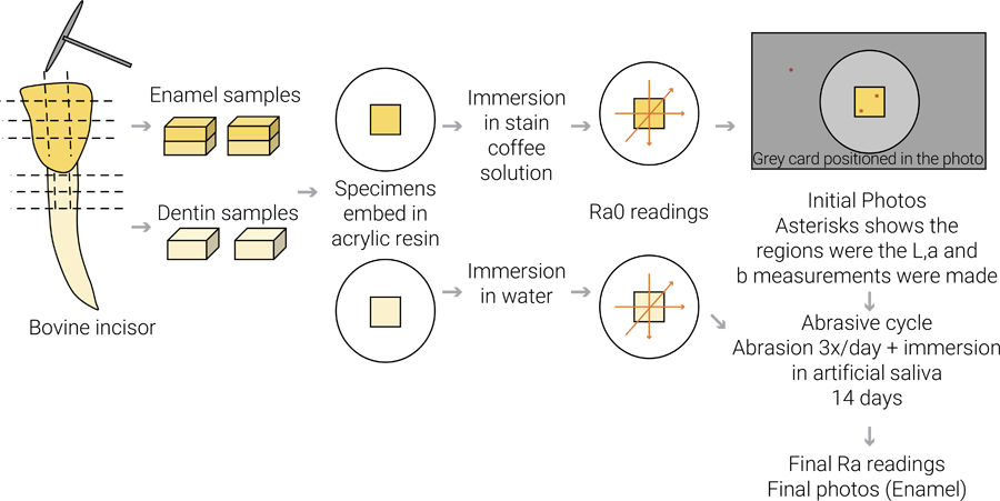
The initial roughness (Ra0) values of each sample were obtained using a contact profilometer (Surftest SJ-410, Mitutoyo, Japan). The parameter Ra was used, and three measurements were obtained from each specimen with a cut-off of 0.25, length of 3.2 mm, and mean speed of 0.5 mm/s, as shown in figure 1. The average of the three measures was considered as the mean Ra value of each specimen.
The values Ra0 values were used to stratify the enamel and dentin samples in 4 groups (n=10) according to the dentifrice tested: Char – Dentifrice containing activated charcoal (3D White Therapy Charcoal – Crest/Oral B); HP – dentifrice containing a low concentration of hydrogen peroxide (Colgate Luminous White Advanced, Colgate Palmolive); Cov – dentifrice containing blue covarine (Close Up White Now, Close Up Unilever); and Cont – dentifrice control without a whitening agent (Colgate Total 12, Colgate Palmolive). The composition of each dentifrice is described in Table 1. Figure 1 shows and schematic drawing of the sample preparation and readings.
Initial color assessment
The color evaluation was made only in the enamel samples, and the colorimetric method used was the one previously described by Bengel21 (2003), Sampaio et al.22 (2019), and Garg et al.23 (2020) using digital photography. Each sample was photographed individually using a digital DSLR camera (Nikon D3200, Japan) coupled with a macro lens (AF-S Macro Nikkor 105 mm, Nikon, Japan) and circular flash (EM 140 DG, Sigma, Japan). The pictures were taken in .raw format using the following parameters: 1/100 s, F 20, ISO 100, flash ETTL, and focal distance set to 60 cm. The camera was positioned on a tripod, with the lens perpendicular to the ground. The samples were placed inside a lightbox with a LED in all directions to minimize shadow formation. A standard gray card (L=55, a=0, and b=0) was placed near the sample for gray balance standardization, as previously described22. The samples were taken from the water, placed over absorbent paper, and the photos were taken immediately after to avoid dehydration.
Color analysis was made using the CIEL*a*b* color system (International Commission on Illumination), where the L* axis indicates the value (lightness or darkness), the a* axis represents the redness (+a*) or the greenness (−a*), and the b* axis demonstrates the yellowness (+b*) or the blueness (−b*). The initial L0, a0, and b0 color coordinates were obtained for each sample with the help of image editing software (Adobe Photoshop v.6) following the parameters described in23. The grey balance was adjusted following the parameters of the grey card in each photo, and the L0, a0, and b0 values were obtained at two points in the sample and averaged. The measurements were made by two previously calibrated researchers blinded to the treatment. The final value for each sample was considered the average between them, and outliers of 10% were measured again by a third researcher. Figure 1 shows the points measured in the sample.
Surface treatment
After the initial Ra and color measurements, the enamel and dentin samples were subjected to an abrasive cycle for 14 days. They were brushed 3x/day using a slurry made from the respective dentifrice of each group and artificial saliva (1:3). Between each brush, they were immersed in artificial saliva. The artificial saliva formulation used was the one described by Eisenburger et al. (Batista et l.24, 2023). Still, once a day, the enamel samples were immersed in the staining coffee solution for 2 minutes, simulating the contact with pigments from the diet. The colorimetric analysis was not made in dentin samples.
The brushing events were made with an electric brush (EDA-10, Techline) with oscillatory movements for 20 seconds, followed by 100 seconds of contact with the dentifrice slurry, resulting in 2 minutes of contact with the dentifrice. The brushing events were performed on a digital scale to standardize the brush weight at 200 grams. A single operator performed the brushing of all samples to avoid bias.
Final roughness (Ra) and color assessment
After the abrasive cycle, the final Raf measurements were measured at the enamel and dentin samples, following the same parameters for the initial measurements. Regarding the color alteration, new photos of all samples were taken following the same parameters adopted for the initial ones. In each picture, the color coordinates (Lf, af, and bf) were measured in the same 2 points used in the initial measurements by two calibrated and blinded operators and averaged. The color change was calculated by the difference between the final and initial values (Δ) for L, a, and b, and the total change was calculated through the delta parameter E (ΔE) using the formula .
Statistical Analysis
Descriptive statistics consisted of calculating the mean and standard deviation of the values of roughness and color alteration and evaluating their normal distribution (Kolmogorov Smirnov test). Roughness data were submitted to repeated measures ANOVA separately for enamel and dentin substrates, followed by Tukey Test. Color alteration (ΔE) was assessed by one-way ANOVA, while the individual parameters (L*a*b*) was assessed by repeated measures Anova, all followed by the Tukey test. In all analyses, the significance level was set at p < 0.05. The analyses were made with the Jamovi software (v. 1.8 - The jamovi project - 2021).
Results
The influence of the dentifrices tested over the tooth surface was evaluated at both enamel and dentin and the repeated measures ANOVA showed significant differences for the time factor (p = 0.026) and dentifrice (p = 0.030) but not for their interaction (p = 0.089) in the enamel. The Tukey test revealed that the dentifrice containing activated charcoal (Char) reduced the enamel roughness and presented lower values than the Cont and Cov dentifrices (Table 2). For dentin, the RM ANOVA showed no significant differences for the time (p = 0.308) and the dentifrices (p = 0.473), but there were for their interaction (p = 0.0002). The Tukey test revealed that the Cont dentifrice increased the dentin Ra, while HP reduced it compared to the initial condition. Char and Cov dentifrices did not change Ra after brushing, compared with the initial condition. Considering the dentifrices, the HP and Cov presented lower Ra values after the abrasive cycle compared to the control, and the Char presented similar values with all others (Table 3).
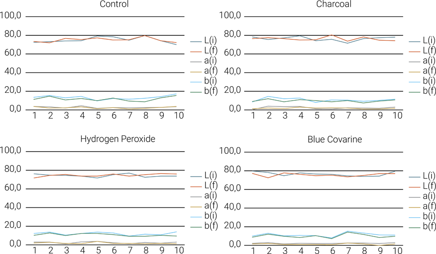
Regarding the color analysis, the one-way ANOVA revealed no significant differences between the dentifrices (p = 0.579) tested for color alteration measured by ΔE (Table 4). The descriptive values of L*, a*, and b* coordinates for all samples are shown in the graphs of Figure 2. Considering the statistical analysis of the coordinates, no significant differences were found between them, with all values of p >0.05 (L: p=0.703; a: p=0.405; b: p=0.851), and the mean values are disposed in table 4.

Descriptive values of the color coordinates (L, a, and b) measured for all samples in the groups tested.
Discussion
The continuous use of the whitening dentifrices tested was able to cause alteration in enamel and dentin roughness, thus denying the first null hypothesis, but the differences found depended on the tooth substrate and the type of dentifrice. For enamel, the dentifrice containing activated charcoal induced a reduction of the surface roughness compared with the initial condition and presented lower Ra values compared with the others, while for dentin, this was not observed (Tables 2 and 3). Although described in the recent literature as an innovation, charcoal has been used since ancient Greece as an oral hygiene product12,13. The rationale behind the use of charcoal relies on its capacity to adsorb pigments and stains25, but evidence shows that its ability to induce tooth whitening is limited or non-existent12,25, as also demonstrated by our results.
Besides not resulting in teeth whitening, in this study, the enamel Ra reduction caused by charcoal-containing dentifrice might indicate a potential to polish the surface, and, although this may cause difficult biofilm formation and deposition of new extrinsic stains, long-term use can lead to significant wear, inducing surface loss. This agrees with previous studies, which found that dentifrices with charcoal do not harm enamel in the short term26,27, but other suggests that they can induce alteration in the enamel mineral content after long exposure12,16,28. For dentin, literature is also controversial29,30, and, in this study, the fact that the roughness changes observed for enamel were not replicated in dentin may be related to differences in the mechanical behavior and composition of both substrates. Despite its lower mineral content, dentin’s higher organic matrix and smear layer formation during brushing may have partially protected the surface from abrasion, masking potential alterations detectable under the tested conditions. These products’ harmful potential is usually related to the type and quantity of abrasives in the formulation, which, in the case of charcoal, depends on the source and the methods used to prepare and mill it12,13, thus possibly explaining the divergent findings regarding their deleterious effects. Also, a few evidence suggest that the charcoal particles are very small and can cause marginal staining of resin composite restorations18, gingival irritation, and inflammation13, and some products available in the market present lower amounts or even no fluorides. Therefore, long-term use of these products shall not be indicated for patients who might benefit from using products that exhibit further protective effects.
Still, these results highlight an alert for the dental community, as charcoal-containing products intended to promote teeth whitening recently flooded the market, and their indication exploded in social media and the internet11. Currently, these platforms are tools with a high capacity for reaching and engaging people, widely used in disseminating and creating content on the most diverse topics. Most of the procedures or products displayed on these platforms are not endowed with any proof or scientific rigor, and the public is usually unaware of their indication or even possible sequelae caused by their use11,31, such as those shown by our results.
Regarding the dentifrice containing hydrogen peroxide tested, it could not induce color alteration, possibly due to the low concentration of the hydrogen peroxide (<1%) available as an active ingredient for whitening and the relatively short exposure compared to the professional options8. Few studies show that these dentifrices can promote tooth color alteration25,32-35, but with no consensus in the literature25,36. In addition, the meta-analysis from Devila et al.37 (2020) indicates that any overall color change resulting from whitening dentifrices use is likely due to stain removal rather than a true bleaching effect. Interestingly, this dentifrice promoted a significant reduction in dentin roughness without altering enamel roughness. The abrasive components of the dentifrice, combined with dentin’s susceptibility to wear, may have removed the smear layer and smoothed the surface, resulting in the observed Ra reduction. This behavior reinforces the need for careful indication of such products in patients with exposed dentin or at high risk for non-carious cervical lesions.
Finally, the dentifrice containing blue covarine also did not promote any significant whitening effect, not being able to improve staining removal or avoid the enamel pigmentation, presenting similar values with the control dentifrice (Table 4). The use of blue covarine or similar pigments in these dentifrices aims to modify the perception of yellowish discoloration by forming a thin and semi-transparent film over tooth surface, thus creating the visual appearance of brighter teeth by shifting the net color towards white25. The literature provides conflicting reports concerning the effectiveness of these pigments in promoting tooth whitening4,38-40 with suggestions that this effect can be achieved with progressive use, which was not evident from our results, as the dentifrices were tested for only 14 days with concomitant staining events were performed, suggesting that they cannot remove the staining properly to form an adequate pigment film. Also, the concentration of this active ingredient is not informed by the fabricant, and it is suggested that it plays an important role in the product’s efficacy4. Still, this dentifrice did not cause a significant alteration of enamel and dentin roughness when compared with initial values, indicating that its abrasiveness is not too high to induce a deleterious effect over enamel or dentin. Despite that, in general, all dentifrices tested were not able to induce satisfactory enamel whitening, and as some might induce adverse effects over tooth tissues, mainly dentin, the use shall be indicated and supervised by the dentists, considering the patient’s risks for caries and non-carious cervical lesions, as well as its periodontal needs.
A limitation of the present study is related to the staining protocol used prior to the brushing cycles. Although the procedure was designed to simulate clinical discoloration and allow comparison among groups, the literature does not provide a standardized methodology for staining in in vitro whitening dentifrice evaluations. There is no consensus regarding the most appropriate staining solution, immersion time, or duration of the staining phase, and these factors can influence the baseline color change, the interaction between pigments and dental substrates, and ultimately the apparent whitening effect of the products. Such methodological variability among studies makes direct comparison of results challenging and may partially explain the conflicting evidence reported in the literature on the efficacy of whitening dentifrices. Also, this study did not calculate ΔE00 or ΔWID values, as these usually are made with spectrophotometric acquisition of CIELAB coordinates, which was not part of the photographic-based protocol employed.
In conclusion, the dentifrices tested were not able to induce a whitening effect over enamel, presenting similar values to the control. Their use shall be cautiously indicated as they can cause surface harm, mainly over dentin, and therefore shall not be used by patients presenting dentin exposure in the oral cavity. The dentifrice containing blue covarine and hydrogen peroxide reduced dentin roughness, indicating possible polishing of the structure, while the one containing charcoal presented this behavior only for enamel.
Acknowledgements
This research was supported by National Council for Scientific and Technological – CNPq/Brazil
REFERENCES
-
1 Joiner A, Luo W. Tooth colour and whiteness: a review. J Dent. 2017 Dec;67S:S3-S10. doi: 10.1016/j.jdent.2017.09.006.
» https://doi.org/10.1016/j.jdent.2017.09.006 -
2 Silva FBD, Chisini LA, Demarco FF, Horta BL, Correa MB. Desire for tooth bleaching and treatment performed in Brazilian adults: findings from a birth cohort. Braz Oral Res. 2018 Mar;32:e12. doi: 10.1590/1807-3107bor-2018.vol32.0012.
» https://doi.org/10.1590/1807-3107bor-2018.vol32.0012 -
3 Hajisadeghi S, Khansari M, Yazdanian M, Keykha E. Effect of whitening dentifrice on discoloration of tooth surface: an updated systematic review and meta-analysis. BMC Oral Health. 2025 Apr;25(1):610. doi: 10.1186/s12903-025-05851-4.
» https://doi.org/10.1186/s12903-025-05851-4 -
4 Tao D, Smith RN, Zhang Q, Sun JN, Philpotts CJ, Ricketts SR, et al. Tooth whitening evaluation of blue covarine containing toothpastes. J Dent. 2017 Dec;67S:S20-S24. doi: 10.1016/j.jdent.2017.10.014.
» https://doi.org/10.1016/j.jdent.2017.10.014 -
5 Borges AB, Zanatta RF, Barros AC, Silva LC, Pucci CR, Torres CR. Effect of hydrogen peroxide concentration on enamel color and microhardness. Oper Dent. 2015 Jan-Feb;40(1):96-101. doi: 10.2341/13-371-L. Epub 2014 Aug 19.
» https://doi.org/10.2341/13-371-L -
6 Jorge OS, Arruda CNF, Torrieri RT, Vivanco RG, Pires-de-Souza FCP. Over-the-counter bleaching agents can help with tooth whitening maintenance. J Esthet Restor Dent. 2022 Mar;34(2):328-34. doi: 10.1111/jerd.12617. Epub 2020 Jul 6.
» https://doi.org/10.1111/jerd.12617 -
7 Greenwall-Cohen J, Francois P, Silikas N, Greenwall L, Le Goff S, Attal JP. The safety and efficacy of 'over the counter' bleaching products in the UK. Br Dent J. 2019 Feb;226(4):271-6. doi: 10.1038/s41415-019-0011-6.
» https://doi.org/10.1038/s41415-019-0011-6 -
8 Freitas MR, Carvalho MM, Liporoni PCS, Fort ACB, Moura RM, Zanatta RF. Effectiveness and adverse effects of over-the-counter whitening products on dental tissues. Front Dent Med. 2021;2:687507. doi: 10.3389/fdmed.2021.687507.
» https://doi.org/10.3389/fdmed.2021.687507 -
9 Silva EMD, Maia JNSMD, Mitraud CG, Russo JES, Poskus LT, Guimarães JGA. Can whitening toothpastes maintain the optical stability of enamel over time? J Appl Oral Sci. 2018 Feb 1;26:e20160460. doi: 10.1590/1678-7757-2016-0460.
» https://doi.org/10.1590/1678-7757-2016-0460 -
10 Demarco FF, Meireles SS, Masotti AS. Over-the-counter whitening agents: a concise review. Braz Oral Res. 2009;23 Suppl 1:64-70. doi: 10.1590/s1806-83242009000500010.
» https://doi.org/10.1590/s1806-83242009000500010 -
11 Bauler LD, Santos CSD, Lima GS, Moraes RR. Charcoal-based dentifrices and powders: analyses of product labels, Instagram engagement, and altmetrics. Braz Dent J. 2021 Mar-Apr;32(2):80-9. doi: 10.1590/0103-6440202104233.
» https://doi.org/10.1590/0103-6440202104233 -
12 Vural UK, Bagdatli Z, Yilmaz AE, Çakir FY, Altundasar E, Gurgan S. Effects of charcoal-based whitening toothpastes on human enamel in terms of color, surface roughness, and microhardness: an in vitro study. Clin Oral Investig. 2021 Oct;25(10):5977-85. doi: 10.1007/s00784-021-03903-x.
» https://doi.org/10.1007/s00784-021-03903-x -
13 Greenwall LH, Greenwall-Cohen J, Wilson NHF. Charcoal-containing dentifrices. Br Dent J. 2019 May;226(9):697-700. doi: 10.1038/s41415-019-0232-8.
» https://doi.org/10.1038/s41415-019-0232-8 -
14 Naidu AS, Bennani V, Brunton JMAP, Brunton P. Over-the-counter tooth whitening agents: a review of literature. Braz Dent J. 2020 Jun;31(3):221-35. doi: 10.1590/0103-6440202003227.
» https://doi.org/10.1590/0103-6440202003227 -
15 Casado BGS, Moraes SLD, Souza GFM, Guerra CMF, Souto-Maior JR, Lemos CAA, et al. Efficacy of dental bleaching with whitening dentifrices: a systematic review. Int J Dent. 2018 Oct 30;2018:7868531. doi: 10.1155/2018/7868531.
» https://doi.org/10.1155/2018/7868531 -
16 Jamwal N, Rao A, Shenoy R, Pal M, Ks A, Br A. Effect of whitening toothpaste on surface roughness and microhardness of human teeth: a systematic review and meta-analysis. F1000Res. 2022 Jan 11;11:22. doi: 10.12688/f1000research.76180.3.
» https://doi.org/10.12688/f1000research.76180.3 -
17 Franco MC, Uehara J, Meroni BM, Zuitton GS, Cenci MS. The effect of a charcoal-based powder for enamel dental bleaching. Oper Dent. 2020 Nov 1;45(6):618-23. doi: 10.2341/19-122-L.
» https://doi.org/10.2341/19-122-L -
18 Bragança GF, Soares PF, Borges JS, Vilela ABF, Santos Filho PC, Soares CJ. Effects of charcoal toothpaste on the surface roughness, color stability, and marginal staining of resin composites. Oper Dent. 2022 Mar 1;47(2):214-24. doi: 10.2341/20-046-L.
» https://doi.org/10.2341/20-046-L -
19 Lima LC, Carvalho AO, Bezerra SJC, Garcia RM, Caneppele TMF, Borges AB, et al. Tooth color change promoted by different whitening toothpastes under alternate cycles of staining and brushing. J Dent. 2023 May;132:104498. doi: 10.1016/j.jdent.2023.104498.
» https://doi.org/10.1016/j.jdent.2023.104498 -
20 Grazioli G, Valente LL, Isolan CP, Pinheiro HA, Duarte CG, Münchow EA. Bleaching and enamel surface interactions resulting from the use of highly-concentrated bleaching gels. Arch Oral Biol. 2018 Mar;87:157-62. doi: 10.1016/j.archoralbio.2017.12.026. Epub 2017 Dec 28.
» https://doi.org/10.1016/j.archoralbio.2017.12.026 -
21 Bengel WM. Digital photography and the assessment of therapeutic results after bleaching procedures. J Esthet Restor Dent. 2003;15 Suppl 1:S21-32; discussion S32. doi: 10.1111/j.1708-8240.2003.tb00315.x.
» https://doi.org/10.1111/j.1708-8240.2003.tb00315.x -
22 Sampaio CS, Atria PJ, Hirata R, Jorquera G. Variability of color matching with different digital photography techniques and a gray reference card. J Prosthet Dent. 2019 Feb;121(2):333-9. doi: 10.1016/j.prosdent.2018.03.009. Epub 2018 Aug 7.
» https://doi.org/10.1016/j.prosdent.2018.03.009 -
23 Garg SA, Chavda SM. Color masking white fluorotic spots by resin infiltration and its quantitation by computerized photographic analysis: a 12-month follow-up study. Oper Dent. 2020 Jan/Feb;45(1):1-9. doi: 10.2341/17-260-T. Epub 2019 Sep 30.
» https://doi.org/10.2341/17-260-T -
24 Batista GR, Zanatta RF, Augusto MG, Arantes GS, Borges AB, Torres CRG. The ability of different formulations of artificial saliva to protect dentin from erosive wear. Braz Dent Sci. 2023;26(2):e3767. doi: 10.4322/bds.2023.e3767.
» https://doi.org/10.4322/bds.2023.e3767 -
25 Vaz VTP, Jubilato DP, Oliveira MRM, Bortolatto JF, Floros MC, Dantas AAR, et al. Whitening toothpaste containing activated charcoal, blue covarine, hydrogen peroxide or microbeads: which one is the most effective? J Appl Oral Sci. 2019 Jan 14;27:e20180051. doi: 10.1590/1678-7757-2018-0051.
» https://doi.org/10.1590/1678-7757-2018-0051 -
26 Viana ÍEL, Weiss GS, Sakae LO, Niemeyer SH, Borges AB, Scaramucci T. Activated charcoal toothpastes do not increase erosive tooth wear. J Dent. 2021 Jun;109:103677. doi: 10.1016/j.jdent.2021.103677.
» https://doi.org/10.1016/j.jdent.2021.103677 - 27 Shaikh M, Sung H, Lopez T, Andra R, McKean B, Jesson J, et al. Effect of charcoal dentifrices on tooth whitening and enamel surface roughness. Am J Dent. 2021 Dec;34(6):295-9.
-
28 Pertivi UI, Eriwati YK, Irawan B. Surface changes of enamel after brushing with charcoal toothpaste. J Phys Conf Ser. 2017.884(1):012002. doi: 10.1088/1742-6596/884/1/012002.
» https://doi.org/10.1088/1742-6596/884/1/012002 - 29 Bowers AN, Tyrakowski CM, Koizumi Y, Shah R, Gopal P, Duarte S. Charcoal dentifrices: a laboratory study of their safety and efficacy. Am J Dent. 2024 Oct;37(5):255-62.
-
30 Vertuan M, Silva JF, Oliveira ACM, Silva TT, Justo AP, Zordan FLS, et al. The in vitro effect of dentifrices with activated charcoal on eroded teeth. Int Dent J. 2023 Aug;73(4):518-23. doi: 10.1016/j.identj.2022.11.001. Epub 2022 Nov 29.
» https://doi.org/10.1016/j.identj.2022.11.001 -
31 Simsek H, Buyuk SK, Cetinkaya E, Tural M, Koseoglu MS. "How I whiten my teeth": YouTube™ as a patient information resource for teeth whitening. BMC Oral Health. 2020 Jul 1;20(1):183. doi: 10.1186/s12903-020-01172-w.
» https://doi.org/10.1186/s12903-020-01172-w -
32 Torres VS, Lima MJP, Valdrighi HC, Campos EJ, Santamaria-Jr M. Whitening dentifrices effect on enamel with orthodontic braces after simulated brushing. Eur J Dent. 2020 Feb;14(1):13-8. doi: 10.1055/s-0039-3403474.
» https://doi.org/10.1055/s-0039-3403474 - 33 Kakar A, Rustogi K, Zhang YP, Petrone ME, DeVizio W, Proskin HM. A clinical investigation of the tooth whitening efficacy of a new hydrogen peroxide-containing dentifrice. J Clin Dent. 2004;15(2):41-5.
-
34 Kim HJ, Jang JH, Choi D, Kim J, Shim JH, Kim DS. Bleaching toothpaste with two different concentrations of hydrogen peroxide: a randomized double-blinded clinical trial. J Dent. 2020 Dec;103:103508. doi: 10.1016/j.jdent.2020.103508.
» https://doi.org/10.1016/j.jdent.2020.103508 - 35 Kleber CJ, Putt MS, Nelson BJ. In vitro tooth whitening by a sodium bicarbonate/peroxide dentifrice. J Clin Dent. 1998;9(1):16-21.
-
36 Alshara S, Lippert F, Eckert GJ, Hara AT. Effectiveness and mode of action of whitening dentifrices on enamel extrinsic stains. Clin Oral Investig. 2014;18(2):563-9. doi: 10.1007/s00784-013-0981-8. Epub 2013 Apr 25.
» https://doi.org/10.1007/s00784-013-0981-8 -
37 Devila A, Lasta R, Zanella L, Agnol MD, Rodrigues-Junior SA. Efficacy and adverse effects of whitening dentifrices compared with other products: a systematic review and meta-analysis. Oper Dent. 2020 Mar/Apr;45(2):E77-E90. doi: 10.2341/18-298-L. Epub 2019 Nov 18.
» https://doi.org/10.2341/18-298-L -
38 Bergesch V, Aguiar FHB, Turssi CP, França FMG, Basting RT, Amaral FLB. Shade changing effectiveness of plasdone and blue covarine-based whitening toothpaste on teeth stained with chlorhexidine and black tea. Eur J Dent. 2017 Oct-Dec;11(4):432-7. doi: 10.4103/ejd.ejd_97_17.
» https://doi.org/10.4103/ejd.ejd_97_17 -
39 Tao D, Sun JN, Wang X, Zhang Q, Naeeni MA, Philpotts CJ, et al. In vitro and clinical evaluation of optical tooth whitening toothpastes. J Dent. 2017 Dec;67S:S25-S28. doi: 10.1016/j.jdent.2017.08.014.
» https://doi.org/10.1016/j.jdent.2017.08.014 -
40 Torres CR, Perote LC, Gutierrez NC, Pucci CR, Borges AB. Efficacy of mouth rinses and toothpaste on tooth whitening. Oper Dent. 2013 Jan-Feb;38(1):57-62. doi: 10.2341/11-360-L. Epub 2012 Jul 7.
» https://doi.org/10.2341/11-360-L
-
Data Availability:
Datasets related to this article will be available upon request to the corresponding author.
Edited by
-
Editor:
Dr. Altair A. Del Bel Cury
Datasets related to this article will be available upon request to the corresponding author.