 Biofabrication breakthroughs: Towards tailor-made hearts and cardiac tissues
Organ failure remains one of the leading causes of death worldwide, posing a formidable challenge for healthcare systems. Conventional organ transplants are limited by the scarcity of donors and the risks associated with immune rejection. In response, 3D bioprinting has emerged as a revolutionary approach with the potential to fabricate tailor-made, patient-specific organs. Central to this technology is the use of bioinks-smart materials that ensure structural integrity, biocompatibility, and the functional performance of printed organs. This review delves into the latest advancements in 3D bioprinting, specifically targeting the heart and cardiac tissues, such as heart valves and entire hearts. It evaluates the capabilities of various bioprinting techniques in creating intricate tissue structures using patient-specific cells, while also addressing the potential for bioprinting to transform the landscape of organ transplantation. Furthermore, the review explores the essential properties of bioinks, the selection of cell sources, and the current progress in clinical trials and patents, offering insights into the future prospects and challenges of cardiac tissue bioprinting. This comprehensive assessment underscores the promise of bioprinting in overcoming the limitations of traditional transplants and advancing personalized medicine.
Keywords:
Biofabrication; 3D printing; Tissue engineering; Bioinks.

INTRODUCTION
3D bioprinting is a method of additive manufacturing that creates tissue or organs layer by layer using a bottom-up technique. It aims to replicate natural cellular architecture by layering ingredients and cells, restoring the normal structure and functionality of complex tissues. This fully automated process allows for accurate cell patterning and regulated ECM structure. The layer-by-layer construction of bioprinted tissues results in interconnected holes for gas and nutrient perfusion and increased intercellular communication, contributing to pre-clinical trials. 3D bioprinting of cardiac tissue, derived from autologous cells, has the potential to address the demand for cardiac tissue constructs for cardiovascular disease treatment. This method is increasingly used to create vasculature scaffolds, as mortality from CVD increases due to a lack of donor organs (Murphy, Skardal, Atala, 2013; Wang et al., 2021). 3D printing technology has revolutionized organ transplantation, offering personalized medicine and improved patient outcomes. The development of 3D-printed hearts holds great promise for preoperative planning, medical education, and complex procedures. These models can incorporate functional elements like integrated blood vasculature, enhancing their utility for studying cardiovascular diseases, testing new therapies, and advancing cardiac medicine. The integration of 3D printing technology in heart transplantation and cardiovascular care promises greater precision, efficacy, and patient-centricity, paving the way for a more personalized and effective healthcare system.
Cardiovascular disease mortality is rising worldwide due to a severe shortage of donor hearts. Since heart tissue cannot regenerate cardiomyocytes, heart transplants are often the only viable treatment. However, the demand for heart transplants far exceeds the supply, resulting in many patients dying while waiting for a donor. From 2005 to 2016, heart transplant listings increased by 57%, while the number of patients on waiting lists surged by 127% (available on the official webpage https://www.organdonor.gov). As of March 24, according to the Health Resources & Services Administration, 3,436 patients were waiting for a heart transplant, but only 812 transplants had been performed (2023). The organ waiting list grows by 3% annually, exacerbating the gap between demand and supply. In addition to the shortage, organ rejection remains a significant issue. Since donated organs come from different individuals, the recipient’s immune system often attacks the new organ, requiring immunosuppressant drugs, which have their own side effects. To address these challenges, “make-to-order” organs are emerging as a promising solution through 3D bioprinting technology. This approach allows for the creation of custom organs using a patient’s own cells, minimizing the risk of rejection and ensuring a perfect fit, representing a breakthrough in the field of regenerative medicine and bioprinting.
Present status for bioprinting of heart and cardiac tissues
3D bioprinting offers promising solutions for heart disease treatments and advancements in drug testing. This technology allows the creation of patient-specific cardiac tissues using their own cells, recreating the complex 3D structure of the heart. It holds significant potential in personalized medicine and regenerative therapies. Cardiac-specific bioinks and improved printing techniques could lead to the development of larger engineered tissue models for more effective testing. The process of bioprinting heart tissue involves three stages: pre-processing, printing, and post-processing. First, a 3D model is generated from clinical imaging techniques such as MRI, CT, and PET scans, allowing for high-fidelity, patient-specific models. Image segmentation is used to customize the 3D model for the specific cardiovascular tissue required. These models are converted into STL files, which guide the printing process to ensure tissue integrity and proper function. Ensuring the printed tissue maintains its shape and functionality is crucial, especially considering the natural rhythm and workload of heart. Strategic collaboration between academia, industry, and regulatory bodies will be key to accelerating the use of 3D bioprinting in clinical and commercial settings (Agarwal et al., 2020; Uccheddu et al., 2018).
Recent advancements in bioprinting technologies have introduced groundbreaking techniques for fabricating heart tissues and blood vessels, offering promising solutions for cardiovascular disease treatment and organ transplantation. Key techniques include extrusion-based bioprinting, inkjet bioprinting, laser-assisted bioprinting, stereolithography (SLA), and digital light processing (DLP)-based bioprinting (Li et al., 2023). Each method employs distinct mechanisms to accurately replicate the structural intricacies of heart tissues and vascular networks. These innovative techniques enable precise deposition of cells and biomaterials, facilitating the development of functional cardiac patches and organoids for therapeutic applications, potentially transforming the future of cardiac tissue engineering and organ replacement.
BIOINKS
3D bioprinting enables the fabrication of functional human tissues and organs, including the heart, liver, skin, bones, and microfluidic models. Essential to this process is bioink, a composite of biomaterials, living cells, and bioactive factors. However, most biomaterials face challenges in terms of vascularization, nutrient and gas exchange, biocompatibility, degradation rate, form stability, and the maintenance of tissue function post-printing. Both natural and synthetic polymers like alginate, gelatin, collagen, and PEG are commonly used for their tunable and biocompatible properties (Xu et al., 2012). Biomaterials used for printing heart tissue and vasculature should possess certain properties to ensure cell viability and integrity of bioprinted construct. Bioprinting cardiac tissue requires high resolution to replicate its complex microarchitecture, balancing fine layer deposition and cell viability. Bioinks must exhibit low shear stress and be easily crosslinkable, resistant to reflow, and suitable for cell culture environments. Challenges like nozzle clogging, phase transitions, and extrusion difficulties hinder the replication of microscale features (Liu et al., 2017). Crosslinking techniques enhance mechanical properties of bioinks, enabling cellular function and mimicking native tissue environments. Hydrogels with adjustable stiffness are crucial for engineering cardiac tissues, requiring elasticity and contractility like natural cardiomyocytes (Lee et al., 2017; Martina, Hutmacher, 2007). Mechanical tests, tensile, compression, and shear, are used to assess structural and functional fidelity. Biocompatibility is crucial for cell adhesion, proliferation, and integration with host tissues, influenced by material chemistry, surface morphology, charge, and mechanical properties, and can be improved by blending synthetic and natural components (McGill, Holland, Kaplan, 2019). Vascularization is equally vital for tissue longevity and function. Strategies such as angiogenic factor inclusion, optimized bioreactor conditions, and bioactive molecule delivery are employed to promote vessel formation and function. Strategies like incorporating angiogenic factors or bioactive molecules are being explored to promote vascularization. To ensure long-term performance, materials must maintain stability under physiological conditions, degrade in sync with tissue regeneration, and produce non-toxic byproducts. Effective ECM remodeling by the cells is essential for forming interconnected, densely packed cardiac tissue (Kim et al., 2019, Serpooshan et al., 2014). Finally, sterilization is crucial for clinical application, and materials must tolerate at least one sterilization method, such as autoclaving, irradiation, or chemical treatments (Axpe, Oyen, 2016).
Bioinks typically consist of biomaterials used to prepare hydrogel, with specific cells and certain biofactors. The cells are either encapsulated within the bioinks or seeded onto bioprinted constructions to enhance the formation of heart tissue.
Biomaterials
Biomaterials come from natural sources like proteins and polysaccharides or synthetic ones. Synthetic materials offer more control but may require biocompatibility modifications. Research now focuses on developing nanocomposite bioinks with specific functionalities as depicted in Figure 1.
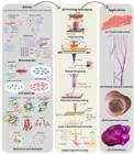
Describes the bioink, 3D printing using different techniques and potential applications with a focus on cardiac patches, vascular channels, and heart valves.
Biomaterials from natural sources
Bioinks, primarily protein-based, are ideal for bioprinting due to their close resemblance to the extracellular matrix. Polysaccharide-based bioinks, like alginate, hyaluronic acid, and chitosan, are preferred for their controllable gelation properties and inherent biocompatibility, making them versatile materials for various applications.
Alginate, a seaweed-derived polysaccharide, is a versatile hydrogel used in biomedical fields due to its biocompatibility, low cytotoxicity, and mild gelation process. Its rapid gelation under physiological conditions makes it an ideal bioink for 3D bioprinting, allowing for the creation of complex structures in various bioprinting methods (Zhang et al., 2015). Yu, et al., 2013 utilized extrusion-based bioprinting to fabricate vascular-like hollow tubes with enhanced mechanical and biological features. Additionally, they employed drop-on-demand inkjet printing to create zigzag tube structures containing fibroblasts, achieving high cell viability. Chitosan is a biocompatible, non-toxic, and antimicrobial agent with hydrating properties. It supports 3D tissue growth, activates macrophages, and promotes cell proliferation. Its haemostatic properties aid in blood clotting and pain reduction. Research by Wang et al. 2018 demonstrated that chitosan scaffolds improve cardiac function, contractility, reduce infarct size, and increase vessel density in cardiac tissues, indicating its potential for cardiac repair applications. Collagen, a key extracellular matrix component, is essential in cardiac tissue engineering for promoting cell adhesion, proliferation, and differentiation. Its biocompatibility and ability to mimic native cardiac environments make it a suitable platform for cell delivery. However, its low Young’s modulus and viscosity limit its printability and mechanical strength (Araña et al., 2014). To overcome these issues, modifications such as methacrylated type I collagen have been developed, allowing UV-induced photopolymerization, tunable stiffness, and incorporation of adhesion molecules. Despite its potential, collagen is often used at low concentrations (≤10 mg/mL) due to weak mechanical properties. When combined with hyaluronic acid, it supports the formation of functional cardiac tissues with aligned sarcomeres and spontaneous contractions (Esser et al., 2023).
Fibrin, a protein essential for blood clot formation and wound healing, has been used by histological engineers to create high-strength hydrogels called natural ECM. Nakamura et al. used fibrin and alginate to fabricate bioprinted constructs exploring inkjet bioprinter (Nakamura, et al., 2010). Scientists developed composite bioink for cartilage tissue engineering using fibrin and collagen, enhancing mechanical properties. The constructs were fabricated in three steps: electrospun PCL, encapsulated chondrocytes in fibrin/collagen, bioprinted, and another PCL layer deposited via electrospinning (Xu et al., 2013). Gelatin, a natural polymer sourced from animal tissues, is commonly used in tissue engineering and bioprinting due to its biocompatibility, biodegradability, low antigenicity, and affordability (Schuurman et al., 2013). Alonzo, et al., 2019 fabricated aortic valve conduits using gelatin-alginate hydrogels based on micro-CT scans, emphasizing that the gelatin-to-alginate ratio was critical for maintaining print quality and structural stability. Wüst et al., 2014 bioprinted 3D tubular structures with gelatin-based hydrogels, though the process was affected by temperature-sensitive gelation. To improve this, gelatin was modified with methacryloyl groups (GelMA), enabling UV-induced cross-linking and offering tunable mechanical properties, making it a promising bioink for cardiac and tissue engineering. Hyaluronic acid (HA), a highly acidic polysaccharide found in ECM, is a key component of connective tissues that helps retain moisture, enables lubrication, and supports cell-matrix interactions. Researchers have used hyaluronic acid, combined with materials like gelatin, to fabricate 3D bioprinted heart valve models that mimic the natural tri-leaflet structure for use in tissue engineering (Duan, et al., 2013).
Biomaterials from synthetic sources
Synthetic polymers are widely used as bioinks in 3D bioprinting due to their superior mechanical strength, stability, and printability. Their physicochemical properties can be adjusted through molecular weight or post-printing treatments, allowing control over degradation rate, stiffness, and functional performance. This adaptability and structural integrity make them ideal for creating complex, durable tissue scaffolds with high precision (Zacchigna, et al., 2011).
Poly ethylene glycol (PEG), a non-cytotoxic and non-immunogenic polymer, is ideal for bioprinting and shape maintenance due to its strong mechanical properties. However, it struggles to attach to cell surfaces, necessitating the use of other biologically active hydrogels. Composites of PEG and natural biomaterials enhance construct degradation (Zacchigna, et al., 2011). Polycaprolactone (PCL) is a biodegradable, biocompatible bioink with successful human implantation history. It promotes tissue growth and new formation, and has shown potential for 3D-printed heart valves due to its flexibility, mechanical strength, and controlled degradation rates (Kabirian, et al., 2018). Polylactic acid (PLA) is a biodegradable polymer used in biomedical applications, particularly in creating vascular scaffolds for cardiovascular implants. It maintains vascular movement and supports adaptive remodelling of the arterial wall, promoting cell viability and extending structural support. PLA is metabolized into harmless byproducts by enzymes like hydrolase and esterase (Puricel et al., 2015).
Biomaterials derived from ECM
Cardiac ECM, composed of fibronectin, collagen I, fibrillin 1, laminin, and sertain issue-specific growth factors. Decellularized cardiac extracellular matrix (dECM), sourced from porcine or ovine tissue, is a biocompatible, thermos-sensitive, and printable platform that supports adhesion, proliferation, migration, and differentiation. In animal models, dECM hydrogels enhance blood vessel formation, activate cardiac-specific genes, and influence apoptosis and fibrosis pathways (Zhu et al., 2017).
Hybrid biomaterials
Hybrid bioinks are 3D printing materials that combine natural and synthetic polymers, offering support, mechanical properties, and customization options. Research shows that grafting hydrophilic segments onto hydrophobic biomaterials can enhance the differentiation of neonatal human cardiac progenitor cells (Colosi et al., 2016). Researchers utilized microfluidic systems to print bioinks, creating support structures and sacrificial components in hollow constructs using PCL as a framework, fibrin-based composite hydrogel as the bioink, and gelatin as the sacrificial material (Wang, et al., 2018).
Novel biomaterials
Researchers developed gold nanorod-incorporated gelatin methacrylate hydrogels that enhance mechanical and electrical properties, improving cell function and supporting synchronized cardiac tissue beating. Gold nanoparticles synthesized within lysozyme nanofibrils were integrated into gelatin-hyaluronic acid hydrogels, enhancing their mechanical strength, conductivity, and antioxidant activity, while maintaining injectability, biocompatibility, and drug release capabilities for myocardial regeneration. Combining carbon nanotubes (CNTs) with PCL in hybrid scaffolds enhances polymer chain alignment, elastic modulus, hardness, and crystallinity. PCL nanocomposites with 1% CNT showed optimal conductivity for H9C2 cells, increasing their proliferation in vitro (Ho et al., 2017).
Cell sources
Cell selection is critical in 3D bioprinting due to requirements such as high proliferation, ease of differentiation, accessibility, and pathogen resistance. Native organ cells play a vital role in maintaining tissue architecture and supporting the stem cell niche (Budharaju, Subramanian Sethuraman, 2021). In cardiac applications, primary cardiomyocytes, stem cells, and hiPSC-derived cardiomyocytes are widely used for regenerative therapies, disease modeling, and drug testing, although immune incompatibility remains a concern (Zhang et al., 2016). Adult cardiomyocytes, known for their calcium handling and terminal differentiation, have been used in bioprinted constructs with polycaprolactone scaffolds to form dense, aligned cardiac fibers responsive to pharmacological agents. Incorporation of PEVA and homologous cells enhanced cadherin and integrin expression, supporting cell maturation and function (Wang et al., 2018). Stem cell types such as iPSCs, ESCs, MSCs, and cardiac progenitors offer high proliferative capacity and differentiation potential, along with lower immune rejection risks. Nonetheless, they present issues like ethical debates, immature cell states, and limited contractility (Agarwal et al., 2021). These cells can be guided toward cardiac lineages using ECM proteins like integrins that trigger key transcriptional pathways. For instance, mouse ESCs seeded on PGA scaffolds demonstrated improved cardiac function in ischemic models, highlighting their therapeutic promise (Lee, 2018). Human induced pluripotent stem cell-derived cardiomyocytes (hiPSC-CMs) are used to create precise cardiac models using GelMA and micro-continuous optical printing. However, their use in adult cardiac tissue engineering is limited due to poor alignment and calcium handling (Ronaldson-Bouchard et al., 2018, Yu et al., 2020). Human pluripotent stem cells (hPSCs), including hESCs and reprogrammed hiPSCs, offer an alternative to primary cardiomyocytes and cardiac progenitor cells, which are scarce. These stem cells can differentiate into multiple cardiac cell types, making them valuable for regenerative therapies, disease models, and drug testing (Takahashi, Yamanaka, 2006). Native cardiac cells like fibroblasts and endothelial cells are crucial for myocardial function, and their incorporation into 3D scaffolds improves cardiomyocyte performance and viability (Arai et al., 2018).
Biofactors and Nutrients
Biofactors are essential proteins that regulate cellular behaviors crucial for tissue regeneration. In 3D bioprinting, their incorporation into bioinks enhances the bioactivity and functionality of printed constructs. Key growth factors such as Vascular Endothelial Growth Factor (VEGF), Basic Fibroblast Growth Factor (bFGF), and Epidermal Growth Factor (EGF) are commonly utilized due to their roles in promoting angiogenesis, proliferation, and tissue repair. VEGF is particularly important for inducing blood vessel formation, facilitating oxygen and nutrient delivery to engineered tissues. Controlled, slow release of VEGF within scaffolds improves vascularization and tissue integration post-implantation (Lee et al., 2019; Mir et al., 2023). Basic Fibroblast Growth Factor (bFGF), also known as FGF-2, enhances cell proliferation, migration, and angiogenesis, accelerating tissue formation and repair. Its combination with VEGF synergistically improves vascular network development, particularly in thick constructs requiring deep nutrient diffusion (Won, et al., 2024). Basic Fibroblast Growth Factor (bFGF), also known as FGF-2, enhances cell proliferation, migration, and angiogenesis, accelerating tissue formation and repair. Its combination with VEGF synergistically improves vascular network development, particularly in thick constructs requiring deep nutrient diffusion (Pajooh et al., 2024). Additionally, bioinks are formulated with essential nutrients, such as amino acids, sugars, and vitamins, embedded in hydrogels like alginate or GelMA to maintain cell viability and function. This nutrient-rich environment supports the maturation of bioprinted tissues for regenerative medicine, disease modeling, and therapeutic applications.
BIOPRINTING TECHNIQUES
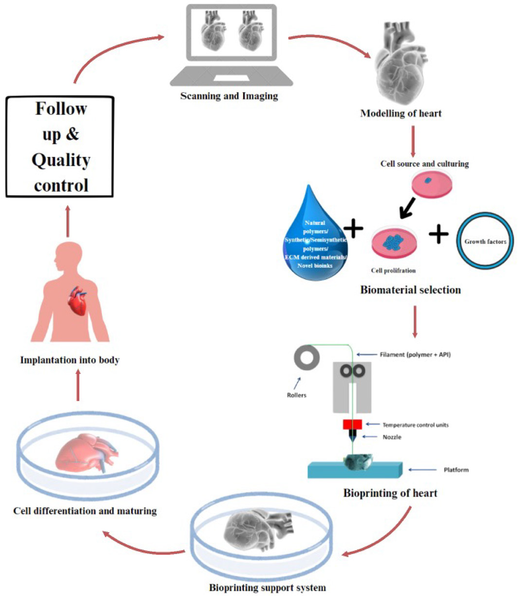
3D bioprinting of the heart involves obtaining detailed imaging data from patients using techniques like MRI or CT scans. This data is then converted into a digital 3D model using specialized software, creating a blueprint for the bioprinting process. The model captures the geometry and anatomical features of the heart, such as chambers, valves, blood vessels, and other structures. Bioprinting requires selecting biomaterials that mimic the properties of natural heart tissue, such as elasticity, conductivity, and biocompatibility. Common biomaterials include hydrogels, bioinks, and scaffold materials. The 3D printer deposits these bioinks and cells layer-by-layer (Chen et al., 2023). The next critical phase in bioprinting is sourcing and culturing cells, where cardiac cells are either harvested from the patient’s own heart or derived from stem cells. These cells are then cultured and combined with bioinks or scaffold materials. During the 3D bioprinting process, advanced techniques like extrusion-based or inkjet bioprinting are used to layer these cell-laden bioinks with precision, mirroring the complex architecture of cardiac tissues. Additionally, vascular networks or supporting scaffolds are printed to ensure structural integrity and facilitate the supply of oxygen and nutrients. After bioprinting, the construct matures in a bioreactor, where the cells differentiate and organize into functional cardiac tissue capable of synchronized contractions (Wu, Zhu, Woo, 2023). Quality control measures ensure the functionality and fidelity of bioprinted heart tissue, which is implanted into patients for therapeutic intervention, drug screening, disease modeling, or further research. Post-implantation, patients are closely monitored and follow-up assessments evaluate the effectiveness and long-term viability tissue (Polonchuk, Gentile, 2021).
Bioprinting, either scaffold-based or scaffold-free, allows cells to grow on a biomaterial matrix, while scaffold-free methods preserve tissue functionality. Additive manufacturing techniques like inkjet, extrusion and laser-assisted methods are used for selective cell and biomaterial patterning as depicted in Figure 1.
Extrusion based bioprinting
Extrusion based bioprinting is a widely used technique where bioink is loaded into a syringe and deposited as continuous strands through mechanical or pneumatic pressure. This method constructs 3D tissues layer-by-layer by extruding biomaterials in the form of cylindrical filaments. It is highly versatile, allowing for the printing of materials with a wide range of viscosities (up to >6 ×10⁷ mPa/s) and offering good structural integrity. However, higher pressure or reduced nozzle diameter can compromise cell viability. While it is a cost-effective platform, extrusion-based bioprinting has lower resolution compared to other methods. Despite this limitation, it has shown promising results in bioprinting complex structures such as myocardium constructs, heart valves, and blood vessels (Alonzo, et al., 2019).
The printing method involves selecting appropriate cells and suspending them in a prepolymer solution. The main nozzle contains a hydrogel containing cells, while the surrounding nozzle contains bioinks like partly crosslinked alginate or a hybrid hydrogel. A composite strand is created using a coaxial needle, combining the core and shell materials. A 3D human chambered cardiac muscle was fabricated using photo-crosslinking native extracellular matrix proteins and human induced pluripotent stem cells (hiPSC)-laden structures allowing continuous action-potential propagation and macroscale beating (Mistry et al., 2017). A 3D bioprinting technique, FRESH v2.0, was used to create a human left ventricle model using collagen type I and hESC-CFs. It offers promise in printing thick myocardial constructs, heart valves, and blood vessels but still suffers from certain challenges like its high cost, lower resolution, slow printing speed, and low cell viability (Alonzo, et al., 2019; Koch et al., 2018). Direct ink writing (DIW) generates 3D architectures layer-by-layer using materials with specific rheological properties, with a minimum printing resolution ranging from hundreds of microns to sub-microns (O’Bryan et al., 2017).
Inkjet based bioprinting
Inkjet bioprinting technology has been adapted for 3D bioprinting (3DBP) by replacing traditional ink cartridges with biological materials and integrating an elevator stage to enable movement along the z-axis for layer-by-layer printing. This method supports cell viability and is compatible with a variety of biomaterials. Using inkjet printing, a non-contact process, 3D aortic tissue constructs have been successfully created by depositing low viscosity bioinks with viable cells onto a substrate. Inkjet bioprinting offers advantages due to its affordability and non-contact nature, reducing contamination risks. Two primary techniques are used: continuous inkjet printing, where a constant stream of droplets is generated, and drop-on-demand (DOD) printing, which creates droplets only as needed. DOD bioprinting can use piezoelectric or thermal mechanisms. Piezoelectric printers use a transducer in a microfluidic chamber, while thermal printers apply electric currents to vaporize bioink, pushing droplets onto the substrate without harming cells. Factors like nozzle size, distance, temperature, and current frequency influence droplet size and placement, allowing for precise multicellular patterns and complex tissue structures (Cui, et al., 2010; Delaney, et al., 2009; Kačarević Ž et al., 2018).
Researchers developed a cardiac patch utilizing aerosol 3D inkjet printing, incorporating a composite of two-dimensional titanium carbide (Ti3C2Tx) MXene and hydrogel. This patch, when seeded with cardiomyocytes, demonstrated enhanced electrical conductivity, and increased gene expression of key cardiac markers, including MYH7, SERCA2, and TNNT2. These improvements suggest that this innovative cardiac patch could be a promising therapeutic approach for myocardial infarction treatment (Kačarević Ž et al., 2018).
Laser assisted bioprinting
Laser-assisted bioprinting (LAB) is a non-contact, direct-writing technique that uses a pulsed laser beam to deposit bio-ink containing cells onto a substrate. The process typically involves three main components: a pulsed laser source, a ribbon coated with bio-ink, and a receiving substrate. The bio-ink, sensitive to heat, is layered onto a target plate, which is then coated with natural polymers or a nutrient medium to support deposition and promote cell growth. LAB encompasses several methods, including Digital Light Processing (DLP), stereolithography (SLA), and Laser-Induced Forward Transfer (LIFT), each contributing to precision and viability in bioprinting processes.
Digital Light Processing (DLP) bioprinting utilizes a digital micro-mirror device (DMV) chip, consisting of millions of micromirrors, to reflect light and project a computer-aided design (CAD) model’s optical pattern onto a photopolymer solution. The unique feature of DLP is that it projects an entire plane of the optical pattern simultaneously, significantly reducing fabrication time. DLP outperforms other bioprinting techniques in terms of resolution, efficiency, and print conditions (Zhang, et al., 2020). Liu et al., 2017 used DLP printing to encapsulate neonatal mouse ventricular cardiac muscles in a hydrogel, demonstrating the rapid method’s ability to create complex cardiac tissue engineering scaffolds. They also created viable cardiac tissue using human embryonic stem cell derived cardiomyocytes and hydrogel, enabling in vitro disease modeling by detecting calcium transients and mechanical force. Similarly, human-induced pluripotent stem cell-derived cardiomyocytes (iPSC-CMs) were successfully bioprinted using DLP, showing expression of mature cardiac marker genes. This indicates potential DLP in fabricating complex, functional cardiac tissue for research and therapeutic purposes.
Stereolithography is a cost-effective, high-resolution, and high-quality method for printing personalized structures for disease modeling, but its slow printing speed (15 mm/s) is a limitation. Chan et al., 2012 used an SLA 3D printer to generate locomotive bio-bots made of hydrogels and cardiomyocytes, powered by a bimorph cantilever construction. The most effective bio-bots used maximum contractile efforts to overcome friction and prevent backward movement. 3D stereolithography technology has been used to create multi-material cantilevers made from PEGDA and acrylic-PEG-collagen mixtures, which enhance cell adhesion and organization without altering elastic modulus. These cantilevers can be used as mechanical sensors and early prototypes for optimal cell-based biohybrid actuator design (Chan et al., 2012).
Laser induced forward transfer (LIFT) is a technique for printing biomolecules, including AFA-LIFT and BioLP, using a laser-absorbing layer at the ribbon-bioink interface. It is computer-controlled and uses a CCD camera for selective cell patterning. LIFT achieves a resolution of 10-50 μm, handles high cell density, and maintains over 95% cell viability post-print. It can select single cells for transfer, making it beneficial for precision tissue construction. LIFT has been used for cardiac regeneration and has shown that hiPSCs are sensitive to biomaterials (Gaebel et al., 2011; Nahmias, et al., 2005).
CURRENT STATUS OF 3D BIOPRINTING OF CARDIAC TISSUES, HEART VALVES AND HEART
3D bioprinting is a rapidly developing technology with the potential to revolutionize the field of cardiac tissue engineering. It offers a promising approach for creating functional heart tissues and even whole hearts for transplantation.
3D bioprinting of cardiac model
A study by Miller et al. has developed a 3D-printed cardiac tissue that mimics the structural and functional complexity of heart tissues. They used an in-house micro-continuous optical printing system to create a cantilever-based tissue scaffold with micropatterns. The 3D microtissues showed high sarcomere organization and synchronous contraction. The force output was quantified through the deflection of an integrated force gauge. The 3D microtissues demonstrated the ability to be maintained over extended durations, generating substantial forces. Their functionality was further validated by testing drug responses across varying doses. This highly adaptable 3D cardiac tissue model holds promise for future applications in high-throughput disease modeling and drug discovery (Miller et al., 2021). Samson et al. created a 3D printing model for bioink extrusion, enabling the study of native cardiac tissue patterns. The 3D printed structure allows cells to grow and proliferate while maintaining the original shape (Samson, Song, 2022). The Lewis group created engineered cardiac tissue using anisotropic organ building blocks (aOBBs) in bioprinting. The bioinks, composed of gelatin and fibrinogen, generate thousands of anisotropic organoid building blocks (aOBBs) with precise control over their aspect ratio and cellular composition. These tissues exhibit a high cellular density and well-regulated alignment, with cardiac macrofilaments displaying greater alignment compared to spheroid-based controls.
Loureiro et al. (2023) explores the potential of GG/ KGM as a bioink for producing stable hydrogel matrices that promote endothelial cell activity as shown in Figure 2. The bioink is a simple and low-cost material-based bioink suitable for 3D printing without toxic solvents. The bioink conjugates two natural polymers gellan gum (GG) and konjac glucomannan (KGM) with complementary characteristics. GG is chosen for its structural and thermoresponsive properties, while KGM increases hydrogel bioactivity. The 3D structures have a rough surface, providing numerous anchorage points for cells, and an interconnected porous network. The hydrogels have suitable mechanical properties, controllable swelling and biodegradation, and biocompatible characteristics, encouraging adhesion to the bioprinted scaffold surface and internalization and migration, which are crucial for cardiac tissue formation. This emerging methodology is essential for developing 3D living functional hydrogels that can overcome current challenges in the cardiac tissue regeneration field (Loureiro et al., 2023).

represents manufacturing, invivo-characterization and in-vivo study of lyophilized 3D printed scaffold. Figure recreated from (Loureiro et al., 2023).
3D bioprinting of heart valves
Valvular heart disease affects 3.1% of adults and is projected to cost 23.6 million lives by 2030. The aortic valve is the most common cause, causing 65.2% of cases. Prosthetic valves, mechanical and bioprosthetic, have limitations, such as lifelong anticoagulation therapy and limited durability. 3D printed heart valves (HVs) offer a solution to anticoagulation therapy and could last a lifetime, but require specific cell infiltration, differentiation, and proliferation, as well as adequate mechanical and haemodynamic functionality. Due to complex regulatory approvals and patient-to-patient variability, 3D printed HVs are yet to reach clinics (Sanz-Garcia et al., 2015; Virani et al., 2021).
Stereolithography (SL) was the first 3D printing technique successfully used to fabricate heart valves. However, subsequent efforts using light-based bioprinting have been limited, primarily due to challenges with material diversity and mechanical properties. For instance, a study employed poly-ethylene glycol-diacrylate (PEGDA) combined with GelMA seeded with cardiac fibroblasts, achieving 80% cell viability after 7 days. Despite this promising result, further advancements have been stalled due to the limited material heterogeneity and relatively low mechanical strength, making it less suitable for broader applications in heart valve development (Lee et al., 2019).
Extrusion-based printing can create anatomically correct 3D printed heart valves using cell-free poly-ethylene glycol-diacrylate (PEGDA) and porcine aortic valve interstitial cells (PAVICs) in 21 days. Studies have shown that both smooth muscle cells (SMCs) and human aortic valvular interstitial cells (HAVICs) remain alive and show increased alpha-smooth muscle actin and vimentin levels after 7 days (Akpek, University, 2018). The extrusion of HAVIC onto a simplified heart valve shape influences cell spreading, mechanical characteristics, and printability. However, initial studies have not addressed extreme mechanical requirements. Various methods to reinforce cell-laden hydrogels have been explored, but none have shown mechanical properties sufficient to match native heart valves (Duan, et al., 2014). The freeform reversible embedding of suspended hydrogels (FRESH) method has been used to create 3D printed human heart valves, allowing for the fabrication of small tissue portions to full adult organs. These valves, made from cell-free collagen-based ink, have multiple anatomical structures and a tri-leaflet design, and have been tested in a physiological flow loop setting (Lee et al., 2019).
Rioux et al. (2022) studied and demonstrates the feasibility of manufacturing sodium-alginate aortic valve scaffolds with a native-like geometry using sugar glass printed molds. This simple and low-cost method could make personalized tissue-engineered heart valves more accessible to patients. Further testing is needed to ensure reliability and repeatability. Further optimizing cell integration, developing viability protocols, and remodelling the scaffold into a tissue-engineered heart valve as shown in Figure 3 (Rioux, et al., 2022).

represents 1. Manufacturing, design, CADD model of 3D Printed aortic valve scaffold 2. Represents injection process and dissolution process of the 3D printed scaffold (Rioux, et al., 2022).
3D bioprinting vascular channels
Blood vessels are crucial for delivering oxygen, nutrients, and blood cells, but synthetic vascular grafts are often used in damaged or diseased cases. However, these grafts face challenges like low patency rates and intimal hyperplasia, which restrict blood flow. Advances in bioprinting and tissue engineering offer promising alternatives to develop biocompatible, durable vascular grafts that mimic natural blood vessel behaviour (Wadey, et al., 2018). Researchers utilized hESC-derived early vascular cells (EVCs) for vascular engineering in cardiac tissues and observed that 3D bioprinting of cardiomyocytes and endothelial-vascular cell (EVC) spheroids promoted vascular differentiation and self-organization, leading to the formation of functional cardiac microtissues with a well-structured microvascular network. This approach holds promise for cell-based treatment of in vivo myocardial infarction testing (Liu et al., 2022). Scientists introduced an innovative bioprinting technique called Sacrificial Writing into Functional Tissue (SWIFT), which utilizes a matrix composed of living organ building blocks (OBBs). This method enables the creation of tissue structures with a high cellular density, incorporating embedded vascular channels that can be perfused. The research demonstrated the ability to integrate these channels into a variety of living matrices formed by OBBs. Researchers have bioprinted functional endothelial-lined channels using human umbilical vein endothelial cells (HUVECs) using a 3D CAD model of a human heart, a method with significant potential for advancing vascularized tissues for regenerative medicine (Skylar-Scott et al., 2019). Wang et al., 2022 created a double-network hydrogel bioink to create hollow constructs mimicking vein and artery tissues. This bioink was used in microfluidic coaxial extrusion bioprinting, offering controllable wall thicknesses, high-throughput production, and minimal waste.
3D bioprinting of heart
Computer-assisted 3D printing is advancing towards the engineering of fully functional whole-heart organs. CAD models of human hearts, developed using CT and MRI scans or sourced from model libraries, are essential tools in this process. However, fabricating a complete 3D heart with accurate geometry remains a challenge due to difficulties in integrating multiple cell types and creating multi-scale structures with different biomaterials. Recently, significant progress has been made with Freeform Reversible Embedding (FRE) printing, which has been used to bioprint elaborate structures that closely replicate the macroscopic anatomy of a heart. Full heart printing has shown promise in 3D structures with heart shape, with high-resolution and accurate bioink deposition. Lee et al., 2019 successfully utilized the FRESH (Freeform Reversible Embedding of Suspended Hydrogels) technique to fabricate a tri-leaflet heart valve, a neonatal-scale collagen heart, and a human cardiac ventricle model, showcasing the precision of the method in material deposition. The printed neonatal-scale collagen heart featured clearly defined leaflets and exhibited sufficient mechanical strength to be manipulated outside of a liquid environment. Noor et al., 2019 printed hearts using two bioinks, Cy5-labeled CMs and ECs, revealing the integrity of the compartments and mechanical qualities of the printed hearts. FRE printing provides a theoretical foundation for creating intricate crafts, making it a viable technique for printing improved tissue scaffolds for various organ system applications.
The Biofabrication of heart tissues and organs progresses, researchers are increasingly filing patents that reflect breakthroughs in both printing technologies and biomaterials. These patents offer insight into innovative methods and bioinks being used to push the boundaries of cardiac tissue engineering. Similarly, clinical trials are critical steps toward validating these innovations, assessing their safety and efficacy in real-world medical settings. Table I presents patents related to 3D printing of cardiac tissues and hearts, showcasing technological advances. They highlight the growing potential of 3D printing in addressing challenges like organ shortages and heart tissue regeneration. The developments in clinical trials in bioprinting of heart and cardiac tissue is compiled in Table II.
Data on clinical trials available on the official website on the 3D printing of cardiac tissue and heart
Clinical trials
CHALLENGES IN BIOPRINTING
3D bioprinting improvements have resulted in efficient and cost-effective technologies, however quality control criteria must be followed prior to transplantation. This comprises developing models, choosing bioink, verifying printing, maturing post-printing, and evaluating product quality. The lack of software for specifying cell locations complicates the printing process significantly (Hollister, 2005). Creating a robust, mechanically rigid 3D framework following transplanting is critical to promoting normal cell development. Proper structural maintenance and mechanical support are required for new tissues to thrive. In vivo vascularization is also required for bioprinted constructions used in tissue engineering to provide growth hormones, oxygen, nutrients, and waste elimination (Kaully, et al., 2009).
A key challenge in 3D bioprinting of cardiac tissue is sourcing high-quality cardiomyocytes and directing their differentiation into various necessary cell types for functional tissue formation. Since the heart lacks a substantial native stem cell population, obtaining cardiomyocytes directly from heart tissue is difficult. Moreover, bioprinted cardiac tissue requires a diverse range of cells, including cardiomyocytes, endothelial cells, and smooth muscle cells, which adds complexity. Embryonic stem cells (ESCs) and induced pluripotent stem cells (iPSCs) offer promise for generating cardiomyocytes, but fully maturing these cells remains challenging. To improve maturation, bioreactor-based methods have been explored, utilizing mechanical, electrical, and biochemical cues to enhance differentiation. Bioprinted cardiac constructs need to replicate the mechanical properties of native heart tissue, such as elasticity and contractility, to support proper dynamic function. For effective blood pumping, bioprinted cardiomyocytes must contract synchronously. Balancing the structural strength and flexibility of the tissue is a key challenge. Biomechanical testing methods, including tensile, compression, and shear tests, are critical for evaluating the mechanical performance of these constructs. Additionally, personalized bioprinting approaches must account for individual variations in tissue stiffness, contractility, and heart geometry to ensure optimal functionality.
3D bioprinting of hearts requires rigorous safety and biocompatibility assessments, requiring interdisciplinary collaboration, specialized facilities, and adherence to Good Laboratory Practice and Good Manufacturing Practice guidelines. Standardization and reproducibility are crucial, but consistency across platforms and labs is challenging. Ethical considerations, informed consent, privacy, transparency, long-term stability, and durability are also essential. Integrating bioprinted hearts with existing medical practices is crucial.
Cost and accessibility pose significant challenges in the widespread adoption of bioprinting technology, as it remains an expensive and resource-intensive process. To encourage broader implementation, it is essential to conduct comprehensive economic evaluations and health technology assessments that compare the long-term financial benefits of bioprinting with traditional heart transplantation and other therapies. Engaging stakeholders, including healthcare providers, regulatory bodies, and patients, will also be key to addressing cost concerns and ensuring that bioprinting solutions are accessible. Demonstrating the cost-effectiveness of bioprinting, particularly its potential to reduce the reliance on scarce donor organs and minimize the risk of rejection, could drive greater acceptance and investment in this revolutionary technology.
FUTURE PROSPECTS
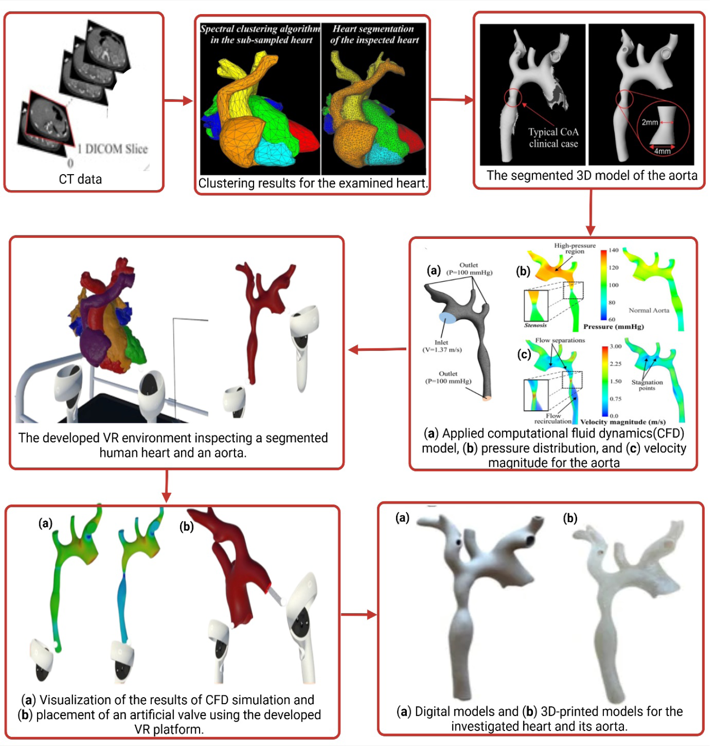
3D cardiac tissue bioprinting has the potential to revolutionize regenerative medicine by replicating complex myocardial architecture and constructing organ-mimetic cellular structures. This technology can enhance cardiac function without relying on transplants and allows for the development of physiologically accurate blood vessel networks (Murphy, Atala, 2014). Combining 3D bioprinting with stem cell technologies, such as induced pluripotent stem cells (iPSCs), allows for the fabrication of functional, patient-specific cardiac models. These models serve as accurate in vivo platforms for drug screening, disease modeling, and personalized treatment strategies. However, challenges remain, particularly regarding tissue thickness and vascularization. Efforts are ongoing to optimize bioinks, particularly hybrid bioinks, to ensure printability, biocompatibility, mechanical stability, biodegradability, and scalability. Four-dimensional (4D) bioprinting, an extension of 3D bioprinting, introduces dynamic constructs that change over time in response to stimuli, potentially leading to highly adaptive tissue systems for therapeutic applications. Advanced computational and imaging technologies are reshaping cardiac research and clinical practice. The integration of 3D-printed cardiovascular models with tools such as Computational Fluid Dynamics (CFD) and Virtual Reality (VR) offers novel insights into heart function, hemodynamics, and disease progression. CFD enables detailed simulation of blood flow and valve dynamics, while VR enhances visualization for surgical planning, medical education, and patient-specific diagnostics as shown in Figure 4 (Charalampous et al., 2023).
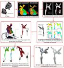
Steps for the 3D reconstruction process of heart and its models using computational fluid dynamics and constructing an VR environment. Figure is recreated from (Charalampous et al., 2023).
In conclusion, while cardiac bioprinting is still in its developmental stages, rapid progress in bioink formulation, printing technologies, and computational modeling paves the way for future clinical applications. These advancements are expected to revolutionize cardiac care through regenerative strategies, personalized therapies, and improved diagnostic tools
CONCLUSION
This review has systematically examined the recent advancements in 3D bioprinting for cardiac tissue engineering, drawing upon an extensive selection of studies published predominantly between 2016 and 2023. The cited literature spans a broad spectrum of experimental, technical, and review-based studies, with a strong emphasis on bioprinting techniques, cardiac tissue constructs, biomaterials, and vascularization strategies. Most works are rooted in in vitro and in vivo experimentation, reflecting the translational potential of current research. The selected studies were carefully curated based on relevance to cardiac tissue engineering, methodological robustness, and technological innovation, with a clear focus on stem cell-based constructs, scaffold design, and vascular integration. The inclusion of both foundational and cutting-edge research ensures a balanced representation of the field’s evolution. This comprehensive analysis underscores the trajectory toward clinically viable, functional cardiac tissues and highlights the need for continued integration of biomaterials science, stem cell biology, and bioprinting technologies to overcome remaining translational challenges.
-
FUNDING
This work is not supported by any fundings agency.
DATA SHARING STATEMENTS
The data can be made available on request to corresponding authors
DATA AVAILABILITY
No datasets were generated or analysed during the current study.
REFERENCES
-
Organ Procurement and Transplantation Network Modernization Initiative: Health Resources & Services Administration; 2023 [1:[Available from: https://www.organdonor.gov/
» https://www.organdonor.gov/ - Agarwal S, Saha S, Balla VK, Pal A, Barui A, Bodhak S. Current developments in 3D bioprinting for tissue and organ regeneration-A review. Front Mech Eng. 2020;6.
- Agarwal T, Fortunato GM, Hann SY, Ayan B, Vajanthri KY, Presutti D, et al. Recent advances in bioprinting technologies for engineering cardiac tissue. Mater Sci Eng. 2021;124(112057.
- Akpek AJJotFoE, University AoG. Analysis of biocompatibility characteristics of stereolithography applied three dimensional (3D) bioprinted artifical heart valves. J Fac Eng Archit Gazi Univ. 2018;33(3).
- Alonzo M, AnilKumar S, Roman B, Tasnim N, Joddar B. 3D Bioprinting of cardiac tissue and cardiac stem cell therapy. Transl Res. 2019;211:64-83.
- Arai K, Murata D, Verissimo AR, Mukae Y, Itoh M, Nakamura A, et al. Fabrication of scaffold-free tubular cardiac constructs using a Bio-3D printer. PLoS One. 2018;13(12):e0209162.
- Araña M, Gavira JJ, Peña E, González A, Abizanda G, Cilla M, et al. Epicardial delivery of collagen patches with adipose-derived stem cells in rat and minipig models of chronic myocardial infarction. Biomaterials. 2014;35(1):143-51.
- Axpe E, Oyen ML. Applications of Alginate-Based Bioinks in 3D Bioprinting. Int J Mol Sci. 2016;17(12).
- Budharaju H, Subramanian A, Sethuraman S. Recent advancements in cardiovascular bioprinting and bioprinted cardiac constructs. Biomater Sci. 2021;9(6):1974-94.
- Chan V, Jeong JH, Bajaj P, Collens M, Saif T, Kong H, et al. Multi-material bio-fabrication of hydrogel cantilevers and actuators with stereolithography. Lab Chip. 2012;12(1):88-98.
- Chan V, Park K, Collens MB, Kong H, Saif TA, Bashir RJSr. Development of miniaturized walking biological machines. Sci Rep. 2012;2(1):857.
- Charalampous P, Kladovasilakis N, Zoumaki M, Kostavelis I, Votis K, Petsios K, et al. Examination of a human heart fabricating its 3D-printed cardiovascular model and employing computational technologies. Appl Sci [Internet]. 2023;13(18).
- Chen XB, Fazel Anvari-Yazdi A, Duan X, Zimmerling A, Gharraei R, Sharma NK, et al. Biomaterials / bioinks and extrusion bioprinting. Bioact Mater. 2023;28(511-36.
- Colosi C, Shin SR, Manoharan V, Massa S, Costantini M, Barbetta A, et al. Microfluidic bioprinting of heterogeneous 3D tissue constructs using low-viscosity bioink. Adv Mater. 2016;28(4):677-84.
- Cui X, Dean D, Ruggeri ZM, Boland T. Cell damage evaluation of thermal inkjet printed Chinese hamster ovary cells. Biotechnol Bioeng. 2010;106(6):963-9.
- Delaney JT, Smith PJ, Schubert US. Inkjet printing of proteins. Soft Matter. 2009;5(24):4866-77.
- Duan B, Hockaday LA, Kang KH, Butcher JT. 3D bioprinting of heterogeneous aortic valve conduits with alginate/gelatin hydrogels. J Biomed Mater Res A. 2013;101(5):1255-64.
- Duan B, Kapetanovic E, Hockaday LA, Butcher JT. Three-dimensional printed trileaflet valve conduits using biological hydrogels and human valve interstitial cells. Acta Biomater. 2014;10(5):1836-46.
- Esser TU, Anspach A, Muenzebrock KA, Kah D, Schrüfer S, Schenk J, et al. Direct 3D-Bioprinting of hiPSC-Derived Cardiomyocytes to Generate Functional Cardiac Tissues. Adv Mater. 2023;35(52):e2305911.
- Gaebel R, Ma N, Liu J, Guan J, Koch L, Klopsch C, et al. Patterning human stem cells and endothelial cells with laser printing for cardiac regeneration. Biomaterials. 2011;32(35):9218-30.
- Garreta E, Oria R, Tarantino C, Pla-Roca M, Prado P, Fernández-Avilés F, et al. Tissue engineering by decellularization and 3D bioprinting. Materials Today. Tissue engineering by decellularization and 3D bioprinting. Mater Today. 2017;20(4):166-78.
- Ho CM, Mishra A, Lin PT, Ng SH, Yeong WY, Kim YJ, et al. 3D printed polycaprolactone carbon nanotube composite scaffolds for cardiac tissue engineering. Macromol Biosci. 2017;17(4).
- Hollister SJ. Porous scaffold design for tissue engineering. Nat Mater. 2005;4(7):518-24.
- Kabirian F, Ditkowski B, Zamanian A, Heying R, Mozafari M. An innovative approach towards 3D-printed scaffolds for the next generation of tissue-engineered vascular grafts. Mater Today: Proc. 2018;5
- Kačarević Ž P, Rider PM, Alkildani S, Retnasingh S, Smeets R, Jung O, et al. An Introduction to 3D Bioprinting: Possibilities, Challenges and Future Aspects. Materials (Basel). 2018;11(11).
- Kaully T, Kaufman-Francis K, Lesman A, Levenberg S. Vascularization--the conduit to viable engineered tissues. Tissue Eng Part B Rev. 2009;15(2):159-69.
- Kim SW, Kim DY, Roh HH, Kim HS, Lee JW, Lee KY. Three-Dimensional Bioprinting of Cell-Laden Constructs Using Polysaccharide-Based Self-Healing Hydrogels. Biomacromolecules. 2019;20(5):1860-6.
- Koch L, Deiwick A, Franke A, Schwanke K, Haverich A, Zweigerdt R, et al. Laser bioprinting of human induced pluripotent stem cells-the effect of printing and biomaterials on cell survival, pluripotency, and differentiation. Biofabrication. 2018;10(3):035005.
- Lee A, Hudson AR, Shiwarski DJ, Tashman JW, Hinton TJ, Yerneni S, et al. 3D bioprinting of collagen to rebuild components of the human heart. Science. 2019;365(6452):482-7.
- Lee RTJC. Adult cardiac stem cell concept and the process of science. Circulation. 2018;138(25):2940-2.
- Lee S, Serpooshan V, Tong X, Venkatraman S, Lee M, Lee J, et al. Contractile force generation by 3D hiPSC-derived cardiac tissues is enhanced by rapid establishment of cellular interconnection in matrix with muscle-mimicking stiffness. Biomaterials. 2017;131(111-20.
- Li W, Wang M, Ma H, Chapa-Villarreal FA, Lobo AO, Zhang YS. Stereolithography apparatus and digital light processing-based 3D bioprinting for tissue fabrication. iScience. 2023;26(2):106039.
- Liu W, Heinrich MA, Zhou Y, Akpek A, Hu N, Liu X, et al. Extrusion Bioprinting of Shear-Thinning Gelatin Methacryloyl Bioinks. Adv Healthc Mater. 2017;6(12).
- Liu Y, Zhang Y, Mei T, Cao H, Hu Y, Jia W, et al. hESCs-derived early vascular cell spheroids for cardiac tissue vascular engineering and myocardial infarction treatment. Adv Sci (Weinh). 2022;9(9):e2104299.
- Loureiro J, Miguel SP, Galván-Chacón V, Patrocinio D, Pagador JB, Sánchez-Margallo FM, et al. Three-dimensionally printed hydrogel cardiac patch for infarct regeneration based on natural polysaccharides. Polymers [Internet]. 2023;15(13).
- Martina M, Hutmacher DW. Biodegradable polymers applied in tissue engineering research: a review. Polym Int. 2007;56(2):145-57.
- McGill M, Holland GP, Kaplan DL. Experimental methods for characterizing the secondary structure and thermal properties of silk proteins. Macromol Rapid Commun. 2019;40(1):e1800390.
- Miller KL, Xiang Y, Yu C, Pustelnik J, Wu J, Ma X, et al. Rapid 3D BioPrinting of a human iPSC-derived cardiac micro-tissue for high-throughput drug testing. Organs-on-a-Chip. 2021;3:100007.
- Mir A, Lee E, Shih W, Koljaka S, Wang A, Jorgensen C, et al. 3D Bioprinting for vascularization. Bioengineering [Internet]. 2023; 10(5).
- Mistry P, Aied A, Alexander M, Shakesheff K, Bennett A, Yang J. Bioprinting using mechanically robust core-shell cell-laden hydrogel strands. Macromol Biosci. 2017;17(6).
- Murphy SV, Atala AJNb. 3D bioprinting of tissues and organs. Nature biotechnology. 3D bioprinting of tissues and organs. Nat Biotechnol. 2014;32(8):773-85.
- Murphy SV, Skardal A, Atala AJJoBMRPA. Evaluation of hydrogels for bio-printing applications. J Biomed Mater Res Part A. 2013;101(1):272-84.
- Nahmias Y, Schwartz RE, Verfaillie CM, Odde DJJB, Laser-guided direct writing for three7dimensional tissue engineering. Biotechnol Bioeng. 2005;92(2):129-36.
- Nakamura M, Iwanaga S, Henmi C, Arai K, Nishiyama YJB. Biomatrices and biomaterials for future developments of bioprinting and biofabrication. Biofabrication. 2010;2(1):014110.
- Noor N, Shapira A, Edri R, Gal I, Wertheim L, Dvir T. 3D Printing of personalized thick and perfusable cardiac patches and hearts. Adv Sci (Weinh). 2019;6(11):1900344.
- O’Bryan CS, Bhattacharjee T, Niemi SR, Balachandar S, Baldwin N, Ellison ST, et al. Three-dimensional printing with sacrificial materials for soft matter manufacturing. MRS Bulletin. 2017;42(8):571-7.
- Pajooh AMD, Tavakoli M, Al-Musawi MH, Karimi A, Salehi E, Nasiri-Harchegani S, et al. Biomimetic VEGF-loaded bilayer scaffold fabricated by 3D printing and electrospinning techniques for skin regeneration. Mater Des. 2024;238(112714.
- Polonchuk L, Gentile C. Current state and future of 3D bioprinted models for cardiovascular research and drug development. ADMET DMPK. 2021;9(4):231-42.
- Puricel S, Arroyo D, Corpataux N, Baeriswyl G, Lehmann S, Kallinikou Z, et al. Comparison of everolimusand biolimus-eluting coronary stents with everolimus-eluting bioresorbable vascular scaffolds. J Am Coll Cardiol. 2015;65(8):791-801.
- Rioux Y, Fradette J, Maciel Y, Bégin-Drolet A, Ruel J. Biofabrication of sodium alginate hydrogel scaffolds for heart valve tissue engineering. Int J Mol Sci. 2022;23(15).
- Ronaldson-Bouchard K, Ma SP, Yeager K, Chen T, Song L, Sirabella D, et al. Advanced maturation of human cardiac tissue grown from pluripotent stem cells. Nature. 2018;556(7700):239-43.
- Samson AAS, Song JMJAMT. Scaffold-free 3D printing for fabrication of biomimetic branched multinucleated cardiac tissue construct: a promising ex vivo model for in situ detection of drug-induced sodium ion channel responses. Appli Mater Today. 2022;27(101416.
- Sanz-Garcia A, Oliver-de-la-Cruz J, Mirabet V, Gandía C, Villagrasa A, Sodupe E, et al. Heart valve tissue engineering: how far is the bedside from the bench? Expert Rev Mol Med. 2015;17(e16.
- Schuurman W, Levett PA, Pot MW, van Weeren PR, Dhert WJ, Hutmacher DW, et al. Gelatin-methacrylamide hydrogels as potential biomaterials for fabrication of tissue-engineered cartilage constructs. Macromol Biosci. 2013;13(5):551-61.
- Serpooshan V, Zhao M, Metzler SA, Wei K, Shah PB, Wang A, et al. Use of bio-mimetic three-dimensional technology in therapeutics for heart disease. Bioengineered. 2014;5(3):193-7.
- Skylar-Scott MA, Uzel SGM, Nam LL, Ahrens JH, Truby RL, Damaraju S, et al. Biomanufacturing of organ-specific tissues with high cellular density and embedded vascular channels. Sci Adv. 2019;5(9):eaaw2459.
- Takahashi K, Yamanaka S. Induction of pluripotent stem cells from mouse embryonic and adult fibroblast cultures by defined factors. Cell. 2006;126(4):663-76.
- Uccheddu F, Carfagni M, Governi L, Furferi R, Volpe Y, Nocerino 3D printing of cardiac structures from medical images: an overview of methods and interactive tools. Int J Interact Design Manuf (IJIDeM). 2018;12
- Virani SS, Alonso A, Aparicio HJ, Benjamin EJ, Bittencourt MS, Callaway CW, et al. Heart Disease and Stroke Statistics-2021 Update: A Report From the American Heart Association. Circulation. 2021;143(8):e254-e743.
- Wadey K, Lopes J, Bendeck M, George S. Role of smooth muscle cells in coronary artery bypass grafting failure. Cardiovasc Res. 2018;114(4):601-10.
- Wang D, Maharjan S, Kuang X, Wang Z, Mille LS, Tao M, et al. Microfluidic bioprinting of tough hydrogel-based vascular conduits for functional blood vessels. Sci Adv. 2022;8(43):eabq6900.
- Wang Z, Lee SJ, Cheng HJ, Yoo JJ, Atala A. 3D bioprinted functional and contractile cardiac tissue constructs. Acta Biomater. 2018;70(48-56.
- Wang Z, Wang L, Li T, Liu S, Guo B, Huang W, et al. 3D bioprinting in cardiac tissue engineering. Theranostics. 2021;11(16):7948-69.
- Won J-E, Moon HK, Kim H-W, Shim JS. Development of multi-layered three-dimensional printed scaffold for sequential delivery of biomolecules. Int J Bioprinting. 2024.
- Wu CA, Zhu Y, Woo YJ. Advances in 3D Bioprinting: Techniques, Applications, and Future Directions for Cardiac Tissue Engineering. Bioengineering [Internet]. 2023; 10(7).
- Wüst S, Godla ME, Müller R, Hofmann S. Tunable hydrogel composite with two-step processing in combination with innovative hardware upgrade for cell-based three-dimensional bioprinting. Acta Biomater. 2014;10(2):630-40.
- Xu C, Chai W, Huang Y, Markwald RR. Scaffold-free inkjet printing of three-dimensional zigzag cellular tubes. Biotechnol Bioeng. 2012;109(12):3152-60.
- Xu T, Binder KW, Albanna MZ, Dice D, Zhao W, Yoo JJ, et al. Hybrid printing of mechanically and biologically improved constructs for cartilage tissue engineering applications. Biofabrication. 2013;5(1):015001.
- Yu C, Miller KL, Schimelman J, Wang P, Zhu W, Ma X, et al. A sequential 3D bioprinting and orthogonal bioconjugation approach for precision tissue engineering. Biomaterials. 2020;258(120294.
- Yu Y, Zhang Y, Martin JA, Ozbolat IT. Evaluation of cell viability and functionality in vessel-like bioprintable cell-laden tubular channels. J Biomech Eng. 2013;135(9):91011.
- Zacchigna M, Cateni F, Drioli S, Bonora GM. Multimeric, Multifunctional Derivatives of Poly(ethylene glycol). Polymers [Internet]. 2011;3(3):[1076-90 pp.].
- Zhang J, Hu Q, Wang S, Tao J, Gou M. Digital Light Processing Based Three-dimensional Printing for Medical Applications. Int J Bioprint. 2020;6(1):242.
- Zhang Y, Yu Y, Akkouch A, Dababneh A, Dolati F, Ozbolat IT. In Vitro Study of Directly Bioprinted Perfusable Vasculature Conduits. Biomater Sci. 2015;3(1):134-43.
- Zhang YS, Arneri A, Bersini S, Shin SR, Zhu K, Goli-Malekabadi Z, et al. Bioprinting 3D microfibrous scaffolds for engineering endothelialized myocardium and heart-on-a-chip. Biomaterials. 2016;110:45-59.
- Zhu K, Shin SR, van Kempen T, Li YC, Ponraj V, Nasajpour A, et al. Gold Nanocomposite Bioink for Printing 3D Cardiac Constructs. Adv Funct Mater. 2017;27(12).
Edited by
-
Associate Editor:
Ruy Beck