Antimicrobial resistance and arsenic tolerance in Enterococcus spp. isolated from free-ranging Leopardus geoffroyi in Candiota, Pampa Biome, Brazil: sentinel indicators of environmental pollution
Abstract
The Pampa Biome harbors a high diversity of endemic species but has experienced substantial pressure from anthropogenic activities that threaten wildlife conservation, including Geoffroy’s cat (Leopardus geoffroyi). Within this context, this study evaluated antimicrobial-resistant and heavy-metal-tolerant enterococci isolated from oral and rectal cavities of 14 free-ranging Geoffroy’s cats (L. geoffroyi) from Seival–Candiota region in Brazilian Pampa as indicators of environmental quality. Isolation was conducted on selective media, species identification was performed using MALDI-TOF mass spectrometry, and antimicrobial susceptibility was assessed by disk diffusion against 12 antimicrobials used in human and veterinary medicine. Antimicrobial resistance genes (msrC, ermB, tetM, and tetL) and heavy-metal tolerance genes (arsA_I, arsA_II, and tcrB) were investigated by PCR. A total of 111 enterococcal isolates were recovered. Enterococcus faecium (37.8 %) and Enterococcus faecalis (29.7 %) predominated, showing distinct distribution between oral and rectal samples. Overall, 74.8 % of isolates were resistant to at least one antimicrobial, mainly rifampicin (41.4 %), erythromycin (34.2 %), and ciprofloxacin (26.1 %). No resistance to gentamicin or vancomycin was detected. Multidrug resistance occurred in 17 isolates (20.5 %). Resistance determinants (msrC, ermB, tetM, and tetL) and arsenic tolerance gene arsA_I were identified, whereas arsA_II and tcrB were not detected. Findings demonstrate the presence of multidrug-resistant and arsenic-tolerant enterococci in free-ranging Geoffroy’s cats, supporting their role as sentinel species of environmental quality and evidencing the influence of anthropogenic activities on animal health.
Keywords:
Anthropogenic impact; environmental bioindicators; wildlife conservation; one health
Resumo
O bioma Pampa abriga uma notável diversidade de espécies endêmicas, mas vem sofrendo intensos impactos decorrentes de atividades antrópicas, que ameaçam a conservação de espécies silvestres, tais como o gato-do-mato-grande (Leopardus geoffroyi). Nesse contexto, este estudo avaliou a resistência a antimicrobianos e a tolerância a metais pesados em enterococos isolados das cavidades oral e retal de 14 gatos-do-mato-grande (L. geoffroyi) de vida livre da região do Seival/Candiota, no bioma Pampa, como bioindicadores da qualidade ambiental. O isolamento foi realizado em meios seletivos, a identificação das espécies foi feita por MALDI-TOF, e a suscetibilidade foi determinada pelo método de disco-difusão frente a 12 antimicrobianos de uso clínico e veterinário. Genes de resistência a antimicrobianos (msrC, ermB, tetM e tetL) e de tolerância a metais pesados (arsA_I, arsA_II e tcrB) foram investigados por PCR. Um total de 111 isolados de enterococos foi recuperado. Enterococcus faecium (37,8 %) e Enterococcus faecalis (29,7 %) predominaram, apresentando distribuição distinta entre amostras orais e retais. No geral, 74,8 % dos isolados apresentaram resistência a pelo menos um antimicrobiano, principalmente à rifampicina (41,4 %), à eritromicina (34,2 %) e à ciprofloxacina (26,1 %). Não foi detectada resistência à gentamicina nem à vancomicina. A resistência a múltiplos antimicrobianos foi observada em 17 isolados (20,5 %). Determinantes de resistência (msrC, ermB, tetM e tetL) e o gene de tolerância ao arsênio (arsA_I) foram identificados, enquanto arsA_II e tcrB não foram detectados. Em conclusão, a presença de enterococos resistentes a múltiplos antimicrobianos e tolerantes ao arsênio em gatos-do-mato-grande de vida livre reforça seu uso como bioindicadores da qualidade ambiental e evidencia o impacto das atividades antrópicas na saúde animal.
Palavras-chave:
Impactos antropogênicos; bioindicadores ambientais; conservação da vida silvestre; saúde única
1. Introduction
The Pampa Biome represents valuable natural and cultural heritage because of its unique biodiversity and ecosystem services. Covering approximately 700,000 km2, this grassland formation extends across southern Brazil, Argentina, and Uruguay (1). In Brazil, the biome occurs exclusively in Rio Grande do Sul State, occupying about 193,836 km2, which corresponds to 69 % of the state's territory and 2.3 % of the national territory (2). The Pampa holds high environmental relevance, as it supports a rich diversity of endemic fauna, including more than 500 bird species and over 100 terrestrial mammal species (3). Despite its importance for biodiversity conservation, the biome has experienced substantial loss of native vegetation over the years. Estimates indicate that original vegetation cover declined from 41.32 % in 2002 to 36.03 % in 2008 (4). Accelerated landscape degradation, mainly driven by agricultural expansion (soybean and rice cultivation) and industrial development (mining and pulp production), has resulted in marked reductions in native fauna and flora.
Within this context, impacts of anthropogenic activities on Pampa ecosystems have been widely documented and monitored (5), reinforcing the urgency of conservation initiatives. The Pampa’s Felines Project was therefore established to evaluate the effects of human pressures on wild felids, operating in collaboration with Geoffroy’s Cat Working Group, an international research and conservation network (6). Project activities are concentrated in Brazilian Pampa, where Geoffroy’s cat (Leopardus geoffroyi) represents one of the flagship species. Although globally classified as Least Concern (7), this species is considered Vulnerable at national (8) and regional levels (9). Major threats include habitat fragmentation, which increases exposure to human-modified landscapes and heightens risks of road mortality, retaliatory hunting, and pathogen transmission from domestic animals (10–12).
Beyond direct conservation threats, detection of subtle environmental impacts on host microbiota represents an additional critical dimension. In this sense, gut microbial communities may reflect host physiological status and environmental pressures, thereby functioning as indicators of ecosystem disturbance and anthropogenic influence (13,14). Among microorganisms used as bioindicators, genus Enterococcus has received particular attention (15–21). Enterococcus species are considered commensal inhabitants of gastrointestinal tract, also being found in the genitourinary tract and genitourinary tracts and oral cavity of humans and animals, but are also widely distributed in environmental matrices such as soil, water, and food (22–28). Molecular and phylogenetic evidence indicates that the genus comprises more than 90 species; among these, E. faecalis, E. faecium, E. hirae, E. durans, E. casseliflavus, E. gallinarum, and E. mundtii are frequently detected in the animal gastrointestinal microbiota (29). In recent years, increasing attention has been directed toward characterization of enterococcal diversity, antimicrobial resistance profiles, and heavy-metal tolerance in isolates obtained from wildlife. These studies have emphasized the potential of enterococci as sentinel organisms for monitoring animal and ecosystem health in potentially contaminated environments (23–28,30–33).
Moreover, despite the ecological importance of Pampa ecosystems and conservation concerns surrounding species such as Geoffroy’s cat (L. geoffroyi), studies characterizing intestinal and oral microbiota from an environmental bioindicator perspective remain limited. Therefore, understanding this interface is essential for elucidating the consequences of human pressures and for supporting conservation strategies aligned with One Health principles. Based on the above, the present study aimed to characterize antimicrobial susceptibility and heavy-metal tolerance in Enterococcus spp. isolated from oral and rectal cavities of free-ranging Geoffroy’s cats captured in the Seival region, in the municipality of Candiota, within the Brazilian Pampa. Ultimately, it also seeks to explore their applicability as bioindicators of environmental quality in natural habitats occupied by this species.
2. Material and methods
2.1 Study site – the Brazilian Pampa biome: municipality of Candiota
The municipality of Candiota is in southern Rio Grande do Sul State, approximately 387 km from the capital, Porto Alegre. It covers a total area of 933.628 km2 and has an estimated population of 10,710 inhabitants, corresponding to a population density of 11.47 inhabitants per km2 (34). Of this area, about 73,234.754 hectares are used for agricultural activities, representing 78.4 % of the municipal territory. Local economic activity is strongly influenced by thermoelectric power generation, supported by extensive coal reserves in underlying geological formations. The municipality currently hosts three major mining and thermoelectric enterprises, in addition to cement production facilities associated with one of the largest global industrial conglomerates in this sector. These activities position Candiota as a strategic energy production hub at national level. Livestock production and crop farming also contribute substantially to local economy, particularly soybean and rice cultivation (35).
2.2 Sampling and collection of biological material from L. geoffroyi
Fourteen free-ranging Geoffroy’s cats (L. geoffroyi) were captured between June 2022 and February 2023 in the Seival region, a district of the municipality of Candiota, Rio Grande do Sul State, Brazil (Supplementary Table 1). Capture procedures were conducted within framework of the Pampa Felines Project and followed methodological protocols described by Tirelli et al (12), as well as guidelines for handling wild mammals proposed by Sikes et al (36)
Capture and sampling activities were authorized by Chico Mendes Institute for Biodiversity Conservation (ICMBio) under license ICMBio/SISBIO-81869-2 for strictly scientific purposes. All animal procedures were previously reviewed and approved by Animal Use Ethics Committee of the Federal University of Rio Grande do Sul (CEUA-UFRGS), according to protocol CEUA/UFRGS-42867.
Oral and rectal swab samples were collected from each individual, immediately placed in Stuart transport medium, and transported under refrigerated conditions (4 °C) to Wildlife Microbiology Laboratory at Institute of Basic Health Sciences, UFRGS. Samples were maintained under refrigeration until microbiological analyses were performed.
2.3 Isolation and phenotypic characterization of Enterococcus spp.
For selective isolation of bacteria belonging to genus Enterococcus, oral cavity (OC) and rectal cavity (RC) swabs were immersed in tubes containing 3 mL of Azide Dextrose Broth (HiMedia®, India) and incubated at 37 °C for 24 h. Subsequently, serial decimal dilutions were prepared in sterile saline solution (0.85 % NaCl). Aliquots of 100 µL from 10−4 and 10−5 dilutions were spread onto Brain Heart Infusion (BHI) agar supplemented with 4.5 % NaCl using surface-spreading technique and incubated at 37 °C for 24 h. After incubation, six colony-forming units (CFU) were randomly selected and subcultured onto Bile Esculin agar plates, followed by incubation under same conditions. Colonies showing esculin hydrolysis, evidenced by medium darkening, were subsequently subcultured onto BHI agar and incubated at 37 °C for 24 h for further analyses. Selected cultures were subjected to phenotypic characterization based on morphophysiological tests, including Gram staining and catalase activity assay, for presumptive identification of Enterococcus spp. Isolates exhibiting phenotypic characteristics associated with the genus were stored in cryotubes containing 1 mL of preservation medium composed of 10% skim milk and 10% glycerol at −20 °C for subsequent molecular identification at the species level (24, 27).
2.4 Identification of Enterococcus species
Species identification was performed using matrix-assisted laser desorption/ionization time-of-flight mass spectrometry (MALDI-TOF MS; Bruker Daltonik GmbH), following procedures described by Sauget et al (37) To differentiate E. gallinarum and E. casseliflavus, species characterized by low-level intrinsic resistance to vancomycin, two complementary biochemical assays were conducted as previously reported by Cartwright et al (38). For pigment production assessment, a colony from each isolate was collected with a sterile swab and examined for yellow pigmentation, which is indicative of E. casseliflavus. Motility testing was additionally performed by inoculating a straight stab into semi-solid Brain Heart Infusion (BHI) agar culture to approximately two-thirds of the medium depth, followed by incubation at 37 °C for 24 h. Under these conditions, E. casseliflavus typically exhibits motility, whereas E. mundtii shows limited or absent motility, thereby enabling reliable differentiation between these species.
2.5 Determination of antimicrobial susceptibility profile
Antimicrobial susceptibility of Enterococcus isolates was assessed using the disk diffusion method on Mueller–Hinton agar plates, following the protocol standardized Kirby–Bauer et al (39) For each isolate, a bacterial suspension was prepared in 5 mL of sterile saline solution (0.85 % NaCl), and turbidity was adjusted to 0.5 McFarland standard (approximately 1.5 × 10⁸ CFU/mL). Standardized inoculum were uniformly spread onto Mueller–Hinton agar plates (Acumedia®, Neogen, Michigan, USA) using sterile swabs. Antimicrobial-impregnated disks were subsequently applied to agar medium surface, and the plates were incubated at 37 °C for 24 h.
After incubation, inhibition zone diameters were measured in millimeters and interpreted according to Clinical and Laboratory Standards Institute (CLSI) criteria(40), Isolates were categorized as susceptible (S), intermediate (I), or resistant (R). Tested antimicrobials included ampicillin (10 µg), vancomycin (30 µg), rifampicin (5 µg), tetracycline (30 µg), ciprofloxacin (5 µg), norfloxacin (10 µg), chloramphenicol (30 µg), nitrofurantoin (300 µg), linezolid (10 µg), erythromycin (15 µg), gentamicin (120 µg), and streptomycin (300 µg), in accordance with CLSI 2021 recommendations.
For analytical purposes, isolates classified as intermediate or resistant were grouped into a single resistant category. Resistance phenotypes were defined as single-drug resistance (SR), double-drug resistance (DR), or multidrug resistance (MDR) when isolates exhibited resistance to one, two, or three or more antimicrobial classes, respectively (41).
2.6 Genomic DNA extraction and detection of genes associated with antimicrobial and heavy-metal resistance
Genomic DNA from Enterococcus isolates was extracted by chemical lysis following protocol described by Bell et al (42). Extracted DNA was stored at −20 °C until use in polymerase chain reaction (PCR) assays for detection of genes associated with antimicrobial resistance and heavy-metal tolerance. PCR conditions were based on previously standardized parameters, as showed in Table 1.
The oligonucleotide primers and the PCR parameters used to detect genes associated with arsenic, copper, erythromycin, and tetracycline in Enterococcus spp.
Detection of antimicrobial resistance genes was performed in isolates phenotypically resistant to erythromycin and tetracycline. These antimicrobial agents are widely used in human and veterinary medicine, and resistance mechanisms in enterococci are frequently associated with mobile genetic elements. PCR was performed on these strains to detect the erythromycin resistance genes ermB and msrC, as well as tetracycline resistance genes tetL and tetM. Enterococcus spp. strain 485 and E. faecium CM5-2 were used as positive controls for ermB and msrC, respectively. E. faecium CM11-3 and CM5-6 served as positive controls for tetL and tetM detection.
To investigate genes associated with heavy-metal tolerance, all isolates were screened for presence of arsA_I and arsA_II, which participate in arsenic resistance, and for tcrB, a determinant linked to copper resistance. E. faecalis ATCC 29212 was used as positive control for arsA_I and tcrB, whereas Listeria monocytogenes ALD11249.1 was employed as positive control for arsA_II detection (21).
3. Results and discussion
To evaluate potential effects of anthropogenic activities on populations of free-ranging Geoffroy’s cats (L. geoffroyi) captured in the Seival region, municipality of Candiota, within Pampa Biome, enterococci isolated from oral and rectal cavities were used as bioindicators.
3.1 Distribution of Enterococcus species in oral and rectal cavities of L. geoffroyi
In this study, a total of 111 Enterococcus spp. isolates were recovered from oral (OC) and rectal (RC) samples obtained from 14 free-ranging Geoffroy’s cats captured in the Seival region between June 2022 and February 2023. From each sample, an average of three to four colonies showing morphology consistent with genus Enterococcus was selected for further characterization. Of total isolates, 50 (45 %) originated from oral swabs and 61 (55 %) from rectal swabs (Supplementary Table 2).
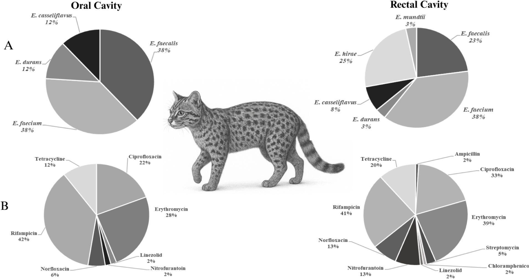
Among identified species, E. faecium (37.8 %; n = 42) and E. faecalis (29.7 %; n = 33) predominated, followed by E. hirae (13.5 %; n = 15), E. casseliflavus (9.9 %; n = 11), E. durans (7.2 %; n = 8), and E. mundtii (1.8 %; n = 2). Species distribution varied according to anatomical sampling site. Oral samples were mainly composed of E. faecalis and E. faecium, whereas rectal samples showed greater species diversity, including higher frequency of E. hirae and exclusive detection of E. mundtii (Figure 1).
Distribution of Enterococcus spp. species. (a) and antimicrobial resistance profile (b) in oral and rectal samples from free-ranging Geoffroy’s cats (Leopardus geoffroyi) captured in the Seival region, municipality of Candiota, Pampa Biome, Brazil, between June 2022 and February 2023.
Diversity of enterococcal species identified in this study appears to reflect the dietary habits of this felid, which primarily preys on small mammals and small birds (48–52). High prevalence of E. faecalis may be associated with rodent consumption, as Lauková et al (53) reported occurrence rates of approximately 70 % for this species in the intestinal microbiota of wild rodents. Similarly, the occurrence of E. faecalis, E. faecium, E. mundtii, E. durans, and E. hirae may be related to avian prey intake, since these species are frequently reported in gut microbiota of wild birds (54) and broiler chickens (55). Overall, enterococcal profile observed in free-ranging Geoffroy’s cats suggests that dietary ecology plays a significant role in shaping colonization patterns of oral and intestinal microbiota.
3.2 Resistance profile of enterococcal isolates from L. geoffroyi
Among 111 isolates evaluated for susceptibility to tested antimicrobials, 83 (74.8 %) exhibited resistance to at least one agent, as detailed in Table 2. Highest resistance frequencies were observed for rifampicin (41.4 %), erythromycin (34.2 %), and ciprofloxacin (26.1 %). Lower resistance rates were recorded for tetracycline (13.5 %), norfloxacin (9.9 %), nitrofurantoin (4.5 %), streptomycin (2.7 %), linezolid (1.8 %), ampicillin (0.9 %), and chloramphenicol (0.9 %). No isolate showed resistance to gentamicin or vancomycin.
Number (%) of antimicrobial resistant Enterococcus spp. isolated from oral and rectal cavities of free-ranging Geoffroy’s cats captured (Leopardus geoffroyi) in the Seival region, municipality of Candiota, Pampa Biome, Brazil, between June 2022 and February 2023.
Enterococci isolated from rectal samples showed greater frequency and complexity of resistance compared with those isolated oral samples. In isolates from oral samples, rifampicin displayed highest resistance rate (42 %), followed by erythromycin (28 %) and ciprofloxacin (22 %). In rectal isolates, resistance frequencies were higher for rifampicin (41 %), erythromycin (39 %), and ciprofloxacin (33 %). Tetracycline resistance occurred at moderate levels in both anatomical sites but was more frequent in rectal isolates (20 % vs. 12 %). Resistance to norfloxacin, linezolid, and nitrofurantoin remained relatively low in both groups, although slightly higher in rectal isolates (Figure 1b). Species such as E. faecium, E. faecalis, E. durans, and E. casseliflavus contributed differentially to these resistance patterns, indicating that rectal microbiota harbored enterococci with higher resistance frequency and broader diversity of multidrug-resistant phenotypes than oral microbiota (Supplementary Tables 3 and 4).
These findings demonstrate that enterococci isolated from oral and rectal cavities of free-ranging Geoffroy’s cats exhibit resistance to antimicrobials widely used in human clinical practice and veterinary medicine, particularly ciprofloxacin, erythromycin, rifampicin, and tetracycline (56–58). The presence of resistant strains in wildlife populations not subjected to antimicrobial treatment suggests indirect acquisition through diet or environmental exposure.
Therefore, detection of resistant enterococci in free-ranging Geoffroy’s cats samples likely reflect contact with anthropogenic pressures within their habitats. The animals studied occur in areas exposed to such activities, including livestock production that may release antimicrobial residues into soil and water bodies, promoting selection and dissemination of resistant bacteria. These observations highlight the influence of human-modified environments on wildlife microbiota and reinforce the value of enterococci as sentinel indicators of environmental quality within integrated One Health surveillance frameworks (58-60).
Among the 83 resistant isolates, 45 (54.2 %) exhibited single-drug resistance (SR), 21 (25.3 %) double-drug resistance (DR), and 17 (20.5 %) multidrug resistance (MDR). Rectal isolates showed higher proportion of MDR phenotypes compared with oral isolates (Figure 2). The presence of MDR enterococci may be associated with ecological overlap between wildlife and human activities, as wild felids and canids often share anthropized environments. Previous studies have reported MDR enterococci in rectal swabs of wild cats from the Pampa Biome and in clinical infections of domestic cats hospitalized in southern Brazil (27, 56). These findings reinforce concerns regarding the selection and dissemination of multidrug-resistant bacteria among human and animal populations.
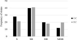
Antimicrobial-resistance profiles of enterococci isolated from oral cavity (OC) and rectal cavity (RC) samples of free-ranging Geoffroy’s cats (Leopardus geoffroyi) captured in the Seival region, municipality of Candiota, Pampa Biome, Brazil, between June 2022 and February 2023. S, susceptible; SR, single-drug resistance; DR, double-drug resistance; MDR, multidrug resistance.
Influence of human activity on occurrence of resistant Enterococcus spp. in wildlife has become increasingly evident, highlighting association between anthropogenic pressure and dissemination of resistance mechanisms. Heck et al (28) evaluated enterococci isolated from free-ranging and captive snakes and reported that animals without human contact were susceptible to all antimicrobials tested. In contrast, isolates obtained from captive snakes exhibited resistance to multiple antimicrobial classes. Similarly, another study reported higher frequency of resistant strains in samples from free-ranging capuchin monkeys (Sapajus nigritus) frequently exposed to human activity, suggesting that anthropized environments may favor selection and spread of these microorganisms (23).
3.3 Detection of genes associated with resistance to tetracycline (tetL and tetM) and erythromycin (msrC and ermB), and tolerance to arsenic (arsA_I and arsA_II) and copper (tcrB)
After antimicrobial susceptibility testing of 111 enterococcal isolates, strains recovered from the same host frequently exhibited similar susceptibility patterns, suggesting possible clonal relatedness. To avoid overestimation in the analyses of antimicrobial resistance and heavy-metal tolerance genes, 81 isolates displaying distinct phenotypic profiles were selected, including 41 from rectal cavity (RC) and 40 from oral cavity (OC).
For detection of resistance determinants, 30 erythromycin-resistant and 18 tetracycline-resistant isolates were subjected to conventional PCR targeting ermB and msrC (erythromycin resistance), and tetL and tetM (tetracycline resistance) (Table 3). Among the erythromycin-resistant isolates, 13 presented resistance genes, of which 12 (40.0 %) were positive for msrC, and one isolate (3.3 %) carried both ermB and msrC genes. Of the msrC-positive isolates, 12 (92.3 %) were identified as E. faecium and one (7.7 %) as E. durans. The isolate harboring both genes was identified as E. faecium and originated from oral cavity. The presence of msrC gene in enterococci from wildlife has been previously reported (23,2) and the literature describes this determinant as species-associated with E. faecium (61), confirming the hypothesis raised in this study, in which 12 samples positive for the gene were from the species. Conversely, 17 erythromycin-resistant isolates (56.6 %) showed no amplification for the genes investigated here, suggesting involvement of additional resistance determinants not evaluated in this study.
Detection of genes conferring resistance to tetracycline, erythromycin, and arsenic in enterococci isolates obtained from oral and rectal cavities of free-ranging Geoffroy’s cats (Leopardus geoffroyi) captured in the Seival region, municipality of Candiota, Pampa Biome, Brazil, between June 2022 and February 2023.
Among the 18 tetracycline-resistant isolates, 5 (27.7 %) were positive for tetM, one (5.5 %) for tetL, and 8 (44.4 %) harbored both genes (Table 3). The tetM gene was most frequently detected in E. faecium (n = 9), followed by E. durans (n = 2), E. faecalis (n = 1), and E. casseliflavus (n = 1), whereas tetL was identified in E. faecium (n = 5), E. durans (n = 3), and E. casseliflavus (n = 1). Four isolates were negative for targeted genes, indicating potential presence of alternative tetracycline resistance mechanisms in these strains.
Detection frequencies of tetL and tetM are consistent with previous investigations reporting these determinants in enterococci from wildlife populations, including study by Araújo (27). The high prevalence of tet resistance genes may be explained by their frequent association with plasmids and mobile or conjugative genetic elements, which facilitate horizontal transfer among bacterial populations (61, 62).
All isolates were additionally PCR-screened for arsA_I and arsA_II, genes associated with arsenic tolerance (Table 3). Thirty-six isolates (44.4 %) were positive for arsA_I, whereas none showed amplification of arsA_II. Screening for tcrB, a determinant linked to copper resistance, yielded negative results in all isolates.
The presence of arsA_I among enterococci recovered from free-ranging felids suggests that arsenic-tolerant microorganisms may be selected under environmental contamination pressures in this habitat. Mining activities and intensive agriculture, both prevalent in study region, represent potential sources of arsenic release into terrestrial and aquatic ecosystems (63). Supporting this interpretation, Mocellin et al (33) reported association between presence of arsA genes in enterococci isolated from seabirds of Abrolhos Archipelago and elevated concentrations of arsenic and other metals (Fe, Mn, Cd, and Pb) detected in blood and/or plumage following Fundão dam disaster. These findings reinforce hypothesis that the occurrence of arsenic tolerance genes in environmental enterococci reflects selective pressures exerted by metal exposure through food webs and/or contaminated habitats.
4. Conclusion
Antimicrobial resistant enterococci were isolated from oral and rectal cavities of free-ranging Geoffroy’s cats (Leopardus geoffroyi) captured in the Seival region, municipality of Candiota, in the Pampa Biome, Rio Grande do Sul State, Brazil. These isolates harbored resistance determinants associated with tetracycline and erythromycin (msrC, tetL, and tetM), as well as arsenic tolerance gene arsA_I. The detection of these strains indicates exposure of wild felids to environments influenced by anthropogenic activities. Thus, this study reinforces the role of enterococci as environmental bioindicators, emphasizing their significance in understanding the interface among animal health, human health, and ecosystem conservation.
Supplementary material
(available only in the online version: https://revistas.ufg.br/vet/article/view/84024)
Supplementary PDF
Table S1. Biological and capture information for free-ranging Geoffroy’s cats (Leopardus geoffroyi) included in the study
Table S2. Distribution of Enterococcus spp. among oral and rectal samples of free-ranging Geoffroy’s cats captured in the Seival region, municipality of Candiota, Pampa Biome, Brazil, between June 2022 and February 2023
Table S3. Number (%) of antimicrobial resistant enterococci isolates recovered from oral cavity samples of free-ranging Geoffroy’s cats captured in the Seival region, municipality of Candiota, Pampa Biome, Brazil, between June 2022 and February 2023
Table S4. Number (%) of antimicrobial resistant enterococci isolates recovered from rectal cavity samples of free-ranging Geoffroy’s cats captured in the Seival region, municipality of Candiota, Pampa Biome, Brazil, between June 2022 and February 2023.
Data availability statement
The complete dataset supporting the results of this study is available in the article and in the Supplementary Material section.
-
Generative AI Use Statement
During the preparation of this manuscript, the authors used ChatGPT to generate the image of the wildcat presented in Figure 1. After using this tool/service, the authors reviewed and edited the content as appropriate and assume full responsibility for the content of this publication.
Acknowledgements
The authors thank the National Council for Scientific and Technological Development (CNPq), the Coordination for the Improvement of Higher Education Personnel (CAPES), Brazil, the Federal University of Rio Grande do Sul (UFRGS), Porto Alegre, Brazil, and the Pampa Wildcats Project.
References
-
1. Bilenca D, Miñarro F. Identificación de Áreas Valiosas de Pastizal (AVPs) en las Pampas y campos de Argentina, Uruguay y sur de Brasil. Argentina; 2004. Available from: http://awsassets.wwfar.panda.org/downloads/libro_avps__bilenca_y_minarro_2004_.pdf
» http://awsassets.wwfar.panda.org/downloads/libro_avps__bilenca_y_minarro_2004_.pdf -
2. Instituto Brasileiro de Geografia e Estatística (IBGE). Biomas e sistema costeiro-marinho do Brasil. Rio de Janeiro: IBGE; 2019. Available from: https://biblioteca.ibge.gov.br/visualizacao/livros/liv101676.pdf
» https://biblioteca.ibge.gov.br/visualizacao/livros/liv101676.pdf -
3. Andrade BO, Dröse W, Aguiar CAD, et al. 12,500+ and counting: biodiversity of the Brazilian Pampa. Front Biogeogr 2023 15.2, e59288. Available from: https://doi.org/10.21425/F5FBG59288
» https://doi.org/10.21425/F5FBG59288 -
4. Centro De Sensoriamento Remoto (CRS), Instituto Nacional De Pesquisas Espaciais (INPE). Relatório técnico do monitoramento do desmatamento do Bioma Pampa 2008-2009. Brasília: Ministério do Meio Ambiente (MMA); 2010. Available from: https://antigo.mma.gov.br/estruturas/sbf_chm_rbbio/_arquivos/relatrio_tcnico_monitoramento_pampa_2008_2009_72.pdf
» https://antigo.mma.gov.br/estruturas/sbf_chm_rbbio/_arquivos/relatrio_tcnico_monitoramento_pampa_2008_2009_72.pdf -
5. Ziliotto M, Kulmann-Leal B, Roitman A, et al. Pesticide pollution in the Brazilian Pampa: Detrimental impacts on ecosystems and human health in a neglected biome. Pollutants 2023;3(2):280–292. Available from: https://doi.org/10.3390/pollutants3020020
» https://doi.org/10.3390/pollutants3020020 -
6. Stockwell S. Working in Brazilian wildcat conservation with Flavia Tirelli 2023; Available from: Available from: https://www.conservation-careers.com/interviews/working-in-brazilian-wildcat-conservation-with-flavia-tirelli/
» https://www.conservation-careers.com/interviews/working-in-brazilian-wildcat-conservation-with-flavia-tirelli/ -
7. International Union For Conservation Of Nature (IUCN). The IUCN Red List of Threatened Species. Version 2025-1. 2025. Available from: https://www.iucnredlist.org/
» https://www.iucnredlist.org/ -
8. Brasil. Ministério do Meio Ambiente. Portaria MMA no. 148, de 7 de junho de 2022. Lista nacional oficial de espécies da fauna ameaçadas de extinção. Brasília, DF: Diário Oficial da União, 2022. Available from: https://in.gov.br/en/web/dou/-/portaria-mma-n-148-de-7-de-junho-de-2022-406272733
» https://in.gov.br/en/web/dou/-/portaria-mma-n-148-de-7-de-junho-de-2022-406272733 -
9. Governador Do Estado Do Rio Grande Do Sul. Decreto no. 51.797, de 8 de setembro de 2014. Espécies da fauna silvestre ameaçadas de extinção do Rio Grande do Sul. Porto Alegre: Governo do Estado do Rio Grande do Sul, 2014. Diário Oficial do Estado do Rio Grande do Sul DOE nº 173, de 09 de setembro de 2014. Available from: https://leisestaduais.com.br/rs/decreto-n-51797-2014-rio-grande-do-sul-declara-as-especies-da-fauna-silvestre-ameacadas-de-extincao-no-estado-do-rio-grande-do-sul Acesso em: 18 fev. 2026
» https://leisestaduais.com.br/rs/decreto-n-51797-2014-rio-grande-do-sul-declara-as-especies-da-fauna-silvestre-ameacadas-de-extincao-no-estado-do-rio-grande-do-sul -
10. Pereira J, Lucherini M, Trigo T. Leopardus geoffroyi The IUCN Red List of Threatened Species [Homepage on the Internet]. IUCN Red List Threat. Species. 2015; Available from: https://www.iucnredlist.org/species/15310/50657011
» https://www.iucnredlist.org/species/15310/50657011 - 11. Peters FB, Mazim F, Favarini, MO, et al. Caça preventiva ou retaliativa de felinos por humanos no extremo sul do Brasil. In: Castaño-Uribe C, editor. Conflictos entre felinos y humanos en América Latina. Bogotá: Instituto de Investigación de Recursos Biológicos Alexander von Humboldt (IAvH), 2016; p. 311–325.
-
12. Tirelli FP, Mazim FD, Crawshaw Jr PG, et al. Density and spatio-temporal behaviour of Geoffroy’s cats in a human-dominated landscape of southern Brazil. Mamm Biol 2019;99(1):128–135. Available from: https://doi.org/10.1016/j.mambio.2019.11.003
» https://doi.org/10.1016/j.mambio.2019.11.003 -
13. Silver S. Bacterial resistances to toxic metal ions: a review. Gene 1996;179(1):9–19. Available from: https://doi.org/10.1016/S0378-1119(96)00323-X
» https://doi.org/10.1016/S0378-1119(96)00323-X -
14. Ienes-Lima J, Prichula J, Abadie M, et al. Anthropic impact on the critically endangered Melanophryniscus admirabilis (Admirable Redbelly Toad): evidence from the presence of multiresistant enterobacteriaceae. Curr Microbiol. 2023;80(11):339. Available from: https://doi.org/10.1007/s00284-023-03433-7
» https://doi.org/10.1007/s00284-023-03433-7 -
15. Byappanahalli MN, Nevers MB, Korajkic A, et al. Enterococci in the environment. Microbiol Mol Biol Rev 2012;76(4):685–706. Available from: https://doi.org/10.1128/MMBR.00023-12
» https://doi.org/10.1128/MMBR.00023-12 -
16. Gaca AO, Lemos JA. Adaptation to adversity: the intermingling of stress tolerance and pathogenesis in enterococci. Microbiol Mol Biol Rev 2019;83(3):10.1128/mmbr. 00008-19. Available from: https://doi.org/10.1128/MMBR.00008-19
» https://doi.org/10.1128/MMBR.00008-19 -
17. Aktan Y, Tan S, Icgen B. Characterization of lead-resistant river isolate Enterococcus faecalis and assessment of its multiple metal and antibiotic resistance. Environ Monit Assess 2013;185(6):5285–5293. Available from: https://doi.org/10.1007/s10661-012-2945-x
» https://doi.org/10.1007/s10661-012-2945-x -
18. Silva VL da, Caçador NC, Silva C dos SF da, et al. Occurrence of multidrug-resistant and toxic-metal tolerant enterococci in fresh feces from urban pigeons in Brazil. Microbes Environ 2012;27(2):179–185. Available from: https://doi.org/10.1264/jsme2.ME11296
» https://doi.org/10.1264/jsme2.ME11296 -
19. De Niederhaeusern S, Bondi M, Anacarso I, et al. Antibiotics and heavy metals resistance and other biological characters in enterococci isolated from surface water of Monte Cotugno Lake (Italy). J Environ Sci Health Part A 2013;48(8):939–946. Available from: https://doi.org/10.1080/10934529.2013.762739
» https://doi.org/10.1080/10934529.2013.762739 -
20. Novais C, Campos J, Freitas AR, et al. Water supply and feed as sources of antimicrobial-resistant Enterococcus spp. in aquacultures of rainbow trout (Oncorhyncus mykiss), Portugal. Sci Total Environ 2018;625:1102–1112. Available from: https://doi.org/10.1016/j.scitotenv.2017.12.265
» https://doi.org/10.1016/j.scitotenv.2017.12.265 -
21. Rebelo A, Mourão J, Freitas AR, et al. Diversity of metal and antibiotic resistance genes in Enterococcus spp. from the last century reflects multiple pollution and genetic exchange among phyla from overlapping ecosystems. Sci Total Environ 2021;787:147548. Available from: https://doi.org/10.1016/j.scitotenv.2021.147548
» https://doi.org/10.1016/j.scitotenv.2021.147548 -
22. Lebreton F, Willems RJ, Gilmore MS. Enterococcus diversity, origins in nature, and gut colonization. In: Gilmore MS, Clewell DB, Ike Y, Shankar N, editors. Enterococci: From Commensals to Leading Causes of Drug Resistant Infection [Internet]. Boston: Massachusetts Eye and Ear Infirmary; 2014–. PMID: 24649513.Available from: https://www.ncbi.nlm.nih.gov/books/NBK190427/
» https://www.ncbi.nlm.nih.gov/books/NBK190427/ -
23. Santestevan NA, Angelis Zvoboda D de, Prichula J, et al. Antimicrobial resistance and virulence factor gene profiles of Enterococcus spp. isolates from wild Arctocephalus australis (South American fur seal) and Arctocephalus tropicalis (Subantarctic fur seal). World J Microbiol Biotechnol 2015;31(12):1935–1946. Available from: https://doi.org/10.1007/s11274-015-1938-7
» https://doi.org/10.1007/s11274-015-1938-7 -
24. Prichula J, Pereira RI, Wachholz GR, et al. Resistance to antimicrobial agents among enterococci isolated from fecal samples of wild marine species in the southern coast of Brazil. Mar Pollut Bull 2016;105(1):51–57. Available from: https://doi.org/10.1016/j.marpolbul.2016.02.071
» https://doi.org/10.1016/j.marpolbul.2016.02.071 -
25. Grassotti TT, Zvoboda DA, Costa LFX, et al. Antimicrobial resistance profiles in Enterococcus spp. isolates from fecal samples of wild and captive black Capuchin Monkeys (Sapajus nigritus) in South Brazil. Front Microbiol 2018;9:2366. Available from: https://doi.org/10.3389/fmicb.2018.02366
» https://doi.org/10.3389/fmicb.2018.02366 -
26. Huff R, Pereira RI, Pissetti C, et al. Antimicrobial resistance and genetic relationships of enterococci from siblings and non-siblings Heliconius erato phyllis caterpillars. PeerJ 2020;8:e8647. Available from: https://doi.org/10.7717/peerj.8647
» https://doi.org/10.7717/peerj.8647 -
27. de Araujo GO, Huff R, Favarini MO, et al. Multidrug resistance in enterococci isolated from wild Pampas Foxes (Lycalopex gymnocercus) and Geoffroy’s Cats (Leopardus geoffroyi) in the Brazilian Pampa biome. Front Vet Sci 2020;7.Available from: https://doi.org/10.3389/fvets.2020.606377
» https://doi.org/10.3389/fvets.2020.606377 -
28. Heck JMS, Prichula J, Huff R, et al. Captive snakes from Brazil as carriers of multidrug-resitant enterococci. Int J Plant Anim Environ Sci 2021;11(3):503–523. Available from: https://doi.org/10.26502/ijpaes.202118
» https://doi.org/10.26502/ijpaes.202118 -
29. Schwartzman JA, Lebreton F, Salamzade R, et al. Global diversity of enterococci and description of 18 previously unknown species. Proc Natl Acad Sci 2024;121(10):e2310852121. Available from: https://doi.org/10.1073/pnas.2310852121
» https://doi.org/10.1073/pnas.2310852121 -
30. Ture M, Altinok I, Alp H. Effects of cage farming on antimicrobial and heavy metal resistance of Escherichia coli, Enterococcus faecium, and Lactococcus garvieae Microb Drug Resist 2018;24(9):1422–1430. Available from: https://doi.org/10.1089/mdr.2018.0040
» https://doi.org/10.1089/mdr.2018.0040 -
31. Vignaroli C, Pasquaroli S, Citterio B, et al. Antibiotic and heavy metal resistance in enterococci from coastal marine sediment. Environ Pollut 2018;237:406–413. Available from: https://doi.org/10.1016/j.envpol.2018.02.073
» https://doi.org/10.1016/j.envpol.2018.02.073 -
32. Prichula J, Van Tyne D, Schwartzman J, et al. Enterococci from wild Magellanic penguins (Spheniscus magellanicus) as an indicator of marine ecosystem health and human impact. Appl Environ Microbiol 2020;86(19). Available from: https://doi.org/10.1128/AEM.01662-20
» https://doi.org/10.1128/AEM.01662-20 -
33. Mocellin RR, Toigo AL, Santos CC dos, et al. Metal tolerance in enterococci isolated from seabirds in Abrolhos Archipelago, Brazil: Evaluating their role as bioindicators of marine pollution. Mar Pollut Bull 2024;207:116866. Available from: https://doi.org/10.1016/j.marpolbul.2024.116866
» https://doi.org/10.1016/j.marpolbul.2024.116866 -
34. Instituto Brasileiro De Geografia e Estatística (IBGE). Censo demográfico 2022: Candiota, Rio Grande do Sul. Rio de Janeiro: IBGE, 2022. Available from: https://www.ibge.gov.br/cidades-e-estados/rs/candiota.html Acesso em: 18 fev. 2026.
» https://www.ibge.gov.br/cidades-e-estados/rs/candiota.html -
35. Prefeitura Municipal de Candiota. A economia local de Candiota. 2023; Available from: https://www.candiota.rs.gov.br/
» https://www.candiota.rs.gov.br/ -
36. Sikes RS, Gannon WL, Animal care and use committee of the american society of mammalogists. Guidelines of the American Society of Mammalogists for the use of wild mammals in research. J Mammal 2011;92(1):235–253. Available from: https://doi.org/10.1644/10-MAMM-F-355.1
» https://doi.org/10.1644/10-MAMM-F-355.1 -
37. Sauget M, Valot B, Bertrand X, et al.Can MALDI-TOF mass spectrometry reasonably type bacteria? Trends Microbiol 2017;25(6):447–455. Available from: https://doi.org/10.1016/j.tim.2016.12.006
» https://doi.org/10.1016/j.tim.2016.12.006 -
38. Cartwright CP, Stock F, Fahle GA, et al.. Comparison of pigment production and motility tests with PCR for reliable identification of intrinsically vancomycin-resistant enterococci. J Clin Microbiol 1995;33(7):1931–1933. Available from: https://doi.org/10.1128/jcm.33.7.1931-1933.1995
» https://doi.org/10.1128/jcm.33.7.1931-1933.1995 - 39. Kirby WM, Bauer AW, Sherris JC, et al. Antibiotic susceptibility testing by a standardized single disk method. Am J Clin Pathol 1966;45(4):493–6.
- 40. Clinical and Laboratory Standards Institute (CLSI). Performance Standards for Antimicrobial Susceptibility Testing. 31st ed. Supplement M100. Wayne, PA: Clinical and Laboratory Standards Institute; 2021.
-
41. Magiorakos AP, Srinivasan A, Carey RB, et al. Multidrug-resistant, extensively drug-resistant and pandrug-resistant bacteria: an international expert proposal for interim standard definitions for acquired resistance. Clin Microbiol Infect 2012;18(3):268–281. Available from: https://doi.org/10.1111/j.1469-0691.2011.03570.x
» https://doi.org/10.1111/j.1469-0691.2011.03570.x -
42. Bell JM, Paton JC, Turnidg EJ. Emergence of vancomycin-resistant Enterococcus in Australia: phenotypic and genotypic characteristics of isolates. Journal of Clinical Microbiology, v.36, p.2187-2190, 1998. J. Clin. Microbiol. 1998;36:2187-2190. Available from: https://doi.org/10.1128/jcm.36.8.2187-2190.1998
» https://doi.org/10.1128/jcm.36.8.2187-2190.1998 -
43. Hasman H, Kempf I, Chidaine B, et al. Copper resistance in Enterococcus faecium, mediated by the tcrB gene, is selected by supplementation of pig feed with copper sulfate. Appl Environ Microbiol 2006;72(9):5784–5789. Available from: https://doi.org/10.1128/AEM.02979-05
» https://doi.org/10.1128/AEM.02979-05 -
44. Sutcliffe J, Grebe T, Tait-Kamradt A, et al. Detection of erythromycin-resistant determinants by PCR. Antimicrob Agents Chemother 1996;40(11):2562–2566. Available from: https://doi.org/10.1128/AAC.40.11.2562
» https://doi.org/10.1128/AAC.40.11.2562 -
45. Werner G, Hildebrandt B, Witte W. The newly described msrC gene is not equally distributed among all isolates of Enterococcus faecium. Antimicrob Agents Chemother 2001;45(12):3672–3673. Available from: https://doi.org/10.1128/AAC.45.12.3672-3673.2001
» https://doi.org/10.1128/AAC.45.12.3672-3673.2001 -
46. Frazzon APG, Gama BA, Hermes V, et al. Prevalence of antimicrobial resistance and molecular characterization of tetracycline resistance mediated by tet (M) and tet (L) genes in Enterococcus spp. isolated from food in Southern Brazil. World J Microbiol Biotechnol 2010;26(2):365–370. Available from: https://doi.org/10.1007/s11274-009-0160-x
» https://doi.org/10.1007/s11274-009-0160-x -
47. Aarestrup FM, Agerso Y, Gerner–Smidt P, et al. Comparison of antimicrobial resistance phenotypes and resistance genes in Enterococcus faecalis and Enterococcus faecium from humans in the community, broilers, and pigs in Denmark. Diagn Microbiol Infect Dis 2000;37(2):127–137. Available from: https://doi.org/10.1016/S0732-8893(00)00130-9
» https://doi.org/10.1016/S0732-8893(00)00130-9 -
48. Manfredi C, Lucherini M, Canepuccia AD, et al. Geographical variation in the diet of Geoffroy’s cat (Oncifelis geoffroyi) in Pampas grassland of Argentina. J Mammal 2004;85(6):1111–1115. Available from: https://doi.org/10.1644/BWG-133.1
» https://doi.org/10.1644/BWG-133.1 -
49. Sousa KS, Bager A. Feeding habits of Geoffroy’s cat (Leopardus geoffroyi) in southern Brazil. Mamm Biol 2008;73(4):303–308. Available from: https://doi.org/10.1016/j.mambio.2007.04.001
» https://doi.org/10.1016/j.mambio.2007.04.001 -
50. Trigo TC, Tirelli FP, Machado LF, et al. Geographic distribution and food habits of Leopardus tigrinus and L. geoffroyi (Carnivora, Felidae) at their geographic contact zone in southern Brazil. Stud Neotropical Fauna Environ 2013;48(1):56–67. Available from: https://doi.org/10.1080/01650521.2013.774789
» https://doi.org/10.1080/01650521.2013.774789 -
51. Migliorini RP, Peters FB, Favarini MO, et al. Trophic ecology of sympatric small cats in the Brazilian Pampa. PLoS One 2018;13(7):e0201257. Available from: https://doi.org/10.1371/journal.pone.0201257
» https://doi.org/10.1371/journal.pone.0201257 - 52. Pereira JA, Aprile G. Gato montés (Oncifelis geoffroyi).In: Felinos de Sudamérica: Manuales de campo. Londaiz Laborde Ediciones. Londaiz Laborde Ediciones. Buenos Aires; 2012. Cap. VII. p. 64-69. Espanhol.
- 53. Lauková A, Strompfová V, Simonová M, et al. Species of Enterococcus faecalis associated with free-living rodents. Folia Microbiol (Praha) 2008;53(2):173–178.
-
54. Poeta P, Costa D, Sáenz Y, et al. Characterization of antibiotic resistance genes and virulence factors in faecal enterococci of wild animals in Portugal. J Vet Med 2005;52(9):396–402. Available from: https://doi.org/10.1111/j.1439-0450.2005.00881.x
» https://doi.org/10.1111/j.1439-0450.2005.00881.x -
55. Cassenego APV, Ellwanger J, Azevedo PA d’, et al. Virulence and biofilm formation by Enterococcus faecalis isolates from cloacal swabs of broilers infected with Eimeria spp. Pesq Vet Bras 2013;33:1433–1440. Available from: https://doi.org/10.1590/S0100-736X2013001200007
» https://doi.org/10.1590/S0100-736X2013001200007 -
56. Silva L da, Grecellé CZ, Frazzon APG, et al. Multidrug-resistant Enterococcus faecium and Enterococcus faecalis isolated from dogs and cats in southern Brazil. Microbiol Res 2024;15(3):1083–1090. Available from: https://doi.org/10.3390/microbiolres15030071
» https://doi.org/10.3390/microbiolres15030071 -
57. Grenni P, Ancona V, Caracciolo AB. Ecological effects of antibiotics on natural ecosystems: A review. Microchem J 2018;136:25–39. Available from: https://doi.org/10.1016/j.microc.2017.02.006
» https://doi.org/10.1016/j.microc.2017.02.006 -
58. Santana CB dos S, Mattos MCC de, Paz RS, et al. Análise do consumo de cloridrato de tetraciclina para uso veterinário no Brasil. Rev Contemp 2023;3(12):26378–26395. Available from: https://doi.org/10.56083/RCV3N12-086
» https://doi.org/10.56083/RCV3N12-086 -
59. Portella DN, Faria EC, Hennequin IS, et al. Impactos causados no meio ambiente por resíduos antimicrobianos de uso veterinário: Impacts caused on the environment use antimicrobial waste. Braz J Anim Environ Res 2023;6(4):3708–3722. Available from: https://doi.org/10.34188/bjaerv6n4-049
» https://doi.org/10.34188/bjaerv6n4-049 -
60. Silva RA da, Luiza VL, Bermudez JAZ, et al. The One Health approach to face bacterial resistance to antibiotics in livestock production. Saúde Em Debate 2025;49:e9713. Available from: https://doi.org/10.1590/2358-289820251449713i
» https://doi.org/10.1590/2358-289820251449713i -
61. Clewell DB, Flannagan SE, Jaworski DD. Unconstrained bacterial promiscuity: the Tn916–Tn1545 family of conjugative transposons. Trends Microbiol 1995;3(6):229–236. Available from: https://doi.org/10.1016/S0966-842X(00)88930-1
» https://doi.org/10.1016/S0966-842X(00)88930-1 -
62. Chopra I, Roberts M. Tetracycline antibiotics: mode of action, applications, molecular biology, and epidemiology of bacterial resistance. Microbiol Mol Biol Rev 2001;65(2):232–260.Available from: https://doi.org/10.1128/MMBR.65.2.232-260.2001
» https://doi.org/10.1128/MMBR.65.2.232-260.2001 -
63. Souza JM, Carneiro MF, Paulelli ACC, et al. Arsenic and rice: toxicity, metabolism, and food safety. Quím Nova 2015; 38(1): 118-127. Available from: https://doi.org/10.5935/0100-4042.20140279
» https://doi.org/10.5935/0100-4042.20140279
Edited by
-
Editor:
Luiz Augusto B. Brito