 ENDOSCOPIC SPINAL SURGERY FOR TREATMENT OF SPINAL EPIDURAL ABSCESS: CASE REPORT
ABSTRACT
Spinal epidural abscess is a pyogenic infection located between the dura mater and the vertebral periosteum, characterized by its severity and potential lethality. Its incidence has been progressively increasing, driven by predisposing factors such as diabetes mellitus and intravenous drug use. Diagnosis is often challenging, requiring imaging tests, especially MRI, to accurately detect the lesion, which can cause spinal cord compression and result in significant neurological deficits. Early intervention is essential to prevent irreversible neurological sequelae and the development of sepsis. The gold standard treatment consists of immediate surgical decompression combined with antibiotic therapy. In this scenario, endoscopic spine surgery has emerged as a promising alternative, as it provides less tissue damage and promotes faster recovery. This study aims to deepen knowledge about the technique and results of the endoscopic approach in the treatment of spinal epidural abscesses by reporting a clinical case in which the patient was successfully treated with uniportal endoscopic surgery. It is important to acknowledge the limitations of this study, which is based on the analysis of a single case. This limitation reinforces the need for studies with larger sample sizes to more robustly validate the efficacy and safety of this surgical technique. Level of Evidence IV; Case report.
Keywords:
Spine; Epidural Abscess; Surgical Procedures, Minimally Invasive; Endoscopic Surgical Procedures
Resumo:
O abscesso epidural espinhal é uma infecção piogênica localizada entre a dura-máter e o periósteo vertebral, caracterizada por sua gravidade e potencial letalidade. Sua incidência tem aumentado progressivamente, impulsionada por fatores predisponentes como diabetes mellitus e o uso de drogas intravenosas. O diagnóstico costuma ser desafiador, exigindo exames de imagem, especialmente a ressonância magnética, para a detecção precisa da lesão, que pode provocar compressão medular e resultar em déficits neurológicos significativos. A intervenção precoce é essencial para prevenir sequelas neurológicas irreversíveis e o desenvolvimento de sepse. O tratamento padrão-ouro consiste na descompressão cirúrgica imediata combinada à antibioticoterapia. Nesse cenário, a cirurgia endoscópica da coluna tem-se destacado como uma alternativa promissora, por proporcionar menor agressão tecidual e favorecer uma recuperação mais rápida. Este trabalho tem como propósito aprofundar o conhecimento sobre a técnica e os resultados da abordagem endoscópica no tratamento do abscesso epidural espinhal, através do relato de um caso clínico, em que a paciente foi tratada com sucesso por cirurgia endoscópica uniportal. É importante reconhecer as limitações deste trabalho, que se baseia na análise de um único caso. Tal restrição reforça a necessidade de estudos com amostras mais amplas, a fim de validar de forma mais robusta a eficácia e segurança dessa técnica cirúrgica. Nível de Evidência IV; Relato de Caso.
Descritores:
Coluna Vertebral; Abscesso Epidural; Procedimentos Cirúrgicos Minimamente Invasivos; Procedimentos Cirúrgicos Endoscópicos
Resumen:
El absceso epidural espinal es una infección piógena localizada entre la dura mater y el periostio vertebral, caracterizada por su gravedad y potencial letalidad. Su incidencia ha aumentado progresivamente, impulsada por factores predisponentes como la diabetes mellitus y el consumo de drogas por vía intravenosa. El diagnóstico suele ser difícil y requiere pruebas de imagen, especialmente resonancia magnética, para detectar con precisión la lesión, que puede causar compresión de la médula espinal y provocar déficits neurológicos significativos. La intervención temprana es esencial para prevenir secuelas neurológicas irreversibles y el desarrollo de sepsis. El tratamiento de referencia consiste en la descompresión quirúrgica inmediata combinada con antibióticos. En este contexto, la cirugía endoscópica de columna se ha convertido en una alternativa prometedora, ya que reduce el daño tisular y promueve una recuperación más rápida. Este estudio busca profundizar en el conocimiento de la técnica y los resultados del abordaje endoscópico en el tratamiento de abscesos epidurales espinales mediante el reporte de un caso clínico en el que el paciente fue tratado exitosamente con cirugía endoscópica uniportal. Es importante reconocer las limitaciones de este estudio, que se basa en el análisis de un solo caso. Esta limitación refuerza la necesidad de estudios con muestras más grandes para validar de forma más robusta la eficacia y seguridad de esta técnica quirúrgica. Nivel de Evidencia IV; Relato de Caso.
Descritores:
Coluna Vertebral; Absceso Epidural; Procedimientos Quirúrgicos Mínimamente Invasivos; Procedimientos Quirúrgicos Endoscópicos
INTRODUCTION
Spinal infections have become more frequent, driven by an increasing prevalence of predisposing factors, including population aging, systemic conditions such as diabetes mellitus, a rise in spinal instrumentation procedures, and intravenous drug use. Greater availability of imaging—particularly magnetic resonance imaging (MRI)—has also contributed to more frequent diagnosis of these conditions.1,2,3
The gold standard treatment for spinal epidural abscess has remained immediate surgical decompression combined with debridement and biopsy for culture, followed by prolonged antibiotic therapy. Although open surgery has been the conventional approach, minimally invasive strategies have gained attention as potential alternatives. In particular, endoscopic techniques have enabled safe and effective decompression with magnified visualization and precise instrument manipulation, reducing surgical trauma and potentially accelerating recovery.4
In this context, we report the case of a patient diagnosed with spinal epidural abscess who underwent surgical treatment using an interlaminar endoscopic technique. This case analysis aimed to expand understanding of this specific approach and the clinical outcomes achieved.
CASE REPORT
Clinical data were obtained retrospectively through a review of the medical records. The participant signed an Informed Consent Form (ICF) in accordance with the ethical principles established for research involving human subjects (study approved by the Research Ethics Committee of Hospital Samaritano under protocol number 3540420.5.0000.5487).
Female patient, 54 years old, with a history of uncontrolled diabetes mellitus, presenting secondary complications of the disease, evidenced by multiple skin lesions with exposure of subcutaneous tissue and absence of adequate healing, had been previously hospitalized for treatment of a urinary tract infection, with antibiotic therapy administered for 21 days.
She returned to the hospital presenting low back pain on palpation, radiating to the lower limbs, more pronounced on the right side, in addition to difficulty walking due to muscle weakness. She also reported diffuse abdominal pain with a colicky pattern, fever, chills, urinary retention, and persistence of the skin lesions. She denied other systemic symptoms.
On neurological examination, she was in fair general condition, alert, and oriented to time and space, with a Glasgow Coma Scale score of 15. Muscle strength was graded according to the Medical Research Council (MRC) scale, demonstrating grade 4 in the lower limbs and grade 5 in the remaining muscle groups. Patellar and Achilles reflexes were bilaterally decreased, and there was pain on palpation of the lumbar paravertebral musculature.
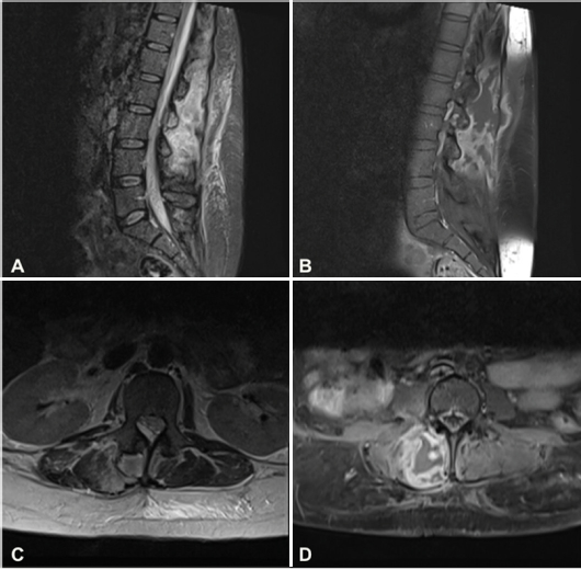
Given the severe pain associated with neurological deficit, magnetic resonance imaging of the lumbar spine was requested, revealing a lesion of probable infectious origin, with an epidural component causing significant compression of the spinal canal. (Figure 1)
Preoperative magnetic resonance imaging revealed a heterogeneous expansive lesion, predominantly hyperintense on T2weighted sequences, with irregular peripheral enhancement, located in the right posterior paravertebral musculature, adjacent to the spinous processes from L1 to L4. The lesion measured approximately 47 × 43 × 142 mm (anteroposterior × laterolateral × craniocaudal). Involvement of the L2–L3 and L3–L4 facet joints was observed, with bone marrow edema in the posterior arch elements of L2 and L3. Extension of the lesion into the spinal canal was evident, situated extramedullary, with caudal identification at the level of the L3 vertebral body. The presence of an epidural collection within the spinal canal resulted in compression of the nerve roots, secondary to canal narrowing at the L1–L2 levels (A, B, C, D).
The diagnostic impression was a collection located in the right posterior paravertebral musculature at the lumbar segment, with marked peripheral enhancement after contrast administration, suggesting an inflammatory/infectious process. The collection extended into the spinal canal, leading to canal narrowing and compression of the neural roots, with possible thoracic involvement (Figure 1).
Surgical drainage and decompression using an interlaminar endoscopic technique were performed. The decision was based on the patient’s clinical history, which included multiple skin lesions with inadequate healing, in addition to poorly controlled diabetes mellitus in an acute phase—factors that would significantly increase the risk of complications in conventional large open procedures, such as wound dehiscence and formation of new collections. The endoscopic approach, in turn, offered reduced tissue trauma, smaller incisions, and a lower risk of complications while maintaining the effectiveness of neural decompression.
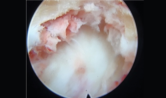
The procedure was performed under general anesthesia, with intraoperative neurophysiological monitoring and the patient positioned prone. Two skin incisions of approximately 7 mm were made: one at L1–L2 and another at L2–L3. During the initial puncture, purulent material was observed, collected, and sent for microbiological culture. An endoscope with a 4.3-mm working channel and 30° angulation was introduced through the working cannula, with continuous saline irrigation maintained at 30 mmHg to prevent increased epidural pressure and dispersion of purulent secretions. A hemi-hemilaminectomy was performed at the treated levels, followed by careful dissection and removal of the ligamentum flavum. Beneath it, amorphous material compressing the dural sac and adjacent neural structures was identified (Figure 2).
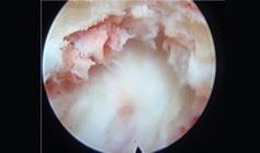
The epidural component was removed using endoscopic instruments, resulting in increased pulsatility of the neural elements and evident decompression. Improvement in motor evoked potentials was recorded by the neurophysiology team during the procedure. At the end, biopsy and extensive debridement were performed, with irrigation of approximately 10 liters of saline solution within the epidural space. All removed tissue was sent for microbiological analysis.
Microbiological culture of the collected material identified methicillin-resistant Staphylococcus aureus (MRSA).
In the immediate postoperative period, the patient presented complete resolution of the pain radiating to the lower limbs, as well as significant improvement in the neurological deficit, with recovery of muscle strength and urinary continence. Intravenous antibiotic therapy was maintained for 6 weeks, according to the treatment protocol for spinal infections.
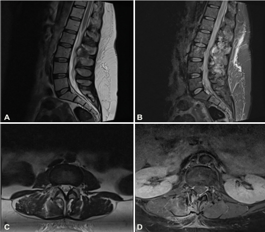
Follow-up imaging, performed six weeks after the surgical intervention and completion of antibiotic therapy, demonstrated a marked reduction of the heterogeneous expansile lesion previously located in the right posterior paravertebral musculature, adjacent to the spinous processes from L1 to L4. Resolution of the epidural component of the lesion, which had previously extended into the vertebral canal, was also observed, indicating a satisfactory therapeutic response and effective neural decompression (Figure 3).
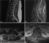
Magnetic resonance imaging obtained six weeks after the surgical procedure demonstrated a significant reduction of the expansile lesion, with complete resolution of its extension into the vertebral canal.
DISCUSSION
Spinal epidural abscess, a rare and potentially severe pyogenic infection located between the spinal dura mater and the vertebral periosteum, was first described by Morgagni in 1761. The pioneering surgical treatment was reported by Barth in 1901. For decades, this condition represented a major therapeutic challenge due to the difficulty of early diagnosis and the limited resources available. Only with advances in imaging techniques, enabling early surgical decompression in combination with systemic antibiotic therapy, was it possible to substantially reduce the morbidity and mortality associated with this disease.1,5
Emergency neurosurgical management is essential to prevent permanent neurological deficits or progression to sepsis.6 Surgical indication is particularly relevant in patients with progressive neurological deterioration, direct spinal cord compression, vascular compromise, mechanical spinal instability, or failure of antibiotic therapy.7 Appropriate and timely management has been decisive for functional and vital prognosis.
The discussion regarding early diagnosis and appropriate therapeutic choice in the management of spinal epidural abscess has therefore become increasingly relevant. Although historically considered a rare condition with a guarded prognosis, the incidence of spinal infections has progressively increased. Recent studies have indicated an occurrence rate of approximately 1 case per 10.000 hospital admissions. On the other hand, mortality associated with epidural abscess has reached up to 16%, highlighting its potentially lethal nature.8
The growing prevalence of highly virulent pathogens, such as methicillin-resistant Staphylococcus aureus (MRSA), as well as other opportunistic organisms, has been attributed to indiscriminate antibiotic use, chronic hemodialysis, and intravenous drug abuse, factors that have directly contributed to increased morbidity related to spinal infections. Moreover, the widespread use of anesthetic techniques such as epidural analgesia and peripheral nerve blocks has been associated with a rise in iatrogenic infections in this region.4,9 The diagnosis of spinal epidural abscess is multifactorial and requires an integrated approach. Initially, the patient’s clinical history, together with fever, localized pain, and neurological signs, guides diagnostic suspicion. Laboratory findings such as leukocytosis, elevated C-reactive protein, and prolactin may reinforce the infectious hypothesis. Identification of the etiological agent through blood cultures, intraoperative samples, or CT-guided biopsies, particularly in cases with paravertebral abscess such as in the psoas muscle, contributes significantly to therapeutic guidance. However, definitive confirmation is achieved through magnetic resonance imaging (MRI), which demonstrates typical features including epidural collection, spinal cord edema, and neural compression.1,5,10
The ideal therapeutic strategy for spinal epidural abscess remains controversial. The main debate concerns early surgical intervention versus conservative antibiotic management.5 Although a large retrospective study did not demonstrate additional benefit from early surgical drainage compared with antibiotics alone11, clinical practice has continued to favor surgery in the presence of progressive neurological decline.6-8 Treatment decisions should therefore be individualized, considering the patient’s clinical status, abscess extent, spinal cord compression, and response to medical therapy.
Discussions in specialized forums, including neurosurgery and neuroradiology congresses, multidisciplinary spine meetings, and collaborative reviews involving infectious disease specialists, orthopedic surgeons, and trauma teams, have reinforced that the optimal approach should remain patient-centered. Surgery has been primarily indicated when source control is required, when there is significant epidural occupation, neural compression, or established neurological deficit.7,11 In such scenarios, minimally invasive techniques, such as uniportal endoscopic surgery, have demonstrated effectiveness, with lower morbidity and faster recovery.
Treatment of spinal epidural abscess has relied on effective drainage of the purulent collection and eradication of the infectious agent. Surgical intervention has been mainly recommended in cases of neurological impairment, spinal cord compression, or failure of medical therapy, and the decision for decompression should be made promptly, ideally within 24 to 36 hours after the onset of neurological deficit, in order to prevent prognostic deterioration. Clinical resolution has usually occurred within 4 to 6 weeks and has been confirmed by follow-up imaging performed between the fourth and eighth weeks after treatment.4
In recent years, minimally invasive techniques have gained prominence in the management of spinal infections, with endoscopic surgery emerging as a promising alternative. This approach has allowed enhanced visualization of contralateral, sublaminar, and foraminal structures, with reduced nerve root manipulation and preservation of the functional integrity of bone and soft tissues. Continuous saline irrigation, a characteristic of the endoscopic method, has contributed to hemostatic control and assisted in reducing local bacterial load.4
Traditionally indicated for disc herniations and spinal stenosis, endoscopic surgery has also proven feasible in cases of epidural abscess associated with early spondylodiscitis, offering a less invasive option with potential to decrease morbidity and accelerate recovery.4 Increasingly, this surgical strategy has been incorporated into the management of spinal infections, enabling accurate bacteriological diagnosis, effective symptomatic relief, and targeted treatment of surgical site infection.12-14
Despite the favorable outcome observed in the present case, the inherent limitations of isolated reports must be acknowledged. Validation of the effectiveness of endoscopic surgery in the treatment of spinal epidural abscess will require more robust investigations, including larger samples and prospectively defined comparison groups, in order to generate consistent and generalizable evidence for clinical practice.
CONCLUSION
The results obtained in this study indicated that the surgical and clinical outcomes of the patient diagnosed with spinal epidural abscess and treated through interlaminar endoscopic spine surgery were satisfactory and met the proposed objectives. The patient experienced favorable clinical progression, without complications or recurrence, culminating in complete resolution of the infectious condition.
The positive outcome observed reinforced the feasibility and safety of endoscopic surgery as a therapeutic alternative in selected cases of spinal epidural abscess. However, prospective studies with larger samples and well-defined comparison groups are required to provide more robust and conclusive evidence.
-
Study conducted by the Clínica Atualli Spine Care. Rua Bela Cintra, 539, 2º andar, Consolação, São Paulo/SP, Brazil.
DATA AVAILABILITY DECLARATION
The contents underlying the research are available in the manuscript.
REFERENCES
-
1 Alam Z, Syed MU, Siddiqui TA, Gunturi A, Reddy B, Alam Z, et al. Spinal lesions: a comprehensive radiologic overview. Front Radiol. 2025;5:1577840. doi: 10.3389/fradi.2025.1577840
» 10.3389/fra-di.2025.1577840 -
2 Dhodapkar MM, Patel T, Rubio DR. Imaging in spinal infections: Current status and future directions. N Am Spine Soc J. 2023;16:100275. doi: 10.1016/j.xnsj.2023.100275
» 10.1016/j.xnsj.2023.1002751 -
3 Carroll R. Vertebral osteomyelitis and epidural abscess. Med Clin North Am. 2025;109(3):601-14. doi: 10.1016/j.mcna.2024.12.007
» 10.1016/j.mcna.2024.12.007 -
4 Kang T, Park SY, Lee SH, Park JH, Suh SW. Spinal epidural abscess successfully treated with biportal endoscopic spinal surgery. Medicine (Baltimore). 2019; 98(50):e18231. doi: 10.1097/MD.0000000000018231
» 10.1097/MD.0000000000018231 -
5 Tuchman A, Pham M, Hsieh PC. The indications and timing for operative management of spinal epidural abscess: literature review and treatment algorithm. Neurosurg Focus. 2014;37(2):E8. doi: 10.3171/2014.6.FOCUS14261
» 10.3171/2014.6.FOCUS14261 -
6 Bureta C, Tominaga H, Yamamoto T, Setoguchi T, Kawamura H, Nagano S, et al. Lumbar spine epidural abscess and facet joint septic arthritis due to Streptococcus agalactiae: a case report. BMC Surg. 2018;18(1):16. doi: 10.1186/s12893-018-0350-2
» 10.1186/s12893-018-0350-2 -
7 Arko IV L, Quach E, Nguyen V, Chang D, Sukul V, Kim BS. Medical and surgical management of spinal epidural abscess: a systematic review. Neurosurg Focus. 2014;37(2):E4. doi: 10.3171/2014.6.FOCUS14127
» 10.3171/2014.6.FOCUS14127 -
8 Ghobrial GM, Beygi S, Viereck MJ, Maulucci CM, Sharan A, Heller J, et al. Timing in the surgical evacuation of spinal epidural abscesses. Neurosurg Focus. 2014;37(2):E1. doi: 10.3171/2014.6.FOCUS14120
» 10.3171/2014.6.FOCUS14120 -
9 Lee JY, Kim JW, Na YJ, Kim T, Han SH. Epidural abscess formation with an atypical pathogen following epidural steroid injection: a case report. Medicine (Baltimore). 2022;101(36):230495. doi: 10.1097/MD.0000000000030495
» 10.1097/MD.0000000000030495 -
10 Hijazi MM, Siepmann T, El-Battrawy I, Aweimer A, Engellandt K, Podlesek D, et al. Diagnostics, Management, and Outcomes in Patients with Pyogenic Spinal Intra-or Epidural Abscess. J Clin Med. 2023;12(24):7691. doi: 10.3390/jcm12247691
» 10.3390/jcm12247691 -
11 Connor Jr DE, Chittiboina P, Caldito G, Nanda A. Comparison of operative and nonoperative management of spinal epidural abscess: a retrospective review of clinical and laboratory predictors of neurological outcome. J Neurosurg Spine. 2013;19(1):119-27. doi: 10.3171/2013.3.SPINE12762
» 10.3171/2013.3.SPINE12762 -
12 Yang SC, Chiu YC, Chen HS, Kao YH, Jou IM, Tu YK. Percutaneous endoscopic debridement and drainage for the treatment of instrumented lumbar spine infection. J Orthop Surg (Hong Kong). 2019;27(3):2309499019863356. doi: 10.1177/2309499019863356
» 10.1177/2309499019863356 -
13 Giordan E, Liu Y, Suvithayasiri S, Ruddo S, Lee C, Hasan GA, et al. Endoscopic treatment of thoracolumbar spondylodiscities: a systematic review and meta-analysis. World Neurosurg. 2024;189:296-306. doi: 10.1016/j.wneu.2024.06.051
» 10.1016/j.wneu.2024.06.051 -
14 Ahn Y. Full-endoscopic spine surgery: its role and limitations. J Korean Neurosurg Soc. 2025;68(5):511-27. doi: 10.3340/jkns.2024.0227
» 10.3340/jkns.2024.0227
Edited by
-
Reviewed by:
Marcelo Risso