 PHEOCHROMOCYTOMA WITH METASTATIC LESION IN THE L4 VERTEBRA AND NEUROLOGICAL DEFICIT: CASE REPORT
ABSTRACT
Objective: To describe a rare single-stage surgical approach for vertebral metastatic pheochromocytoma in a middle-aged adult and review perioperative management strategies. Introduction Pheochromocytomas are rare neuroendocrine tumors (incidence 0.2–0.9/100,000/year), and about 10% develop metastases. Bone involvement— especially in the spine —is common in malignant cases. Spinal metastases from pheochromocytoma present hemodynamic challenges due to catecholamine secretion, and no consensus exists on treatment.
Case presentation: A 38-year-old man with right adrenal pheochromocytoma on doxazosin reported severe low back pain and an L4 sensorimotor deficit. CT and MRI revealed an osteolytic lesion at L4 with epidural extension and additional metastatic foci at T11 and L1. Declining staged procedures, he underwent single-stage oblique anterolateral L4 corpectomy and fusion with an expandable cage. Preoperative preparation: α-blockade (doxazosin 2 mg/day), β-blockade, and fluoroscopic tumor embolization.
Discussion: Vertebral metastasis from pheochromocytoma is rare, with male predominance and frequent involvement of the thoracic spine. Diagnosis is based on adrenergic symptoms, neurological deficits, elevated metanephrines, and imaging (CT/MRI). Biopsy is avoided due to bleeding risk. Surgical preparation includes α/β-adrenergic blockade and embolization. For management, en bloc resection with stabilization is preferred, with a challenging postoperative period requiring intensive care due to hemodynamic instability.
Conclusion: Single-stage vertebral resection for metastatic pheochromocytoma is feasible when accompanied by adequate adrenergic blockade, preoperative embolization, and multidisciplinary support. Despite aggressive intervention, recurrence and neurological decline are common, underscoring the importance of early diagnosis and individualized perioperative planning. Level of Evidence IV; Case-Report.
Keywords:
Pheochromocytoma; Spinal Metastasis; Lumbosacral; Decompression; Therapeutic Embolization; Vertebral Corpectomy
RESUMO:
Objetivo: Descrever uma abordagem cirúrgica rara em tempo único para feocromocitoma metastático vertebral em um adulto de meia-idade e revisar estratégias de manejo perioperatório.
Introdução: Feocromocitomas são tumores neuroendócrinos raros (incidência de 0,2–0,9/100.000/ano), com cerca de 10% evoluindo com metástases. O acometimento ósseo — especialmente da coluna vertebral — é comum nos casos malignos. Metástases espinhais por feocromocitoma apresentam desafios hemodinâmicos devido à secreção de catecolaminas, e não há consenso sobre o tratamento.
Apresentação do caso: Homem de 38 anos com feocromocitoma adrenal direito em uso de doxazosina relatava dor lombar intensa e déficit sensitivo-motor em L4. Tomografia e ressonância revelaram lesão osteolítica em L4 com extensão epidural e focos metastáticos adicionais em T11 e L1. Após recusar cirurgia em estágios, foi submetido a corpectomia oblíqua ântero-lateral de L4 e artrodese com uso de cage expansível. A preparação pré-operatória incluiu bloqueio alfa (doxazosina 2 mg/dia), bloqueio beta e embolização tumoral fluoroscópica.
Discussão: A metástase vertebral de feocromocitoma é rara, com predomínio em homens e acometimento frequente da coluna torácica. O diagnóstico baseia-se em sintomas adrenérgicos, déficit neurológico, metanefrinas elevadas e imagem (TC/RM). A biópsia é evitada pelo risco de sangramento. O preparo cirúrgico inclui bloqueio α/β-adrenérgico e embolização. Para o manejo, a ressecção em bloco com estabilização é preferível, com pós-operatório difícil exigindo cuidados intensivos devido à instabilidade hemodinâmica.
Conclusão: A ressecção vertebral em tempo único para feocromocitoma metastático é viável quando acompanhada de bloqueio adrenérgico adequado, embolização pré-operatória e suporte multidisciplinar. Apesar da intervenção agressiva, a recorrência e o declínio neurológico são comuns, ressaltando a importância do diagnóstico precoce e do planejamento perioperatório individualizado. Nível de Evidência IV; Relato de Caso.
Descritores:
Feocromocitoma; Metástase Espinhal; Lombossacral; Descompressão; Embolização Terapêutica; Corpectomia Vertebral
RESUMEN
Objetivo: Describir un abordaje quirúrgico raro en un solo tiempo para feocromocitoma metastásico vertebral en un adulto de mediana edad y revisar estrategias de manejo perioperatorio.
Introducción: Los feocromocitomas son tumores neuroendocrinos raros (incidencia de 0,2–0,9/100.000/año), y aproximadamente el 10% desarrolla metástasis. El compromiso óseo —especialmente de la columna vertebral— es común en los casos malignos. Las metástasis espinales por feocromocitoma presentan desafíos hemodinámicos debido a la secreción de catecolaminas, y no existe un consenso sobre el tratamiento.
Discusión: La metástasis vertebral por feocromocitoma es rara, con predominio en hombres y afectación frecuente de la columna torácica. El diagnóstico se basa en síntomas adrenérgicos, déficit neurológico, metanefrinas elevadas e imágenes (TC/RM). La biopsia se evita por riesgo de sangrado. La preparación quirúrgica incluye bloqueo adrenérgico α/β y embolización. Para el manejo, se prefiere la resección en bloque con estabilización, con un postoperatorio complejo que requiere cuidados intensivos por inestabilidad hemodinámica.
Resultados: Pérdida sanguínea estimada de 5.000 mL; se transfundieron tres unidades de concentrado de hematíes. La histopatología confirmó feocromocitoma metastásico con tinción positiva para cromogranina A, inhibina, sinaptofisina y TFE3. En el posoperatorio, presentó taquicardia supraventricular, shock séptico y trombosis venosa profunda. Posteriormente, una estenosis traqueal requirió tubo en T de Montgomery. Estudios de control mostraron recidiva en L4 y progresión en T3, T11 y L1, por lo que se inició radioterapia paliativa. Evolucionó con empeoramiento de la debilidad en extremidades inferiores e incontinencia urinaria, requiriendo cuidados intensivos prolongados.
Conclusión: La resección vertebral en un solo tiempo para feocromocitoma metastásico es factible cuando se acompaña de un bloqueo adrenérgico adecuado, embolización preoperatoria y apoyo multidisciplinario. A pesar de una intervención agresiva, son frecuentes la recurrencia y el deterioro neurológico, lo que subraya la importancia del diagnóstico temprano y de una planificación perioperatoria individualizada. Nivel de Evidencia IV; Reporte de Caso.
Descriptores:
Feocromocitoma; Metástasis Espinal; Lumbosacra; Descompresión; Embolización Terapéutica; Corpectomía Vertebral
INTRODUCTION
Pheochromocytomas and paragangliomas are neuroendocrine tumors of chromaffin cells: pheochromocytomas arise in the adrenal medulla, and paragangliomas originate in sympathetic ganglia of the thorax, abdomen, and pelvis1. The annual incidence of pheochromocytoma is 0.2 – 0.9/100 0002 and approximately 10 % become malignant3. Up to 71% of those develop bone metastases — mainly in the spine, sacrum, and pelvis — with 38% presenting synchronously4.
Diagnosis is based on plasma or urinary metanephrine levels, followed by CT or MRI (sensitivities of 89% and 98%, respectively), although specificity is limited5,6. If imaging does not allow for a definitive diagnosis, positron emission tomography - computed tomography (PET-CT) with metaiodobenzylguanidine (MIBG) or fluorodeoxyglucose (FDG) is indicated7.
Primary treatment includes α/β-blockade and surgical resection. In spinal metastases, vertebroplasty or open surgery with preoperative embolization and neoadjuvant therapy are common, but no consensus exists3,5. We report a vertebral metastatic pheochromocytoma in an adult male to expand the literature and discuss perioperative management.
CASE PRESENTATION
History and Examination
A 31-year-old man with right adrenal pheochromocytoma on doxazosin and prior L4 decompression with L3–L5 fusion reported one year of severe low back pain and progressive L4 sensorimotor deficit (paresis, hypoesthesia) without sphincter involvement.
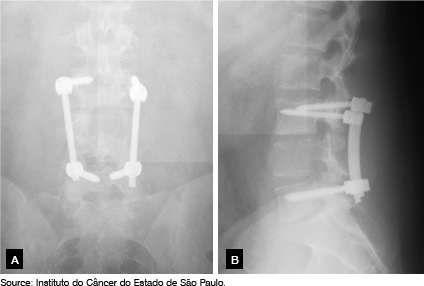
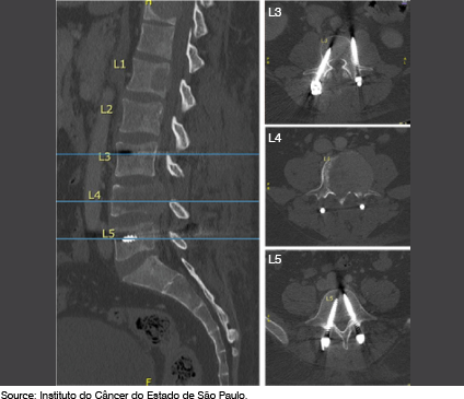
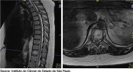
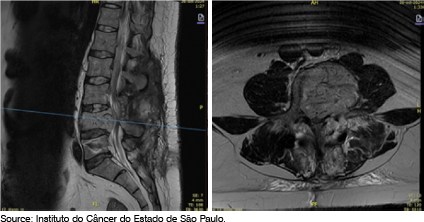
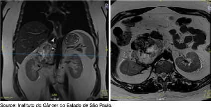
Radiograph demonstrated L3–L5 fusion with bilateral pedicle screws and rods (Figure 1). Non-contrast CT revealed an expansile osteolytic lesion at L4 with left epidural extension, infiltrative margins, intralesional necrosis, and soft-tissue attenuation, without significant periosteal reaction (Figure 2). MRI showed infiltration of the right posterior element at T3; T11 retropulsion with cord compression (Figure 3); a pathologic L4 fracture with < 50 % lateral height loss and cauda equina compression to L5 (Figure 4); and signs of prior L4 laminectomy and left medial facetectomy. Abdominal MRI showed an 8.5 × 7.3 cm right adrenal mass with retrohepatic tumor thrombus and greater than 180° renal artery encasement. (Figure 5)
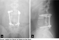
Anteroposterior (A) and lateral (B) radiographs of the lumbar spine obtained postoperatively following decompressive surgery performed at an outside facility.
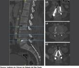
Sagittal and axial non-contrast computed tomography (CT) showing an expansile, osteolytic lesion with ill-defined margins in the L4 vertebral body, along with signs of prior instrumentation at L3 and L5 using bilateral pedicle screws and rods.
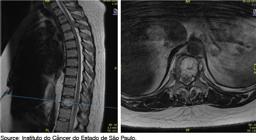
Preoperative sagittal and axial T2-weighted magnetic resonance imaging revealing vertebral metastases and spinal cord compression at the T11 level.
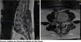
Preoperative sagittal and axial T2-weighted magnetic resonance imaging revealing vertebral metastases and spinal cord compression at the L4 level.
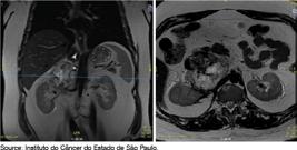
Coronal (A) and axial (B) T2-weighted magnetic resonance imaging showing a right perirenal expansive lesion with hepatorenal and right renal artery invasion.
Procedural Planning and Surgery
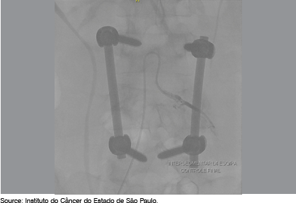
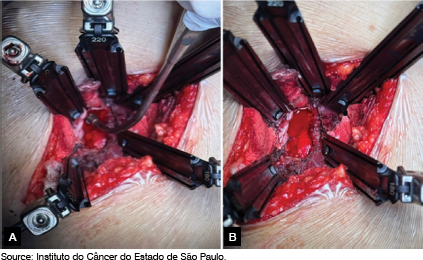
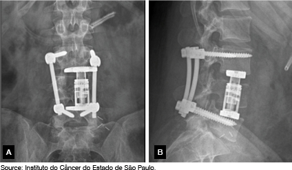
The patient declined staged procedures; a single-stage approach was chosen. Preoperatively, he received α-blockade (doxazosin 2 mg/day) and β-blockade, followed by fluoroscopic tumor embolization (Figure 6). With the patient supine, an oblique anterolateral approach was chosen. A 4 cm incision was made between the anterior superior iliac spine and umbilicus. Intermuscular dissection, retroperitoneal mobilization, and partial psoas retraction were performed (Figure 7), displacing vessels to expose L4 under fluoroscopy with vascular surgery assistance. L4 corpectomy and interbody fusion with an expandable cage were performed (Figure 8). Estimated blood loss was 5,000 mL, requiring three units of packed red blood cells.
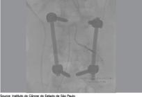
Anteroposterior fluoroscopy during total preoperative embolization of the tumor at L4 level.
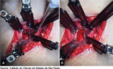
Intraoperative image of the window created for intralesional tumor resection (A) and after corpectomy of the L4 vertebra (B).
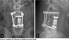
Postoperative radiographs in anteroposterior view (A) and lateral view (B) showing an expandable cage replacing the L4 vertebra and prior L3-L5 arthrodesis with pedicle screws positioned at an external facility.
Pathology
Histology confirmed metastatic pheochromocytoma, showing epithelioid cells with moderate-to-high cytologic atypia in an organoid-trabecular pattern and positive immunostaining for chromogranin A, inhibin, synaptophysin, and TFE-3.
Postoperative Course
In the ICU, he experienced supraventricular tachycardia requiring three cardioversions and developed pulmonary septic shock treated with piperacillin-tazobactam. On the ward, he developed left lower extremity deep vein thrombosis managed with enoxaparin and compression. At 45 days, he presented with dyspnea and inspiratory stridor due to tracheal stenosis 2 cm below the vocal cords. A Montgomery T-tube tracheostomy was placed. Imaging then showed L4 recurrence, progression at T3 and T11, and new L1 involvement; palliative radiotherapy (20 Gy per segment) was initiated. One month later, he developed worsening lower extremity weakness and urinary incontinence from cord compression and remains in the ICU for symptom management.
DISCUSSION
Epidemiology
Visani et al.8 systematically reviewed 15 articles on spinal metastatic paragangliomas (44 patients, 73 lesions). Of these, 77% were male. Metastases involved the thoracic spine in 54%, lumbar spine in 22%, cervical spine in 12%, and sacral region in 12%. Primary lesions affected 79.5% of adrenal sites (equivalent to pheochromocytomas), with a mean age of 42 years (range 21–69). Our patient—a 38-year-old man with thoracic (T11) and lumbosacral (L1, L4) metastases—matches the male predominance and lesion distribution described by Visani and by Ayala et al.4, although he is younger than the average.
Diagnostic Investigation
Classic pheochromocytoma symptoms include headache, palpitations, fatigue, pruritus, sweating, and paroxysmal hypertension. In this case, the patient presented with hypertension and tachycardia, warranting plasma or urinary metanephrine measurement and confirmation by CT or MRI (Table 1). In spinal metastases, neurological deficits vary by level: cervical lesions cause bilateral paresis/paresthesia of upper and lower extremities and may impair respiratory function; thoracolumbar lesions produce low back pain with corticospinal and spinothalamic deficits affecting lower limbs; lumbosacral involvement can lead to sphincter dysfunction, radiculopathy, proprioceptive deficits, and postural control issues6. The lesions at T11, L1, and L4 caused catecholamine-related symptoms (controlled with α-blockade) and compressive symptoms consistent with the anatomical level involved, such as low back pain and neurological deficits.
On imaging, paragangliomas are known as “chameleon tumors” due to varied appearances and mimicry of other benign and malignant neoplasms. Contrast-enhanced CT may show hyperdense lesions with cystic change, necrosis, calcification, and post-contrast enhancement, though one-third exhibit late-phase washout. T2-weighted MRI reveals moderate-to-intense signal and gadolinium enhancement (“lightbulb” appearance).8-12 Liu et al.13 described spinal metastases as osteolytic with contrast enhancement and absent calcifications in 18 patients. In our case, CT and MRI findings were diagnostic when integrated with the patient’s symptomatology and prior diagnosis of adrenal pheochromocytoma, obviating the need for nuclear imaging such as PET-CT with MIBG or FDG.7
As a result, in patients with known primary disease, positive biochemical testing, and confirmatory imaging, a biopsy may be avoided due to bleeding and dissemination risks.
Preoperative Treatments
Adrenergic receptor blockers reduce postoperative mortality. Competitive α-blockers (doxazosin or prazosin, 6–20 mg/day) are preferred over phenoxybenzamine because they cause less reflex hypotension and tachycardia and have a shorter half-life, simplifying postoperative dose adjustments as catecholamine levels fall after tumor resection. Although α-blockade lowers hypertensive crises, it can mask post-resection hypotension and does not fully prevent intraoperative blood pressure spikes.14 β-blockers should be initiated only after α-blockade to avoid unopposed α-receptor stimulation and hypertensive crisis. The target blood pressure is below 130/80 mmHg to ensure hemodynamic stability and minimize perioperative risk.6
Preoperative embolization of segmental arteries significantly reduces tumor blood supply and improves intraoperative hemodynamics, with a single case of massive intraoperative hemorrhage reported.15 Therefore, α-blockade and embolization appear safe and should be considered in surgical planning to minimize blood loss and improve hemodynamic stability. In our patient, these strategies effectively prevented a substantially more severe hemorrhagic event.
Surgical Treatment
Both primary and spinal metastatic lesions can store and secrete catecholamines during manipulation, creating a major hemodynamic challenge that directly influences treatment strategy1.
Visani et al.8 noted that seven patients undergoing minimally invasive treatments (percutaneous cement injection and biopsy) experienced disease progression. Although this systematic review of case reports and series is limited by small sample size and selection/publication bias, there are no robust comparative studies; hence, minimally invasive approaches are reserved for selected patients with prohibitive surgical risk.
Kaloostian et al.15 evaluated en bloc tumor resections and found no recurrence in 27 of 29 cases, suggesting en bloc resection is preferable to piecemeal removal and superior to minimally invasive techniques. Minimally invasive approaches should be reserved for patients with high risk (poor functional status, hemodynamic instability, extensive disease).
Moreover, vertebral instrumentation is often required for mechanical stability. Liu et al.13 emphasize that osteolytic lesions necessitate stabilization. In our patient, CT revealed malpositioned L3 pedicle screws penetrating the lateral cortex without compromise of the spinal canal, from prior instrumentation at an outside facility. We initially planned metastasis resection followed by implant revision, but the patient refused a second surgery. Consequently, we preserved the existing L3–L5 fusion and placed an expandable cage at L4 after corpectomy.
Postoperative Treatments
Management of hemodynamic perturbations, pain, and glycemic control requires ICU care with invasive arterial monitoring for at least 24–48 hours postoperatively. Kaloostian et al.15 reported significant morbidity associated with en bloc resection, and immediate postoperative hyperinsulinism — due to loss of catecholamine inhibition on pancreatic β-cells — necessitates glucose monitoring and metabolic support.
CONCLUSION
This case exemplifies the complexity of managing vertebral metastatic pheochromocytoma, integrating clinical, radiological, and surgical aspects to achieve therapeutic success. After preoperative preparation, corpectomy with vertebral decompression and resection helps prevent tumor progression and relieve paroxysmal and compressive symptoms. Postoperative intensive care is essential to manage hemodynamic instability and prevent life-threatening complications.
-
Study conducted by the Instituto do Câncer do Estado de São Paulo (ICESP), São Paulo, Brazil.
REFERENCES
- 1 Fishbein L. Pheochromocytoma and Paraganglioma: Genetics, Diagnosis, and Treatment. Hematol Oncol Clin North Am. 2016;30(1):135-150.
- 2 Singh A, Santangelo G, Ellens N, Kohli G, Pranaat R, Bender MT. Preoperative embolization and en bloc resection of a metastatic pheochromocytoma of the cervical spine. J Cerebrovasc Endovasc Neurosurg. 2024;26(3):331-337.
- 3 Liu S, Zhou X, Huo Z, Yao S, Wang Y, Liu Y. Clinical features and prognosis analysis of metastatic spinal pheochromocytoma: A single center retrospective study. J Bone Oncol. 2020;24:100312.
- 4 Ayala-Ramirez M, Palmer JL, Hofmann MC, et al. Bone metastases and skeletal-related events in patients with malignant pheochromocytoma and sympathetic paraganglioma. J Clin Endocrinol Metab. 2013;98(4):1492-1497
- 5 Lenders JWM, Duh QY, Eisenhofer G, et al. Pheochromocytoma and paraganglioma: an Endocrine Society clinical practice guideline. J Clin Endocrinol Metab. 2014;99(6):1915–1942.
- 6 Blake MA, Boland G. Adrenal imaging. 2009. ISBN: 9781934115862.
- 7 Timmers HJLM, Taïeb D, Pacak K, Lenders JWM. Imaging of pheochromocytomas and paragangliomas. Endocr Rev. 2024;45(3):414–434.
- 8 Visani J, Mongardi L, Cultrera F, de Bonis P, Lofrese G, Ricciardi L, et al. Surgical treatment of metastatic pheochromocytomas of the spine: a systematic review. J Integr Neurosci. 2021;20:499–507.
- 9 Blake M, Kalra M, Maher M, et al. Pheochromocytoma: an imaging chameleon. Radiographics. 2004;24 Suppl 1:S87–99.
- 10 Itani, M., & Mhlanga, J. (2019). Imaging of Pheochromocytoma and Paraganglioma. In R. Mariani-Costantini (Ed.), Paraganglioma: A Multidisciplinary Approach. Codon Publications.
- 11 Čtvrtlík F, Koranda P, Schovánek J, Škarda J, Hartmann I, Tüdös Z. Current diagnostic imaging of pheochromocytomas and implications for therapeutic strategy. Exp Ther Med. 2018;15(4):3151-3160.
- 12 Herr K, Muglia VF, Koff WJ, Westphalen AC. Imaging of the adrenal gland lesions. Radiol Bras. 2014;47(4):228–39.
- 13 Liu S, Zhou X, Song A, et al. Successful treatment of malignant pheochromocytoma with sacrum metastases: a case report. Medicine (Baltimore). 2018;97(35):e12184.
- 14 Bruynzeel H, Feelders RA, Groenland TH, et al. Risk factors for hemodynamic instability during surgery for pheochromocytoma. J Clin Endocrinol Metab. 2010;95(2):678–685.
- 15 Kaloostian PE, Zadnik PL, Awad AJ, McCarthy E, Wolinsky JP, Sciubba DM. En bloc resection of a pheochromocytoma metastatic to the spine for local tumor control and for treatment of chronic catecholamine release and related hypertension. J Neurosurg Spine. 2013;18(6):611–616.