 Senecionine from Senecio brasiliensis reduces the expression of interferon-induced genes in corpus luteum cells in vitro
ABSTRACT:
This study investigated the effects of senecionine, a pyrrolizidine alkaloid found in plants of the genus Senecio spp., particularly Senecio brasiliensis, which causes significant damage in dairy and beef cattle production. Bovine luteal cells cultured in vitro were used as an experimental model to explore this. These cells were exposed to two concentrations of senecionine (10 and 100 ng/mL) for 24 hours, followed by the addition of interferon tau (IFNT), a precursor protein involved in maternal recognition of pregnancy in ruminants. Gene expression analyses were performed using the RT-qPCR technique, focusing on genes responsive to IFNT, such as ISG15, MX1, MX2, and OAS1. Genes related to cellular processes were also evaluated, including GADD45B, CCDN2, and CTGF, an important precursor in the Hippo signaling pathway. The integrity of the cell culture model was confirmed by analyzing the expression of the 3B-HSD gene and measuring progesterone concentration. This study provided evidence of the effects of senecionine on bovine luteal cells, revealing its ability to interfere with the response of these cells to IFNT. These findings contributed to understanding the underlying mechanisms of senecionine’s toxic effects in animals. They may assist in developing prevention and control strategies for the damage caused by this plant in production animals.
Key words:
senecionine; luteal cells; bovine; interferon tau; pyrrolizidine alkaloid
RESUMO:
Este estudo investiga os efeitos da senecionina, um alcaloide pirrolizidínico encontrado em plantas do gênero Senecio spp., particularmente Senecio brasiliensis, que causa danos significativos na produção de gado leiteiro e de corte. Para explorar isso, foram utilizadas células luteais bovinas cultivadas in vitro como modelo experimental. Essas células foram expostas a duas concentrações de senecionina (10 e 100 ng/mL) por 24 horas, seguidas pela adição de interferon tau (IFNT), uma proteína precursora envolvida no reconhecimento materno da gestação em ruminantes. Análises de expressão gênica foram realizadas utilizando a técnica de RT-qPCR, focando em genes responsivos ao IFNT, como ISG15, MX1, MX2 e OAS1. Adicionalmente, foram avaliados genes relacionados a processos celulares, incluindo GADD45B, CCDN2 e CTGF, um importante precursor na via de sinalização Hippo. A integridade do modelo de cultura celular foi confirmada pela análise da expressão do gene 3B-HSD e pela medição da concentração de progesterona. Este estudo fornece evidências dos efeitos da senecionina em células luteais bovinas, revelando sua capacidade de interferir na resposta dessas células ao IFNT. Esses achados contribuem para a compreensão dos mecanismos subjacentes aos efeitos tóxicos da senecionina em animais e podem auxiliar no desenvolvimento de estratégias de prevenção e controle dos danos causados por essa planta em animais de produção.
Palavras-chave:
senecionina; células luteais; bovinos; interferon tau; alcaloide pirrolizidínico
INTRODUCTION
Seneciosis is a major plant-induced poisoning and a leading cause of cattle mortality in southern Brazil (RIET-CORREA et al., 2013; TUSTIN et al., 2001) This intoxication results from the ingestion of Senecio spp., particularly Senecio brasiliensis, a plant widely distributed across the country, occurring in all Brazilian biomes and 12 states (TELES et al., 2023). The genus Senecio contains pyrrolizidine alkaloids (PAs), which, upon metabolism, generate highly reactive pyrrolic derivatives that induce cytotoxicity and liver damage (RIET-CORREA et al., 2013). The economic losses attributed to Senecio spp. intoxication are substantial, particularly in chronically exposed cattle that may not exhibit overt clinical signs but suffer from compromised reproductive performance (PANZIERA et al., 2018). While low reproductive efficiency in intoxicated cattle has traditionally been attributed to weight loss and hepatic dysfunction, emerging evidence suggests that PAs also have direct effects on ovarian tissue (KRABBE et al., 2015; PANZIERA et al., 2018). These findings indicated that Senecio spp. poisoning may contribute to subfertility by altering ovarian function and disrupting maternal-embryonic communication (SANTOS et al., 2008). Maternal-embryonic communication is a crucial physiological process that ensures the establishment and maintenance of pregnancy in ruminants. This communication relies on the secretion of interferon-tau (IFNT) by the developing embryo, which interacts with luteal cells to stimulate interferon-stimulated genes (ISGs) such as ISG15, MX1, MX2, and OAS1 (ANTONIAZZI et al., 2013; SPENCER et al., 2007). The activation of ISGs is essential for pregnancy recognition and maintenance, preventing premature luteolysis (BOTT et al., 2010). In addition to pregnancy recognition, IFNT regulates genes involved in luteal cell proliferation and survival. Specifically, GADD45B and CCDN2 play roles in apoptosis resistance and cell cycle progression, respectively (MIHM et al., 2008; SHERR & ROBERTS, 1995). Disruption of these processes can lead to reduced progesterone synthesis and increased luteal regression, compromising pregnancy maintenance (MANN & LAMMING, 2001). Furthermore, the Hippo signaling pathway, a critical regulator of cell proliferation and tissue homeostasis, is involved in luteal cell function (HUH et al., 2019). When activated, this pathway suppresses the expression of CTGF, a downstream effector of YAP1/TAZ, impairing cell proliferation (JUSTICE et al., 1995; WANG et al., 2016). Given the documented alterations in the luteal cells of cattle intoxicated by Senecio spp., it is crucial to determine whether the pyrrolizidine alkaloid senecionine (SN) affects IFNT-mediated gene expression and the functional integrity of luteal cells. This study investigated the impact of SN on the expression of IFNT-induced genes and key regulators of cellular health in bovine corpus luteum cells in vitro. Understanding these mechanisms will provide insights into the reproductive consequences of Senecio spp. intoxication and inform potential mitigation strategies in livestock management.
MATERIALS AND METHODS
Chemicals
Unless otherwise stated, chemicals and reagents were purchased from Sigma Chemical Company (Sigma-Aldrich, St. Louis, MO, USA).
Cell culture
Bovine ovaries were collected from a abattoir located in Santa Maria, Rio Grande do Sul, Brazil, and transported to the laboratory in 0.9% NaCl saline solution containing penicillin (100 IU/mL) and streptomycin (100 μg/mL) at 4 °C. The ovaries were washed in the laboratory with 0.9% NaCl saline solution and 70% ethanol. For this experiment, early and intermediate CLs were selected, measuring 15-25 mm in diameter, with luteal tissue visible on the ovarian surface, a bloodstained surface, and a pink, brown, or orange color with a compact and soft consistency. Luteal cell culture was adapted from a previous protocol published by ANTONIAZZI et al. (2013). The CLs were classified according to their morphological characteristics as early (1-6 days post-ovulation), intermediate (8-12 days post-ovulation), and late (15-17 days post-ovulation), as previously described (MIYAMOTO et al., 2000). The connective tissue adjacent to the corpus luteum was mechanically removed, cut into small pieces with a scalpel blade, and placed in a tube containing DMEM-F12 medium supplemented with 0.5% bovine serum albumin (BSA) at 37 °C for 10 minutes. The supernatant was then discarded, and the sedimented luteal tissue was dissociated in a solution of 0.1% type I collagenase and DMEM-F12 supplemented with 0.5% BSA, subjected to agitation for 1 minute every 10 minutes over 40 minutes at 37 °C. The cell suspension was then supplemented with DMEM-F12 containing 10% fetal bovine serum (FBS) to inactivate the collagenase. The cells were filtered through a 70 µm nylon filter, resuspended in 2 mL of Red Blood Cell Buffer, and centrifuged at 1,000 rpm for 10 minutes. The pellet was resuspended, washed, and centrifuged three times for 10 minutes at 1,000 rpm with DMEM-F12 solution supplemented with 10% FBS, 100 IU/mL penicillin, and 100 μg/mL streptomycin. Cell viability was assessed using the Trypan Blue exclusion method, with viability exceeding 85% in all replicates. The cells were seeded in 24-well plates at a density of 2 × 10^5 cells/well and cultured in DMEM-F12 supplemented with 5% FBS, 100 IU/mL penicillin, and 100 μg/mL streptomycin for 12 hours at 37 °C in 5% CO₂. Initially, the cells were incubated with the following treatments: Group 1: DMEM-F12 (control), Group 2: DMEM-F12, Group 3: DMEM-F12 with 10 ng/mL SN, Group 4: DMEM-F12 with 100 ng/mL SN. After 24 hours of culture, the medium was replaced with fresh DMEM-F12 culture medium, free of FBS and supplemented with 100 IU/mL penicillin and 100 μg/mL streptomycin. The cells were cultured under starvation conditions for 6 hours at 37 °C in 5% CO₂. After this period, the cells received the following treatments: Group 1: DMEM-F12 (control), Groups 2, 3, and 4: DMEM-F12 with 1 ng/mL of roIFNT. The cells were incubated for 6, 12, and 24 hours at 37 °C in 5% CO₂. After incubation, cells and culture medium were collected and stored at -80 °C for subsequent analysis. Three independent replicates were performed on different days, using five corpora lutea from different cows for each in vitro experiment.
RNA Extraction, reverse transcription, and real-time PCR
Total mRNA from luteal cells was extracted following the protocol recommended by the manufacturer using the PureLink™ RNA Mini Kit (Thermo Fisher Scientific, MA, USA). The extracted mRNA was quantified by measuring optical density using a NanoDrop™ spectrophotometer (Thermo Fisher Scientific, MA, USA) at a wavelength of 260 nm. The purity of the mRNA was assessed by the OD260/OD280 absorption ratio, and values below 1.8 were not used. Reverse transcription was performed using the iScript cDNA Synthesis Kit (Bio-Rad Laboratories, CA, USA) according to the manufacturer’s recommendations. RT-qPCR was carried out using the CFX384TM Real-Time System (Bio-Rad Laboratories, CA, USA) with GoTaq® DNA Polymerase (Promega, WI, USA) and specific primers (Table 1). After an initial denaturation step at 95 °C for 3 minutes, 40 cycles were performed at 95 °C for 10 seconds, followed by 1 minute at 60 °C to amplify each transcript. Target gene amplification Ct was normalized to the average expression level of RPL19, and GAPDH housekeeping genes, according to the ratio R = housekeeping ECt/target ECt, where E is the amplification efficiency for each primer pair. The abundance analysis of interferon-stimulated genes such as ISG15 (JOHNSON et al., 1999), MX1, MX2 (OTT et al., 1998), and OAS1 (MIRANDO et al., 1991) were performed, along with genes involved in proliferation and survival such as GADD45B and CCDN2 (MIHM et al., 2008), and the steroidogenic enzyme 3β-HSD. The quantification of relative abundance was carried out using the Pfaffl method (PFAFFL, 2001) (Table 1).
Analysis of progesterone
The progesterone concentration in the culture medium of luteal cells was measured in a specialized clinical analysis laboratory using an electrochemiluminescence kit (ADVIA Centaur®, Siemens Healthcare, Munich, Germany), with a sensitivity of 0.05 ng/mL of progesterone.
Statistical analysis
The analysis of variance was conducted using JMP software (SAS Institute), with treatment as the main factor and replicates as the random variable. Differences between means were evaluated using Tukey’s multiple comparison test. Homogeneity of variance was assessed using the O’Brien test. When the data did not follow a normal distribution (Shapiro-Wilk test), they were transformed to a logarithmic scale. The results are presented as mean ± standard error of the mean (SEM), and a value of P < 0.05 was considered statistically significant.
RESULTS AND DISCUSSION
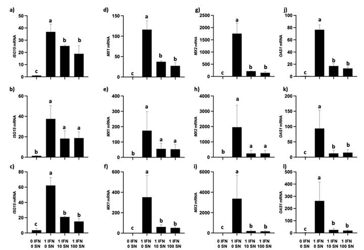
Interferon-stimulated genes (ISGs), particularly ISG15, are proteins expressed in the uterine endometrium during early pregnancy in response to the paracrine action of IFNT derived from the embryo in ruminants (BOTT et al., 2010). When IFNT was added to the corpus luteum (CL) culture, a dose-dependent response was observed in ISG15, MX1, MX2, and OAS1. This response is consistent with studies suggesting that the abundance of ISG15 in luteal cells increases dose-dependently in response to recombinant ovine or bovine IFNT up to 1 ng/mL (ROBERTS et al., 2008). Our treatment with one ng/mL of IFNT increased the mRNA abundance of these ISGs at all time points in cells without SN (Figure 1, panels a-l). Conversely, groups intoxicated with 10 or 100 ng/mL of SN showed significant decreases in ISG15 and other ISGs compared to groups without SN, indicating that SN can significantly reduce the mRNA abundance of ISGs in CL and negatively impact the maternal recognition mechanism of pregnancy. Recent studies have associated GADD45B and CCDN2 with ovarian function and dominant follicle development (MIHM et al., 2008; SHERR & ROBERTS, 1995). GADD45B, a protein rapidly induced by stress factors, possesses anti-apoptotic properties (DE SMAELE et al., 2001) When IFNT was added to the CL, higher GADD45B mRNA abundance was observed compared to the group without any addition, suggesting that IFNT acts like a protector of the CL. This protection is likely due to GADD45B’s role in DNA damage repair and cell survival (Figure 2, panels a-c) (SHEIKH et al., 2000). In groups intoxicated with 10 or 100 ng/mL of SN, a reduced abundance of GADD45B mRNA was observed, supporting the hypothesis that GADD45B helps protect cells against oxidative damage-induced cell death (YU et al., 2013). CCDN2 regulates the G1-to-S transition in the cell cycle, promoting proliferation (SHERR & ROBERTS, 1995). The CL treated with IFNT showed higher CCDN2 mRNA abundance compared to the untreated group, indicating that IFNT may enhance cell cycle functionality (Figure 3, panels d-f). However, groups treated with 10 or 100 ng/mL of SN exhibited significant decreases in CCDN2 mRNA, suggesting that SN might induce apoptosis by disrupting the cell cycle. The drastic reduction in proliferative and pro-survival factors observed in SN-treated groups raises questions regarding cell viability. Although, initial viability assessed by Trypan Blue exclusion was high, the possibility remains that prolonged exposure to SN may lead to increased cell death. However, it is important to highlight that the morphology of the cultured cells remained unchanged across all groups without evidence of corpus luteum cell cluster disaggregation. This suggested that the treatment did not cause widespread cell death. Thus, the observed decrease in proliferation and survival gene expression could be attributed more to transcriptional regulation by SN rather than to direct cytotoxic effects. Future studies should include additional viability assessments, such as annexin V/propidium iodide staining or caspase activity assays, to clarify if SN induces apoptosis or necrosis in luteal cells (BRABSON et al., 2021; LEE et al., 2018; XU & OUYANG, 2022). The Hippo pathway, which regulates tissue growth, differentiation, proliferation, and apoptosis, primarily targets CTGF (connective tissue growth factor) (JUSTICE et al., 1995; SHOME et al., 2020). In the group treated only with IFNT, there was no statistical difference in CTGF mRNA abundance compared to the untreated group, suggesting an inactive Hippo pathway. However, groups treated with 10 or 100 ng/mL of SN showed significantly reduced CTGF mRNA, indicating Hippo pathway activation and the YAP1/TAZ-TEAD complex’s inability to regulate CTGF mRNA. This suggested that SN inhibits cell proliferation by activating the Hippo pathway. Interferon-tau (IFNT) increases the mRNA abundance of ISGs, GADD45B, and CCDN2 in the corpus luteum (CL), enhancing cell survival and proliferation. However, SN significantly reduces these gene expressions, negatively impacting cell function and the maternal recognition of pregnancy. SN activates the Hippo pathway, inhibiting cell proliferation and decreasing the abundance of 3B-HSD mRNA, which may impair progesterone synthesis; although, it was not detected in this experiment. Future research should focus on determining whether SN-induced toxicity directly leads to apoptosis or necrosis and assess the broader implications of SN exposure on reproductive health in cattle.

Effects of senecionine (SN) and interferon-tau (IFNT) on interferon-stimulated gene (ISG) expression in bovine luteal cells. Relative mRNA abundance of ISG15 (a-c), MX1 (d-f), MX2 (g-i), and OAS1 (j-l) at 6, 12, and 24 hours post-treatment. Luteal cells were exposed to SN (10 or 100 ng/mL) for 24 hours, followed by treatment with IFNT (1 ng/mL). Gene expression was analyzed using RT-qPCR. Results are presented as mean ± SEM. Different letters indicate statistically significant differences among groups (P < 0.05).
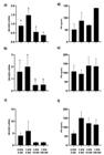
Expression of 3β-HSD and progesterone (P4) synthesis in bovine luteal cells exposed to senecionine (SN) and interferon-tau (IFNT). (A-C) Relative mRNA expression of 3β-HSD at 6, 12, and 24 hours. (D-F) Progesterone concentration (ng/mL) in culture media at corresponding time points. IFNT induced an increase in 3β-HSD expression and P4 levels, while SN (10 or 100 ng/mL) reduced both parameters. Results are shown as mean ± SEM. Different letters represent statistically significant differences (P < 0.05).

Impact of senecionine (SN) and interferon-tau (IFNT) on mRNA expression of genes related to apoptosis, proliferation, and Hippo signaling in bovine luteal cells. Relative mRNA abundance of GADD45B (a-c), CCDN2 (d-f), and CTGF (g-i) were measured at 6, 12, and 24 hours post-treatment. IFNT (1 ng/mL) increased the expression of GADD45B and CCDN2, while SN exposure (10 or 100 ng/mL) significantly reduced their expression. SN also downregulated CTGF, suggesting Hippo pathway activation. Data are shown as mean ± SEM. Bars with different letters indicate significant differences (P < 0.05).
CONCLUSION
In summary, senecionine from Senecio brasiliensis impairs the response of bovine luteal cells to interferon-tau, reducing the expression of interferon-stimulated genes, as well as markers of cell survival, proliferation, and steroidogenesis. These effects likely involve activation of the Hippo pathway and occur without evident cytotoxicity, suggesting transcriptional modulation. The findings highlighted a potential mechanism by which pyrrolizidine alkaloids compromise reproductive function in cattle, underscoring the need for further studies on their long-term effects and mitigation strategies.
ACKNOWLEDGMENTS
This study was supported by Coordenação de Aperfeiçoamento de Pessoal de Nível Superior (CAPES - funding code 001), Fundação de Amparo à Pesquisa do Estado do Rio Grande do Sul (FAPERGS), and Conselho Nacional de Desenvolvimento Científico e Tecnológico (CNPq). Special thanks to Dr. Fuller Bazer from Texas A&M University for providing the interferon tau used in this study. Thanks to Frigorífico Silva for providing the bovine ovaries used in this study.
REFERENCES
-
ANTONIAZZI, A. Q. et al. Endocrine delivery of interferon tau protects the corpus luteum from prostaglandin F2 alpha-induced luteolysis in ewes. Biology of Reproduction, v.88, n.6, 2013. Available from: <Available from: https://academic.oup.com/biolreprod/article/88/6/144%2C%201-12/2514293?utm_source=.com >. Accessed: May, 20, 2023. doi: 10.1095/biolreprod.112.105684.
» https://doi.org/10.1095/biolreprod.112.105684.» https://academic.oup.com/biolreprod/article/88/6/144%2C%201-12/2514293?utm_source=.com -
BOTT, R. C. et al. Uterine vein infusion of Interferon Tau (IFNT) extends luteal life span in ewes. Biology of Reproduction, v.82, n.4, p.725-735, 2010. Available from: <Available from: https://academic.oup.com/biolreprod/article/82/4/725/2557911?utm_source=.com >. Accessed: May, 22, 2023. doi: 10.1095/biolreprod.109.079467.
» https://doi.org/10.1095/biolreprod.109.079467.» https://academic.oup.com/biolreprod/article/82/4/725/2557911?utm_source=.com -
BRABSON, J. P. et al. Epigenetic regulation of genomic stability by Vitamin C. Frontiers in Genetics, v.12, 2021. Available from: <Available from: https://www.frontiersin.org/journals/genetics/articles/10.3389/fgene.2021.675780/full?utm_source=.com >. Accessed: May, 26, 2023. doi: 10.3389/fgene.2021.675780.
» https://doi.org/10.3389/fgene.2021.675780.» https://www.frontiersin.org/journals/genetics/articles/10.3389/fgene.2021.675780/full?utm_source=.com -
DE SMAELE, E. et al. Induction of gadd45β by NF-κB downregulates pro-apoptotic JNK signalling. Nature, v.414, n.6861, p.308-313, 2001. Available from: <Available from: https://www.nature.com/articles/35104560?utm_source=.com >. Accessed: May, 26, 2023. doi: 10.1038/35104560.
» https://doi.org/10.1038/35104560.» https://www.nature.com/articles/35104560?utm_source=.com -
HUH, H. D. et al. Regulation of TEAD transcription factors in cancer biology. Cells, v.8, n.6, 2019. Available from: <Available from: https://www.mdpi.com/2073-4409/8/6/600 >. Accessed: Jun. 02, 2023. doi: 10.3390/cells8060600.
» https://doi.org/10.3390/cells8060600.» https://www.mdpi.com/2073-4409/8/6/600 -
JOHNSON, G. A. et al. Expression of the interferon Tau Inducible Ubiquitin Cross-Reactive protein in the ovine uterus, Biology of Reproduction, v.61, n.1, p.312-318, 1999. Available from: <Available from: https://academic.oup.com/biolreprod/article/61/1/312/2734935?logintrue >. Accessed: Jun. 02, 2023. doi: 10.1095/biolreprod61.1.312.
» https://doi.org/10.1095/biolreprod61.1.312.» https://academic.oup.com/biolreprod/article/61/1/312/2734935?logintrue -
JUSTICE, R. W. et al. The Drosophila tumor suppressor gene warts encodes a homolog of human myotonic dystrophy kinase and is required for the control of cell shape and proliferation. Genes & Development, v.9, n.5, p.534-546, 1995. Available from: <Available from: https://genesdev.cshlp.org/content/9/5/534 >. Accessed: Jun. 05, 2023. doi: 10.1101/gad.9.5.534.
» https://doi.org/10.1101/gad.9.5.534.» https://genesdev.cshlp.org/content/9/5/534 -
KRABBE, A. A. et al. Megalocitose de células luteínicas grandes de vacas prenhes com seneciose crônica. Pesquisa Veterinaria Brasileira, v.35, n.1, p.33-38, 2015. Available from: <Available from: https://www.scielo.br/j/pvb/a/GqdBhZ6MYshzYfGnrxfHVvD/?lang=pt&utm_source.com >. Accessed: Jun. 05, 2023. doi: 10.1590/S0100-736X2015000100008.
» https://doi.org/10.1590/S0100-736X2015000100008.» https://www.scielo.br/j/pvb/a/GqdBhZ6MYshzYfGnrxfHVvD/?lang=pt&utm_source.com -
LEE, J. J. et al. Gene discovery and polygenic prediction from a genome-wide association study of educational attainment in 1.1 million individuals. Nature Genetics, v.50, n.8, p.1112-1121, 2018. Available from: <Available from: https://www.nature.com/articles/s41588-018-0147-3?utm_source=.com >. Accessed: Jun. 06, 2023. doi: 10.1038/s41588-018-0147-3.
» https://doi.org/10.1038/s41588-018-0147-3.» https://www.nature.com/articles/s41588-018-0147-3?utm_source=.com -
MANN, G. E.; LAMMING, G. E. Relationship between maternal endocrine environment, early embryo development and inhibition of the luteolytic mechanism in cows. Reproduction, v.121, n.1, p.175-180, 2001. Available from: <Available from: https://rep.bioscientifica.com/view/journals/rep/121/1/175.xml?utm_source=.com >. Accessed: Jun. 06, 2023. doi: 10.1530/rep.0.1210175.
» https://doi.org/10.1530/rep.0.1210175.» https://rep.bioscientifica.com/view/journals/rep/121/1/175.xml?utm_source=.com -
MIHM, M. et al. Differentiation of the bovine dominant follicle from the cohort upregulates mRNA expression for new tissue development genes. Reproduction, v.135, n.2, p.253, 2008. Available from: <Available from: https://rep.bioscientifica.com/view/journals/rep/135/2/253.xml?utm_source=.com >. Accessed: Jun. 07, 2023. doi: 10.1530/REP-06-0193.
» https://doi.org/10.1530/REP-06-0193.» https://rep.bioscientifica.com/view/journals/rep/135/2/253.xml?utm_source=.com -
MIYAMOTO, Y. et al. Is tumor necrosis factor α a trigger for the initiation of endometrial prostaglandin F2α release at luteolysis in cattle? Biology of Reproduction, v.62, n.5, p.1109-1115, 2000. Available from: <Available from: https://academic.oup.com/biolreprod/article/62/5/1109/2734600?utm_source=t.com&login=true >. Accessed: Jun. 08, 2023. doi: 10.1095/biolreprod62.5.1109.
» https://doi.org/10.1095/biolreprod62.5.1109.» https://academic.oup.com/biolreprod/article/62/5/1109/2734600?utm_source=t.com&login=true -
MIRANDO, M. A. et al. Stimulation of 2′,5′-oligoadenylate synthetase activity in sheep endometrium during pregnancy, by intrauterine infusion of ovine trophoblast protein-1, and by intramuscular administration of recombinant bovine interferon-αI1.m Reproduction, v.93, n.2, p.599-607, 1991. Available from: <Available from: https://rep.bioscientifica.com/view/journals/rep/93/2/jrf_93_2_041.xml >. Accessed: Jun. 08, 2023. doi: 10.1530/jrf.0.0930599.
» https://doi.org/10.1530/jrf.0.0930599.» https://rep.bioscientifica.com/view/journals/rep/93/2/jrf_93_2_041.xml -
OTT, T. L. et al. Effects of the Estrous Cycle and Early Pregnancy on Uterine Expression of Mx Protein in Sheep (Ovis aries), Biology of Reproduction, v.59, n.4, p.784-794, 1998. Available from: <Available from: https://academic.oup.com/biolreprod/article/59/4/784/2740851?login=true >. Accessed: Jun. 10, 2023. doi: 10.1095/biolreprod59.4.784.
» https://doi.org/10.1095/biolreprod59.4.784.» https://academic.oup.com/biolreprod/article/59/4/784/2740851?login=true -
PANZIERA, W. et al. Poisoning of cattle by Senecio spp. In Brazil: A review. Pesquisa Veterinaria Brasileira, v.38, n.8, p.1459-1470, 1 ago. 2018. Available from: <Available from: https://www.pvb.com.br/portal/download_artigo/MjYxMXwyMDI1MTAyMDA5MzM0MA >. Accessed: Jun. 10, 2023. doi: 10.1590/1678-5150-PVB-5795.
» https://doi.org/10.1590/1678-5150-PVB-5795.» https://www.pvb.com.br/portal/download_artigo/MjYxMXwyMDI1MTAyMDA5MzM0MA -
RIET-CORREA, F. et al. Mycotoxicoses of ruminants and horses. Journal of Veterinary Diagnostic Investigation, v.25, n.6, p.692-708, 2013. Available from: <Available from: https://journals.sagepub.com/doi/10.1177/1040638713504572?utm_source=.com >. Accessed: Jun. 12, 2023. doi: 10.1177/1040638713504572.
» https://doi.org/10.1177/1040638713504572.» https://journals.sagepub.com/doi/10.1177/1040638713504572?utm_source=.com -
ROBERTS, R. M. et al. Interferons and the maternal-conceptus dialog in mammals. Seminars in Cell & Developmental Biology, v.19, n.2, p.170-177, 2008. Available from: <Available from: https://www.sciencedirect.com/science/article/abs/pii/S1084952107001656?utm_source=.com >. Accessed: Jun. 12, 2023. doi: 10.1016/j.semcdb.2007.10.007.
» https://doi.org/10.1016/j.semcdb.2007.10.007.» https://www.sciencedirect.com/science/article/abs/pii/S1084952107001656?utm_source=.com -
SANTOS, J. C. A. et al. Patogênese, sinais clínicos e patologia das doenças causadas por plantas hepatotóxicas em ruminantes e eqüinos no Brasil 1. PesquisaVeterinária Braileira, v.28, n.1, p.1-14, 2008. Available from: <Available from: https://pvb.org.br/portal/pesquisa?term=Santos+J.C.A > Accessed: Jun. 15, 2023. doi: 10.1590/S0100-736X2008000100001.
» https://doi.org/10.1590/S0100-736X2008000100001.» https://pvb.org.br/portal/pesquisa?term=Santos+J.C.A -
SHEIKH, M. S. et al. Role of Gadd45 in apoptosis. Biochemical Pharmacology, v.59, n.1, p.43-45, 2000. Available from: <Available from: https://pubmed.ncbi.nlm.nih.gov/10605933/ >. Accessed: Jun. 16, 2023. doi: 10.1016/S0006-2952(99)00291-9.
» https://doi.org/10.1016/S0006-2952(99)00291-9.» https://pubmed.ncbi.nlm.nih.gov/10605933/ -
SHERR, C. J.; ROBERTS, J. M. Inhibitors of mammalian G1 cyclin-dependent kinases. Genes and development, v.9, n.10, p.1149-1163, 1995. Available from: <Available from: https://scispace.com/pdf/inhibitors-of-mammalian-g1-cyclin-dependent-kinases-38eef2u6ay.pdf >. Accessed: Jun. 18, 2023. doi: 10.1101/gad.9.10.1149.
» https://doi.org/10.1101/gad.9.10.1149.» https://scispace.com/pdf/inhibitors-of-mammalian-g1-cyclin-dependent-kinases-38eef2u6ay.pdf -
SHOME, D. et al. The HIPPO transducer YAP and its targets CTGF and Cyr61 drive a Paracrine Signalling in cold atmospheric Plasma-Mediated wound healing. Oxidative Medicine and Cellular Longevity, v.2020, n.1, p.4910280, 2020. Available from: <Available from: https://pubmed.ncbi.nlm.nih.gov/32104533/ >. Accessed: Jun. 20, 2023. doi: 10.1155/2020/4910280.
» https://doi.org/10.1155/2020/4910280.» https://pubmed.ncbi.nlm.nih.gov/32104533/ -
SPENCER, T. E. et al. Fetal-maternal interactions during the establishment of pregnancy in ruminants. Society of Reproduction and Fertility supplement, v.64, p.379-396, 2007. Available from: <Available from: https://pubmed.ncbi.nlm.nih.gov/17491160/ >. Accessed: Jun. 21, 2023. doi: 10.5661/RDR-VI-379.
» https://doi.org/10.5661/RDR-VI-379.» https://pubmed.ncbi.nlm.nih.gov/17491160/ -
TELES, A. M. et al. Senecio in Flora e Funga do Brasil. Jardim Botânico do Rio de Janeiro, 2023. Available from: <Available from: https://floradobrasil.jbrj.gov.br/FB27354 >. Accessed: Mar. 18, 2025.
» https://floradobrasil.jbrj.gov.br/FB27354 -
TUSTIN, R. C. et al. Plantas tóxicas do Brasil, C.H. Tokarnia, J. Döbereiner and P.V. Peixoto: book review. Journal of the South African Veterinary Association, v.72, n.2, p.107-108, 2001. Available from: <Available from: https://journals.jsava.aosis.co.za/index.php/jsava/article/view/632?utm_source=.com >. Accessed: Jun. 22, 2023. doi: 10.10520/EJC99400.
» https://doi.org/10.10520/EJC99400.» https://journals.jsava.aosis.co.za/index.php/jsava/article/view/632?utm_source=.com -
WANG, K.-C. et al. Flow-dependent YAP/TAZ activities regulate endothelial phenotypes and atherosclerosis. Proceedings of the National Academy of Sciences, v.113, n.41, p.11525-11530, 2016. Available from: <Available from: https://www.pnas.org/doi/10.1073/pnas.1613121113?utm_source=.com >. Accessed: Jun. 23, 2023. doi: 10.1073/pnas.1613121113.
» https://doi.org/10.1073/pnas.1613121113.» https://www.pnas.org/doi/10.1073/pnas.1613121113?utm_source=.com -
XU, W.; OUYANG, F. The application of AI technologies in STEM education: a systematic review from 2011 to 2021. International Journal of STEM Education, v.9, n.1, p.59, 2022. Available from: <Available from: https://stemeducationjournal.springeropen.com/articles/10.1186/s40594-022-00377-5?utm_source=.com >. Accessed: Jun. 24, 2023. doi: 10.1186/s40594-022-00377-5.
» https://doi.org/10.1186/s40594-022-00377-5.» https://stemeducationjournal.springeropen.com/articles/10.1186/s40594-022-00377-5?utm_source=.com -
YU, Y. et al. GADD45β mediates p53 protein degradation via Src/PP2A/MDM2 pathway upon arsenite treatment. Cell Death & Disease, v.4, n.5, p.e637-e637, 2013. Available from: <Available from: https://www.nature.com/articles/cddis2013162?utm_source=.com >. Accessed: Jun. 28, 2023. doi: 10.1038/cddis.2013.162.
» https://doi.org/10.1038/cddis.2013.162.» https://www.nature.com/articles/cddis2013162?utm_source=.com
-
CR-2024-0570.R2
-
DATA AVAILABILITY STATEMENT
The raw data is available directly with the author.
-
DECLARATION OF USE OF ARTIFICIAL INTELIGENCE
For the linguistic review of the manuscript, we used artificial intelligence tools with the purpose of enhancing the clarity and coherence of the text. We emphasize that the authors carefully supervised all modifications to ensure that the scientific content remained unchanged. This use is declared to ensure full transparency.
Edited by
-
ASSOCIATE EDITOR:
Rudi Weiblen (0000-0002-1737-9817)
-
SCIENTIFIC EDITOR:
Gustavo Desire Antunes Gastal (0000-0002-5317-2207)
The raw data is available directly with the author.