 Clinical, epidemiological, pathological features and molecular analysis of sheep-associated malignant catarrhal fever infection in cattle in Rio Grande do Sul, Brazil
ABSTRACT:
Malignant catarrhal fever (MCF) is a severe, multisystemic, and usually fatal disease. In Brazil, MCF is frequently diagnosed in cattle and associated with OvHV-2. This study described the clinical and epidemiological characteristics, necropsy findings, and genetic identification of OvHV-2. Two outbreaks of MCF were identified in cattle in the western region of Rio Grande do Sul, Brazil, where only one animal was affected on each farm. The clinical signs observed were apathy, anorexia, bilateral corneal opacity, motor incoordination, decubitus, and pedaling movements. Macroscopic alterations included secretions and crusts in the nasal and ocular regions, the udder and vulva, and corneal opacity. Molecular analysis confirmed the wide distribution of OvHV-2 in the tissues of the affected animals. Sequencing of the amplified fragment showed that the two samples are genetically similar to others identified in cattle, grouping them with viruses of North American and Brazilian origin. The findings contribute to our knowledge of the disease and the molecular identity of the samples in the two outbreaks diagnosed.
Key words:
cattle; macavirus; vasculitis; neurological disease; malignant catarrhal fever
RESUMO:
A febre catarral maligna (FCM) é uma doença severa, multissistêmica e geralmente fatal. No Brasil, a FCM é frequentemente diagnosticada em bovinos e associada com o OvHV-2. Esse estudo descreve as características clínicas, epidemiológicas, achados de necropsia e da identificação genética do OvHV-2. Foram identificados dois surtos de FCM em bovinos na região oeste do Rio Grande do Sul, Brasil, onde apenas um animal adoeceu em cada propriedade. Os sinais clínicos observados foram apatia, anorexia, opacidade bilateral de córnea, incoordenação motora, decúbito e movimentos de pedalagem. As alterações macroscópicas cursaram com secreções e crostas na região nasal e ocular, crostas na região do úbere e vulva e opacidade de córnea. A análise molecular confirmou a ampla distribuição do OvHV-2 nos tecidos dos animais afetados. O sequenciamento do fragmento amplificado mostrou que as duas amostras são geneticamente semelhantes entre si e a outras identificadas em bovinos, agrupando-se com vírus de origem americana e brasileira. Os achados contribuem para o conhecimento da doença e da identidade molecular das amostras presente nos dois focos diagnosticados.
Palavras-chave:
bovino; macavirus; vasculite; doença neurológica; diagnóstico molecular
INTRODUCTION
Malignant catarrhal fever (MCF) is a multisystemic disease that is usually fatal disease of cattle (LI et al., 2014; O’TOOLE & LI, 2014). Different viral species of the genus Macavirus, subfamily Gammaherpesvirinae, and family Orthoherpesviridae are associated to MCF (GATHERER et al., 2021). Viral replication is associated with proliferation and dysregulation of lymphocyte activity and vascular lesions (LI et al., 2014). In Brazil, sheep-associated malignant catarrhal fever (SA-MCF), caused by Ovine gammaherpesvirus 2 (OvHV-2), recently renamed Macavirus ovinegamma2 is the only form diagnosed to date, and bovine species are the most affected (HEADLEY et al., 2020). In SA-MCF, sheep are the asymptomatic carriers of OvHV-2, and cattle are considered terminal hosts with no ability to transmit the virus. Outbreaks are characterized by low morbidity and high mortality (HEADLEY et al., 2020; O’TOOLE & LI, 2014; RECH et al., 2005).
In cattle, the incubation period ranges from three to ten weeks, and the most common clinical signs are anorexia, hyperthermia, conjunctivitis, corneal opacity, ocular and nasal discharge, salivation, diarrhea, cutaneous exanthema, lymphadenopathy, and motor incoordination (HEADLEY et al., 2020). The manifestation in cattle occurs in isolated or epizootic cases, where several animals are affected (HEADLEY et al., 2020; RECH et al., 2005). Microscopic lesions are characterized by diffuse vasculitis with an accumulation of mononuclear cells in the adventitia of vessels, fibrinoid necrosis of the vascular endothelium, proliferation, and infiltration of mononuclear cells, infiltration and necrosis of mucous membranes and skin (HEADLEY et al., 2020; O’TOOLE & LI, 2014; RECH et al., 2005). Diagnosis of suspected cases is confirmed by histopathological evaluation of tissues and/or molecular identification of OvHV-2 by PCR (HEADLEY et al., 2020; LI et al., 2014).
In Rio Grande do Sul State, extensive and mixed sheep and cattle farming is common, and since 1973, MCF has been diagnosed with sporadic and isolated outbreaks (RECH et al., 2005). Therefore, this stud described the clinical-epidemiological-pathological characteristics and the presence of OvHV-2 genetic material in the two outbreaks of SA-MCF in cattle in Rio Grande do Sul, Brazil.
We analyzed epidemiological and clinical-pathological information and the viral genetic basis of OvHV-2 samples present in two outbreaks (LV27/17 and LV37/18) of MCF was investigated. Epidemiological and clinical information was collected from producers and/or veterinarians involved in the cases. Necropsy examinations on MCF-affected cattle were carried out, and tissue samples were fixed in 10% formalin and processed for routine histopathological evaluation. Tissue fragments were also frozen (-20 oC) for extraction of total DNA using the phenol/chloroform method (GREEN & SAMBROOK, 2012). The DNA extracted from the tissues (250 - 500 ng) was used for semi-nested PCR amplification of the tegument protein gene of the OvHV-2 (ORF75) using the primers (556 5’-AGTCTGGGTATATGAATCCAGATGGCTCTC -3’; 555 5’-TTCTGGGGTAGTGG CGAGCGAAGGC TTC-3’; 755 5’-AAGATAAGCACCAGTTATGCATCTGATAAA -3’) and conditions described previously (BAXTER et al., 1993).The PCR products were electrophoresed in a 1.5% agarose gel, stained with a non-mutagenic dye, and visualized under ultraviolet light. The PCR products of two positive tissues from each outbreak were sequenced in duplicate by the Sanger method, using the same amplification primers (AB-3500 - Applied Biosystems, ACTGene Análises Molecular Ltda, Alvorada, Rio Grande do Sul, Brazil). The nucleotide sequences obtained were analyzed using the BLAST program <http://www.ncbi.nlm.nih.gov/BLAST> to confirm viral identity. Consensus sequences of each sequenced sample were compared with reference strains and other sequences from the GenBank database. For this, all sequences (from this study and from GenBank) were edited, aligned (ClustalW multiple alignment) using the Bio Edit Alignment Editor software suite, version 7.7.1 (HALL, 1999). Phylogenetic analysis was conducted using the Molecular Evolutionary Genetics Analysis (MEGA) software version 11 (TAMURA et al., 2021; STECHER et al., 2020). The evolutionary history was inferred by using the maximum-likelihood method based on the Jukes-Cantor model (JUKES & CANTOR, 1969), using 1000 bootstrap replicates.
Case 1 (LV27/17) was identified in a six-year-old lactating Holstein cow in the municipality of Quaraí in June 2017 (winter). There were also dairy herd (~40 lacting cow) beef cattle (~250) and sheep (~300) on the farm, which occasionally shared pastures and facilities. Only this cow was affected. The cow was kept on cultivated pasture (oat - Avena strigosa and ryegrass - Lolium multiflorum) and native grassland and received supplementation based on feed and silage. Initially, the affected animal developed anorexia, apathy, edema with bilateral corneal opacity, vocalization, and repetitive head and eyelid movements, as well as seromucous nasal discharge. The following day, the animal was recumbent, evolving to pedal movements. Death occurred 72 hours after the onset of the predominantly neurological clinical signs. The entire head was removed and sent to evaluation, together with a fragment of the liver, kidneys, and lungs.
Macroscopically, a marked bilateral corneal opacity and multiple white foci on the capsular surface of the kidneys were observed. Microscopically, the frontal cortex, parietal cortex, cerebellum, medulla oblongata, thalamus, bulb, pons, obex and rete mirabile showed lesions characteristic of MCF. The lesions were characterized by a moderate to marked mononuclear inflammatory infiltrate of multifocal to coalescent lymphocytes in the vessels of the leptomeninges and the neuropil, predominantly in the muscular layer and adventitia of the vessels. Some vessels showed moderate fibrinoid necrosis and random necrosis of neuronal bodies, especially in the region of the cerebral cortex.
Case 2 (LV37/18) was diagnosed on a farm in the municipality of Alegrete in September 2018 (spring). The herd was composed of 27 beef cattle, and approximately 100 sheep, all animals shared the natural and cultivated pastures (oat - Avena strigosa and ryegrass - Lolium multiflorum). Only one three-year-old Brangus cow, postpartum recently, was clinically affected. It developed apathy, anorexia, and aggression for approximately ten days. It was followed by corneal opacity and bilateral purulent nasal discharge, and the cow was euthanized 25 days after the first signs (Figure 1). We performed a complete necropsy and tissue collection Macroscopically, hyperemic lesions, hemorrhages, crusts, and ulcers were observed on the oral and nasal mucosa, pharynx, esophagus, and trachea. Ulcers in the pre-stomach, abomasum, and intestine were also observed. There was generalized enlargement of the lymph nodes. The histological lesions were characterized by vasculitis with fibrinoid degeneration and/or necrosis of the vessel walls, with a perivascular infiltrate of mononuclear cells in the central nervous system (frontal cortex, obex, thalamus, cerebellum and spinal cord) (Figure 1).

Cow, farm 2. Showed corneal opacity with ocular secretion, and catarrhal discharge in the nostrils (A). At necropsy, multifocal erosions and ulcers on the esophageal mucosa (B). Cross-section of arterial vessels in the rete mirabile region of bovine 1. There is marked lymphohistioplasmocytic fibrinoid arteritis with marked distension of the middle layer of the artery. Note the hyperemia and intense infiltrate in the vascular lumen. Hematoxylin and eosin (100x) (C). The gray matter of the telencephalic cortex of bovine from farm 2. Moderate lymphohistioplasmocytic vasculitis, hyperemia and slight edema in the Virchow-Robin space in a vessel of the neuropil. Hematoxylin and eosin (200x) (D).
The presence of OvHV-2 was confirmed by the amplification of a 238 bp fragment of the ORF75 gene in the second round of the reaction in different tissues of the animals. In the animal from LV 27/17, it was possible to detect the presence of OvHV-2 in the cerebral hemispheres, thalamus, and medulla oblongata. In LV 37/18, it was possible to assess viral distribution and confirm the presence of the agent in the brain, brainstem, cerebral cortex, sclera, cornea, retina, medulla, tonsils, vulvar mucosa, udder skin, bladder, and lymph nodes (pre-scapular, submandibular). The virus DNA was not detected in the pre-cural and mesenteric lymph nodes, skin of the nasal region, nasal turbinate, esophagus, kidney, spleen, and small intestine.
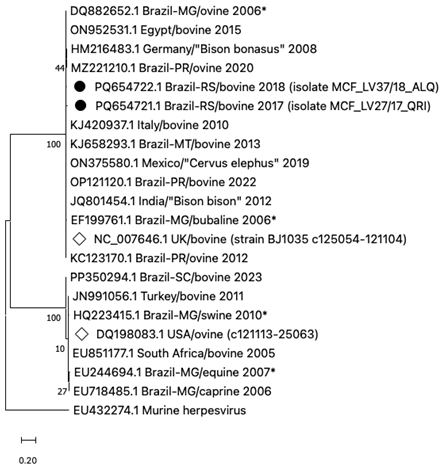
The PCR products of the thalamus and spinal cord (LV27/17) and the frontal cortex and cornea (LV 37/18) were sequenced. The consensus sequences obtained from the two tissue samples were identical, and identified as MCF_LV27/17_QRI and MCF_LV37/18_ALQ and deposited in GenBank with the accession numbers PQ654722.1 and PQ654721.1, respectively. Nucleotide BLAST analysis confirmed the identity of the sequences, showing 98% to 100% identity with other sequences in GenBank. Phylogenetic analysis of the nucleotide sequence revealed that the two samples were grouped together in the same cluster, along with the reference (NC_007646.1) identified in the United Kingdom and other OvHV-2 sequences from bovines identified in Brazil (Mato Grosso, Paraná), Italy and Egypt (Figure 2). The two clinical and epidemiological presentations described were consistent with SA-MCF.
Phylogenetic analysis based on 22 nucleotide sequences of the Macavirus ovinegamma 2 (OvHV-2) tegument protein gene. The evolutionary history was inferred by using the Maximum Likelihood method and the evolutionary distances were determined based on the Jukes-Cantor model, with 1000 bootstrap replicates. The percentage of trees in which the associated taxa clustered together is shown below the branches. Sequences from this study are identified (⏺), as well the reference complete genome sequences (◊). GenBank accession number, origins/hosts and collection date (or submission date*) of the OvHV-2 nucleotide sequences are available (MG-Minas Gerais; MT-Mato Grosso; PR-Paraná; RS-Rio Grande do Sul; SC-Santa Catarina; UK-United Kingdom; USA-United States of America).
The necropsy and histopathological findings, associated with the amplification of an OvHV-2 genome sequence, unequivocally confirmed the presence of the viral agent. MCF has been described in Brazil since 1956, and in Rio Grande do Sul (RS) since 1973, and several aspects still need to be investigated (HEADLEY et al., 2020; RECH et al., 2005). In RS, most of the diagnosed cases occurred in the central and southern regions of the state (RECH et al., 2005). The two cases described were recorded in the western region, characterized by extensive mixed production of cattle and sheep. Extensive farming systems are predominant in this region, and the herds are mostly beef cattle and sheep for wool and/or meat production (SILVA et al., 2013). These animals are often raised together, facilitating pathogen transmission between species. This situation favors the spread of infectious diseases since there is more significant contact between animals and they share common areas. Therefore, the region meets the epidemiological characteristics for the occurrence of infection and disease.
The lack of case reports in this state region may be associated with various factors such as poor access to laboratory diagnosis, the reduced number of affected animals, the sporadic occurrence of the disease, as well as producers’ and veterinarians’ lack of knowledge about the disease (HEADLEY et al., 2020; RECH et al., 2005; SILVA et al., 2013). These factors, alone or together, make it difficult to confirm cases and report the disease in the region and can lead to underreporting. MCF should also be considered a differential diagnosis for other diseases, especially those affecting cattle’s neurological and respiratory systems (O’TOOLE & LI, 2014).
In both outbreaks, only one animal was affected, and both affected farms kept cattle and sheep in close contact. In the SA-MCF form, sheep are considered the natural hosts of OvHV-2 and the main source of infection for other species. The excretion of OvHV-2 in nasal secretions and direct transmission to cattle appears to be the main form of infection (LI et al., 2014; O’TOOLE & LI, 2014). Carrier sheep excrete the virus at certain times, usually associated with periods of stress, such as postpartum. It was not possible to determine when the ewes on the property lambed. However, the lambing season in the region’s flocks occurs mainly from June until October (SILVA et al., 2013). The best form of MCF prevention is to avoid or reduce contact between these two species, especially during the period of greatest transmission (RUSSELL et al., 2009). The identification of sheep flocks infected with OvHV-2 by detection of viral DNA or by serology, followed by segregation of these animals, is suggested as an alternative for the prevention of the disease (ELOI et al., 2017; LI et al., 2014).
The amplification of a fragment of the ORF75 gene encoding a tegument protein made it possible to quickly and unequivocally confirm the involvement of OvHV-2 in both cases. In addition, phylogenetic analysis of the fragment showed that the OvHV-2 present in the two cases is similar to other viruses identified in cattle from different regions of Brazil and to a sample from the United States, considered a reference (HEADLEY et al., 2020). A complete analysis of the OvHV-2 genome would be necessary to enable more remarkable inferences about the characteristics of the samples present in the outbreak. In addition, serological studies to determine the infection rate in cattle and sheep, in specific cases and in other herds, could produce complementary information relevant to understanding the epidemiology of OvHV-2 in the region.
Despite the scarcity of information on the circulation of OHV-2 in flocks of sheep and herds of cattle in Brazil, as well as the sporadic occurrence of the disease, the increase in animal density per area, the intensification of production, and the mechanisms of viral latency may favor the emergence of new cases and aggravate the losses. In conclusion, two cases of SA-MCF were diagnosed in the western region of RS. The clinical signs, epidemiological data, lesions, and microscopic findings confirmed the disease and demonstrated the presence of OvHV-2 in this region. Genetic analysis of the samples indicated similarities with other viruses already identified in Brazil. Virological and serological studies to investigate the presence and circulation of the agent in the region’s herds are necessary to understand the circulation of OvHV-2 in cattle and sheep and determine the importance of the infection. Furthermore, veterinarians and producers should be alert, consider SA-MCF in suspected cases, and send material for laboratory diagnosis.
ACKNOWLEDGMENTS
JCJS is the recipient of a scholarship for a doctorate from Coordenação de Aperfeiçoamento de Pessoal de Nível Superior (CAPES). This research was financed with resources from Laboratório de Virologia/UNIPAMPA, Pró-Reitoria de Pós-graduação, Pesquisa e Inovação, Universidade Federal do Pampa (PROPPI/UNIPAMPA), and by the Coordenação de Aperfeiçoamento de Pessoal de Nível Superior (CAPES), Brazil - Finance code 001.
REFERENCES
-
BAXTER, S. I. et al. PCR detection of the sheep-associated agent of malignant catarrhal fever. Archives of Virology, v.132, n.1-2, p.145-159, 1993. Available from: <Available from: https://link.springer.com/article/10.1007/BF01309849 >. Accessed: Mar. 18, 2020. doi: 10.1007/BF01309849.
» https://doi.org/10.1007/BF01309849.» https://link.springer.com/article/10.1007/BF01309849 -
ELOI, R. S. A. et al. Ovine herpesvirus type 2 (OvHV-2) infection rate in sheep herds of the Federal District, Brazil. Pesquisa Veterinária Brasileira, v.37, n.7, p.657-661, 2017. Available from: <Available from: https://www.scielo.br/j/pvb/a/PCGbhrHm86dMyL3V6mvNwsq/abstract/?lang=en >. Accessed: Mar. 18, 2020. doi: 10.1590/S0100-736X2017000700001.
» https://doi.org/10.1590/S0100-736X2017000700001.» https://www.scielo.br/j/pvb/a/PCGbhrHm86dMyL3V6mvNwsq/abstract/?lang=en -
GATHERER, D. et al. ICTV Virus Taxonomy Profile: Herpesviridae 2021. Journal of General Virology, v.102, n.10, p.001673, 2021. Available from: <Available from: https://www.microbiologyresearch.org/content/journal/jgv/10.1099/jgv.0.001673 >. Accessed: Aug. 15, 2024. doi: 10.1099/jgv.0.001673.
» https://doi.org/10.1099/jgv.0.001673.» https://www.microbiologyresearch.org/content/journal/jgv/10.1099/jgv.0.001673 - GREEN, M. R.; SAMBROOK, J. Molecular Cloning: A Laboratory Manual. Cold Spring Harbor Laboratory Press; 4th revised edition. 2028 pg. 2012.
- HALL, T. A.; BioEdit: a user-friendly biological sequence alignment editor and analysis program for Windows 95/98/ NT. Nucleic Acids Symposium Series, 41, p.95-98, 1999.
-
HEADLEY, S. A. et al. A review of the epidemiological, clinical, and pathological aspects of malignant catarrhal fever in Brazil. Brazilian Journal of Microbiology, v.51, n.3, p.1405-1432, 2020. Available from: <Available from: https://link.springer.com/article/10.1007/s42770-020-00273-6 >. Accessed: Aug. 15, 2024. doi: 10.1007/s42770-020-00273-6.
» https://doi.org/10.1007/s42770-020-00273-6.» https://link.springer.com/article/10.1007/s42770-020-00273-6 - JUKES, T. H.; CANTOR C. R. Evolution of protein molecules. In MUNRO, H. N. editor, Mammalian Protein Metabolism, Academic Press: New York, p.21-132, 1969.
-
LI, H. et al. Malignant catarrhal fever: inching toward understanding. Annual Review Animal Biosciences, v.2, p.209-233, 2014. Available from: <Available from: https://www.annualreviews.org/content/journals/10.1146/annurev-animal-022513-114156 >. Accessed: Aug. 15, 2024. doi: 10.1146/annurev-animal-022513-114156.
» https://doi.org/10.1146/annurev-animal-022513-114156.» https://www.annualreviews.org/content/journals/10.1146/annurev-animal-022513-114156 -
O’TOOLE, D.; LI, H. The pathology of malignant catarrhal fever, with an emphasis on ovine herpesvirus 2. Veterinary Pathology, v.51, n.2, p.437-452, 2014. Available from: <Available from: https://journals.sagepub.com/doi/full/10.1177/0300985813520435 >. Accessed: Mar. 18, 2020. doi: 10.1177/0300985813520435.
» https://doi.org/10.1177/0300985813520435.» https://journals.sagepub.com/doi/full/10.1177/0300985813520435 -
RECH, R. R. et al. Malignant catarrhal fever in cattle in Rio Grande do Sul, Brazil: epidemiology, clinical signs and pathology. Pesquisa Veterinária Brasileira, v.25, n.2, p.97-105, 2005. Available from: <Available from: https://www.scielo.br/j/pvb/a/snHFypBVPZqwt6QcD8gwMVD/abstract/?lang=en >. Accessed: Aug. 15, 2024. doi: 10.1590/S0100-736X2005000200006.
» https://doi.org/10.1590/S0100-736X2005000200006.» https://www.scielo.br/j/pvb/a/snHFypBVPZqwt6QcD8gwMVD/abstract/?lang=en -
RUSSELL, G. C. et al. Malignant catarrhal fever: a review. The Veterinary Journal, v.179, p.324-335, 2009. Available from: <Available from: https://www.sciencedirect.com/science/article/pii/S1090023307003899?via%3Dihub >. Accessed: Aug. 15, 2024. doi: 10.1016/j.tvjl.2007.11.007.
» https://doi.org/10.1016/j.tvjl.2007.11.007.» https://www.sciencedirect.com/science/article/pii/S1090023307003899?via%3Dihub -
SILVA, A. P. S. P. et al. Sheep industry in the State of Rio Grande do Sul, Brazil: description of the production system and the main health and reproductive aspects. Pesquisa Veterinária Brasileira, v.33, n.12, p.1441-1446, 2013. Available from: <Available from: https://www.scielo.br/j/pvb/a/t8TQ9k7TmHk9DVmJ7wP5JvJ/abstract/?lang=en >. Accessed: Aug. 15, 2024. doi: 10.1590/S0100-736X2013001200010.
» https://doi.org/10.1590/S0100-736X2013001200010.» https://www.scielo.br/j/pvb/a/t8TQ9k7TmHk9DVmJ7wP5JvJ/abstract/?lang=en -
STECHER, G. et al. Molecular Evolutionary Genetics Analysis (MEGA) for macOS. Molecular Biology and Evolution, v.37, p.1237-1239, 2020. Available from: <Available from: https://academic.oup.com/mbe/article/37/4/1237/5697095 >. Accessed: Jan. 20, 2025. doi 10.1093/molbev/msz312.
» https://doi.org/10.1093/molbev/msz312» https://academic.oup.com/mbe/article/37/4/1237/5697095 -
TAMURA K. et al. MEGA 11: Molecular Evolutionary Genetics Analysis Version 11. Molecular Biology and Evolution, 2021. Available from: <Available from: https://academic.oup.com/mbe/article/38/7/3022/6248099?login=true >. Accessed: Jan. 20, 2025. doi: 10.1093/molbev/msab120.
» https://doi.org/10.1093/molbev/msab120.» https://academic.oup.com/mbe/article/38/7/3022/6248099?login=true
-
CR-2025-0150.R1
-
BIOETHICS AND BIOSECURITY COMMITTEE APPROVAL
For all due purposes, the authors of the article “Molecular diagnosis of cases of sheep-associated malignant catarrhal fever in cattle in Rio Grande do Sul, Brazil” declare that the project that gave rise to the data of the same was not submitted for evaluation to the Ethics Committee of the Universidade Federal do Pampa. However, we are aware of the content of the resolutions of the National Council for the Control of Animal Experimentation - CONCEA <http://www.mct.gov.br/index.php/content/view/310553.html> if it involves animals. Thus, the authors assume full responsibility for the data presented and are available for possible questioning, should they be required by the competent bodies.
-
DATA AVAILABILITY STATEMENT
All datasets generated and/or analyzed in the current study are available and may also be made available by the corresponding author upon reasonable request.
-
DECLARATION OF USE OF ARTIFICIAL INTELLIGENCE
The authors declare that no generative AI was used to write, analyze, or interpret the data in this manuscript.
Edited by
-
ASSOCIATE EDITOR
Rudi Weiblen (0000-0002-1737-9817)
All datasets generated and/or analyzed in the current study are available and may also be made available by the corresponding author upon reasonable request.