 Short-term effects of platelet- rich plasma on the synovial fluid of horses with induced synovitis
ABSTRACT:
Platelet-Rich Plasma (PRP) directly and indirectly modifies the composition of synovial fluid in horses. This study evaluated the early effects of intra-articular PRP on the synovial fluid of horses with experimentally induced synovitis. Twelve horses were divided into two groups: a control group that received no treatment, and a treatment group that received intra-articular PRP. Five time points were established: T0, T3, T6, T24, and T48. At T0, all horses received an intra-articular injection of lipopolysaccharide from Escherichia coli. At T3, synovial fluid samples were collected from all horses, and the treatment group received PRP via the same joint access. Additional synovial fluid samples were collected at T6, T24, and T48 for evaluation. The PRP preparation protocol used in this study resulted in an adequate platelet concentration that did not affect the physicochemical and cytological characteristics of the synovial fluidor the concentrations of IL-1RA, MMP-9, and MMP-2 in horses with induced synovitis.
Key words:
cell therapy; joint disease; PRP; sports medicine
RESUMO:
Sabe-se que ouso intra-articular do plasma rico emplaquetas (PRP) modifica direta ou indiretamente a composição do líquido sinovial de equinos. O objetivo deste estudo foi avaliar os efeitos do PRP a curto prazo no líquido sinovial de equinos com sinovite induzida. Foram utilizados 12 equinos distribuídos em dois grupos: grupo controle, que não recebeu tratamento, e o grupo tratamento, que recebeu PRP intra-articular. Foram estabelecidos cinco momentos experimentais: T0, T3, T6, T24 e T48. No momento T0, todos os animais receberam uma aplicação de lipopolissacarídeo de Escherichia coli intra-articular. No momento T3, foram colhidas novas amostras de líquido sinovial de todos os animais e o mesmo acesso articular foi utilizado para aplicar PRP nos animais do grupo de tratamento. Nos momentos T6, T24 e T48 foram colhidas as amostras subsequentes de líquido sinovial e essas submetidas a avaliação. O protocolo de preparação do PRP utilizado no presente estudo é capaz de produzir um concentrado de plaquetas adequado, que não altera as características físico-químicas e citológicas, bem como as concentrações de IL-1RA, MMP-9 e MMP-2 do líquido sinovial de equinos com sinovite induzida.
Palavras-chave:
medicina esportiva; doença articular; PRP; terapia celular
INTRODUCTION
Platelet-rich plasma (PRP) has been widely used as a treatment for various orthopedic injuries in horses, with its intra-articular application showing promising results in the treatment of osteoarthritis (OA) (GARBIN et al., 2022; MORAES et al., 2015; TEXTOR & TABLIN, 2013). In vitro studies have demonstrated that PRP can mitigate the catabolic effects of inflammatory cytokines, such as interleukin-1 (IL-1) and matrix metalloproteinases (MMPs) (XIE et al., 2014; XU et al., 2021), which are key molecules in joint inflammation.
OA results from injuries to multiple articular and periarticular structures, such as the synovial membrane, cartilage, subchondral bone, and cortical bone. While there is ongoing debate regarding which tissue structures undergo the initial changes in OA, increasing evidence has suggested that synovitis plays a significant early pathogenic role in joint inflammation, rather than merely a consequence of joint damage (ATUKORALA et al., 2016). Synovial inflammation, even without joint injury or instability, has been shown to induce articular cartilage degradation in horses (MCILWRAITH & SICKLE, 1981) and is associated with both early and late OA in human patients (BENITO et al., 2005; SANCHEZ-LOPEZ et al., 2022), as well as contributing to OA progression in mice (LIAO et al., 2020). Additionally, synovitis is known to produce pain and increase the production of inflammatory mediators, contributing to the progression of articular cartilage degeneration in horses (MCILWRAITH et al., 2016).
IL-1 is the primary cytokine associated with degenerative changes in synovial joints, and due to its highly inflammatory nature, blocking its activity is a key focus in the treatment of various inflammatory diseases. The natural antagonist of IL-1, the interleukin-1 receptor antagonist protein (IL-1RA), has become a central focus in studies on cellular therapies (KANEKO et al., 2019; MORAES et al., 2015).
In turn, MMPs, particularly MMP-2 and MMP-9, are proteases capable of degrading the components of the extracellular matrix of articular cartilage. These enzymes are among the main proteases expressed in the synovial fluid (SF) of animals with OA (MEHANA et al., 2019).
Intra-articular administration of PRP is known to directly or indirectly modify the chemical, physical, and cytological composition of SF in horses (MORAES et al., 2015). Previous studies have shown that PRP administration in healthy equine joints induces a mild to moderate inflammatory response in the SF, which does not harm the articular environment (GARBIN et al., 2022; MORAES et al., 2015; TEXTOR & TABLIN, 2013). However, few studies have specifically evaluated the effects of intra-articular PRP application on the SF of horses with synovitis (SMIT et al., 2019).
Therefore, this study produced PRP with enriched platelet concentration while maintaining low counts of erythrocytes and leukocytes and to evaluate the early effects of intra-articular PRP on the SF of horses with experimentally induced acute synovitis.
MATERIALS AND METHODS
Platelet - rich plasma preparation
Twelve healthy horses, both male and female, of mixed breeds were used in this study. The animals underwent a comprehensive clinical and hematological examination. Additionally, the carpal joint was evaluated through inspection, palpation, radiography (EcoRay model Orange® 1060HF; Fujifilm® FCR Capsula X), and ultrasonography (Logic E, GE®, linear transducer of 7 to 12 MHz) to ensure joint health.
After local antisepsis, approximately 100 mL of blood was collected from the jugular vein of each horse into 25 sodium citrate tubes, each with a capacity of 4.5 mL (BD Vacutainer® Sodium Citrate 3.2%). One tube was used for a total blood platelet count, and the remaining tubes were used for PRP preparation. The blood samples were homogenized and centrifuged (Centribio® Model 80-2B) at 145 x g for 10 minutes. In a laminar flow hood, all centrifuged plasma was collected and transferred to dry tubes, which were then centrifuged at 2325 x g for 10 minutes. After the second centrifugation, approximately 80% of the supernatant plasma was discarded, and the remaining fraction (PRP) was homogenized in a shaker before being transferred to a sterile syringe. Platelet counts for whole blood and PRP were performed using an automated hematology analyzer.
Experimental time points
The horses were divided into two groups, each consisting of six animals: the control group (CG: C1, C2, C3, C4, C5, and C6) and the treatment group (TG: P1, P2, P3, P4, P5, and P6). Five experimental time points were established: T0, T3, T6, T24, and T48 (MORAES et al., 2015) (Figure 1).
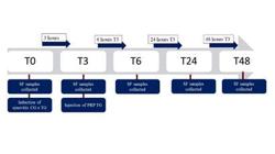
Graphical representation of the experimental timeline for synovitis induction, synovial fluid collection, and PRP injection in 12 horses.
At time point T0, all animals underwent arthrocentesis via access to the lateral recess of the radiocarpal joint (MOYE et al., 2007) for synovial fluid (SF) collection and induction of synovitis. Synovitis was induced by administering 0.25 ng of LPS (Lipopolysaccharide from Escherichia coli 055: B5, Sigma Aldrich®) diluted in 1 mL of sterile phosphate-buffered saline (PBS) solution intra-articularly (HUNT et al., 2019; LOON et al., 2012; LUCIA et al., 2013; NEUENSCHWANDER et al., 2019).
At time point T3, three hours after T0, new SF samples were collected from all animals. At the same time, the TG animals received an injection of 4 mL of PRP. At time points T6, T24, and T48, corresponding to 6, 24, and 48 hours after T3; respectively, SF samples were collected from all animals (MORAES et al., 2015), totaling 48 samples.
Clinical evaluation
The animals were clinically evaluated before each experimental time point through static and dynamic visual inspection and palpation of the radiocarpal joint. The parameters assessed included pain on palpation, heat, effusion, and the degree of lameness determined according to the American Association of Equine Practitioners (AAEP) Lameness Classification System.
Synovial fluid analysis
The SF samples were subjected to physical, chemical, and cytological analysis. The parameters evaluated included color, viscosity, mucin clot test, total protein (TP) concentration, total and differential nucleated cell count (TNCC), and erythrocyte count. Additionally, the concentrations of IL-1RA, MMP-2, and MMP-9 in the SF were determined. For these evaluations, the sandwich ELISA method was used for IL-1RA determination (Human Interleukin 1 Receptor Antagonist, Elabscience®) and MMP-2 and MMP-9 (Matrix Metalloproteinase 2, Matrix Metalloproteinase 9, Elabscience®) using commercial kits, following the manufacturers’ recommendations. Previous studies have validated similar kits for determining IL-1RA (FRISBIE et al., 2002), MMP-2, and MMP-9 (FIETZ et al., 2008) concentrations in equine SF.
Statistical analysis
Data were analyzed using GraphPad Prism software. Quantitative data were subjected to the Kolmogorov-Smirnov test to assess normality. For comparisons between groups at different time points, an unpaired t-test was used for parametric data, and the Mann-Whitney test was used for non-parametric data. For paired data, the Wilcoxon test was applied. To compare time points within the same group, ANOVA was used for parametric data, and the Friedman test was used for non-parametric data. A significant level of 5% was adopted for all tests.
RESULTS
The PRP production yielded significant variation in platelet increment and total leukocyte count (Table 1). No differences in lameness were noted between groups. Mild lameness occurred following synovitis induction (T3 and T9) across all animals, with either mild or no lameness observed at later times (T24 and T48). Palpation of the joint region post-synovitis induction, compared to the contralateral limb, revealed increased temperature, pain, and effusion in all animals, with no distinctions among groups.
Post-synovitis, there was an increase in nucleated cells in the synovial fluid (SF), predominantly neutrophils, alongside elevated protein levels and reduced viscosity and mucin clot quality across all 12 animals. SF color variations included light yellow (4.16%), orange (14.59%), and reddish (81.25%) (Figure 2). Most samples exhibited decreased viscosity (58.33%) and poor mucin clot formation (66.66%). Density escalated at each measurement time post-induction in both groups, with significantly higher values at T6 in the treatment group (TG). No significant changes in total protein concentration were seen between groups post-T3, though variations were noted over time within the control group (CG) and TG (Table 2).
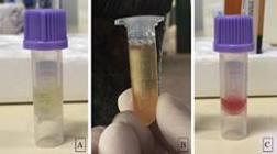
Classification of synovial fluid coloration obtained via arthrocentesis from the radiocarpal joint in horses with induced synovitis: light yellow (A), orange (B), and reddish (C).
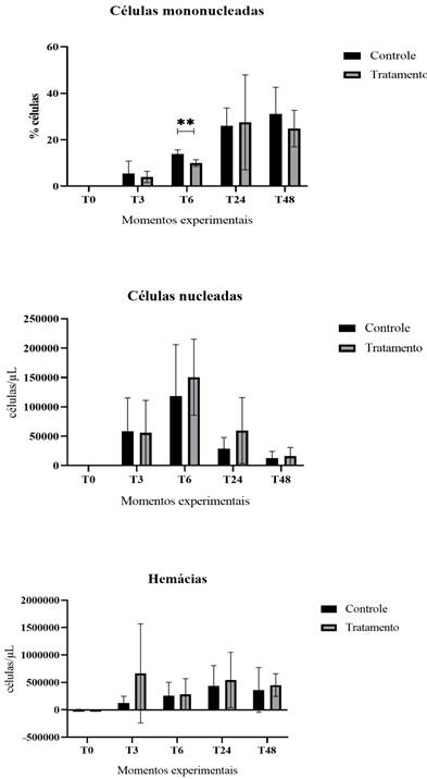
Total nucleated cell count (TNCC) and differential counts of neutrophils, lymphocytes, eosinophils, mononuclear cells, and erythrocytes showed no significant temporal differences between groups, except for mononuclear cells at T6, where the CG presented higher values (P < 0.01) compared to the TG (Figure 3). Neutrophil differential counts remained consistent across groups.
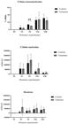
Graphical representation of the means and standard deviations of nucleated cells, mononuclear cells, and erythrocyte counts in the synovial fluid from the radiocarpal joint of equines with induced synovitis, treated or untreated with platelet- rich plasma, at five experimental time points. ** indicates a statistical difference between time points according to the Wilcoxon test, with P < 0.01.
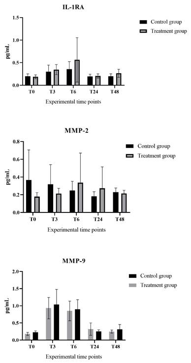
No statistical differences were detected in IL-1RA levels among the groups across five experimental time points. Increases in MMP-2 and MMP-9 concentrations were noted at all assessed times in both groups, albeit without significant differences (Figure 4).
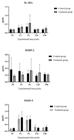
Graphical representation of the means and standard deviations of interleukin-1 receptor antagonist protein and matrix metalloproteinases 2 and 9 concentrations in the synovial fluid from the radiocarpal joint of equines with induced synovitis, treated or untreated with platelet- rich plasma, at five experimental time points.
DISCUSSION
The synovitis induction model, utilizing 0.25 ng of LPS as described in prior studies (HUNT et al., 2019; LOON et al., 2012; LUCIA et al., 2013; NEUENSCHWANDER et al., 2019), successfully induced transient joint inflammation in equines. This was evidenced by an increase in nucleated cells in the synovial fluid (SF), predominantly neutrophils, along with elevated protein levels and reduced viscosity and mucin clot quality, observed three hours post-induction in all 12 animals.
The PRP preparation method aimed to produce plasma enriched with platelets while maintaining lower levels of total leukocytes and erythrocytes compared to the animal’s plasma. This protocol yielded a concentration high in platelets and low in leukocytes and erythrocytes, as intended.
Despite higher leukocyte counts in the PRP of animals P2, P4, and P5, the clinical signs of joint inflammation mirrored those in animals receiving PRP with lower leukocyte levels. The erythrocyte concentration remained low across all PRP samples, which is beneficial since high erythrocyte levels in the joint can lead to synoviocyte damage, cell death, and articular cartilage degradation (BRAUNE et al., 2014; MORAES et al., 2015).
Platelet concentration is deemed crucial for PRP efficacy and protocol selection, although there is no standardization for this parameter (CASTILLO et al., 2011; ROSSI et al., 2019; SÁNCHEZ et al., 2010; SINGLA et al., 2017). Variability in platelet counts was observed, with increments ranging from 2.05 to 13 times that of whole blood. Particularly, PRP from animals P4, P5, and P6 showed higher increments, reaching six, seven, and 13 times, respectively. However, this variability did not affect the clinical or laboratory indicators of joint inflammation.
Most samples displayed a reddish hue persisting until T24, potentially attributable to repeated arthrocentesis, limb movement during procedures, or needle repositioning (STEEL, 2008). After T24, erythrocyte numbers decreased, likely reflecting the resolution of transient synovitis induced by LPS and the inflammation from repeated arthrocentesis (RINNOVATI et al., 2017). Thus, PRP seemed not to impact erythrocyte counts.
Following synovitis induction, an expected increase in total protein (TP) concentration occurred due to inflammation (CHEN et al., 2017; KNOP et al., 2016). At T3, TP concentrations in both groups surpassed reference values, peaking at T6, and remaining elevated until T48. Although, differences were not statistically significant, TP levels were notably higher in the treatment group (TG), possibly due to PRP application, which enhances protein levels and enzyme release (BROSSI et al., 2015; MORAES et al., 2015). From T6 onward, TP concentrations gradually decreased, aligning in both groups as the transient synovitis resolved (BRAMA et al., 2004).
In the analyzed samples, viscosity was reduced, and mucin clots were deemed poor following synovitis induction, indicative of decreased hyaluronic acid (HA) concentration due to inflammation (STEEL, 2008). This decrease is typically facilitated by hyaluronidase activity within the extracellular matrix of the inflamed environment, leading to HA breakdown. Although, PRP is known to stimulate HA production in synoviocytes via growth factors (KNOP et al., 2016), the study observed predominantly poor-quality clots in both groups post-induction, suggesting no significant impact of PRP on mucin clot quality within the short evaluation period (STEEL, 2008).
Total nucleated cell count (TNCC) and differential counts of neutrophils, lymphocytes, eosinophils, mononuclear cells, and erythrocytes showed no temporal differences between groups, except for an elevation in mononuclear cells at T6 in the control group, potentially due to enhanced migration triggered by the inflammatory response (FERNANDES et al., 2020). Although, there were no statistically significant differences, TNCC increases were more pronounced in the treatment group (TG) at T6 and T24, likely due to cytokine release from platelet α-granules enhancing inflammatory cell recruitment (WU et al., 2016). After T6, TNCC trends aligned between groups, with a decline in counts suggesting a resolution of synovitis. This pattern indicated that PRP did not significantly affect nucleated cell reduction beyond this point.
The interleukin-1 receptor antagonist protein (IL-1RA) plays a crucial anti-inflammatory role in equines with synovitis (CARMONA & PRADES, 2009). Peak inflammation occurred three hours post-synovitis induction, corresponding with elevated IL-1RA levels at T3 and T6 in both groups, mirroring findings from another study comparing joints treated and untreated with HA (NIEMELÄ et al., 2019). Despite the lack of significant differences between groups, higher IL-1RA levels were noted in the PRP-treated group, possibly reflecting a stronger stimulus for endogenous synthesis or a higher concentration within the PRP (ZIEGLER et al., 2019).
Matrix metalloproteinases (MMPs), notably involved in osteoarthritis (OA) progression, degrade collagen in articular cartilage and are elevated in the synovium even during early OA stages (ARAKI et al., 2016; DAVIDSON et al., 2006; YAO et al., 2023). Although, MMP-2 and MMP-9 levels did not differ significantly, MMP-9 concentrations were notably higher at T3 and T6, likely influenced by increased pro-inflammatory cytokines during these stages (TURLO et al., 2022).
The anticipated pro-inflammatory potential of intra-articular PRP, as noted in other studies (MACHADO et al., 2019; MORAES et al., 2015; SMIT et al., 2019; TEXTOR & TABLIN, 2013), was not significant in this experiment. This suggested that PRP-induced inflammation might be negligible in animals already experiencing synovitis, possibly due to the low leukocyte count in the produced PRP correlating with a milder inflammatory response within the joint.
CONCLUSION
The PRP preparation protocol employed in this study effectively generates a concentration with high platelet levels while maintaining low counts of erythrocytes and leukocytes, which does not worsen the clinical signs or laboratory indicators of synovitis. Over a short duration, PRP does not affect the physicochemical or cytological properties of the synovial fluid, nor does it alter the concentrations of IL-1RA, MMP-9, and MMP-2 in equines with experimentally induced acute synovitis.
ACKNOWLEDGMENTS
The authors thank the Programa de Pós-graduação em Ciência Animal da Universidade Federal de Goiás (UFG), the Escola de Veterinária e Zootecnia da Universidade Federal de Goiás and the Conselho Nacional de Desenvolvimento Científico e Tecnológico (CNPq).
REFERENCES
-
ARAKI, Y. et al. Histone m ethylation and STAT-3 differentially regulate Interleukin-6-Induced Matrix Metalloproteinase Gene Activation in Rheumatoid Arthritis Synovial Fibroblasts. Arthritis and Rheumatology, v.68, n.5, p.1111-1123, 2016. Available from: <Available from: https://acrjournals.onlinelibrary.wiley.com/doi/epdf/10.1002/art.39563 >. Accessed: Mar. 15, 2023. doi: 10.1002/art.39563.
» https://doi.org/10.1002/art.39563.» https://acrjournals.onlinelibrary.wiley.com/doi/epdf/10.1002/art.39563 -
ATUKORALA, I. et al. Synovitis in knee osteoarthritis: a precursor of disease? Annals of the Rheumatic Diseases, v.75, n.2, p.390-395, 2016. Available from: <Available from: https://ard.bmj.com/content/75/2/390.long >. Accessed: Feb. 18, 2024. doi: 10.1136/annrheumdis-2014-205894.
» https://doi.org/10.1136/annrheumdis-2014-205894.» https://ard.bmj.com/content/75/2/390.long -
BENITO, M. J. et al. Synovial tissue inflammation in early and late osteoarthritis. Annals of the Rheumatic Diseases, v.64, n.9, p.1263-1267, 2005. Available from: <Available from: https://ard.bmj.com/content/64/9/1263.long >. Accessed: Feb. 18, 2024. doi: 10.1136/ard.2004.025270.
» https://doi.org/10.1136/ard.2004.025270.» https://ard.bmj.com/content/64/9/1263.long -
BRAMA, P. A. J. et al. Collagenase-1 (MMP-1) activity in equine synovial fluid: Influence of age, joint pathology, exercise and repeated arthrocentesis. Equine Veterinary Journal, v.36, n.1, p.34-40, 2004. Available from: <Available from: https://beva.onlinelibrary.wiley.com/doi/abs/10.2746/0425164044864705?sid=nlm%3Apubmed >. Accessed: Mar. 15, 2023.doi: 10.2746/0425164044864705.
» https://doi.org/10.2746/0425164044864705.» https://beva.onlinelibrary.wiley.com/doi/abs/10.2746/0425164044864705?sid=nlm%3Apubmed -
BRAUNE, S. et al. Changes in platelet morphology and function during 24 hours of storage. Clinical Hemorheology and Microcirculation, v.58, n.1, p.159-170, 2014. Available from: <Available from: https://content.iospress.com/articles/clinical-hemorheology-and-microcirculation/ch1876 >. Accessed: Mar. 18, 2023. doi: 10.3233/CH-141876.
» https://doi.org/10.3233/CH-141876.» https://content.iospress.com/articles/clinical-hemorheology-and-microcirculation/ch1876 -
BROSSI, P. M. et al. Platelet-rich plasma in orthopedic therapy: A comparative systematic review of clinical and experimental data in equine and human musculoskeletal lesions. BMC Veterinary Research, v.11, n.1, p.1-17, 2015. Available from: <Available from: https://pmc.ncbi.nlm.nih.gov/articles/PMC4449579/ >. Accessed: Mar. 15, 2023. doi: 10.1186/s12917-015-0403-z.
» https://doi.org/10.1186/s12917-015-0403-z.» https://pmc.ncbi.nlm.nih.gov/articles/PMC4449579/ -
CARMONA, J. U.; PRADES, M. The pathophysiology of osteoarthritis pain. Compedium Equine, v.4, n.1, p.28-40, 2009. Available from: <Available from: https://www.vetfolio.com/learn/article/pathophysiology-of-osteoarthritis >. Accessed: Mar. 15, 2023.
» https://www.vetfolio.com/learn/article/pathophysiology-of-osteoarthritis -
CASTILLO, T. N. et al. Comparison of growth factor and platelet concentration from commercial platelet-rich plasma separation systems. American Journal of Sports Medicine, v.39, n.2, p.266-271. 2011. Available from: <Available from: https://journals.sagepub.com/doi/abs/10.1177/0363546510387517?journalCode=ajsb >. Accessed: Mar. 15, 2023. doi: 10.1177/0363546510387517.
» https://doi.org/10.1177/0363546510387517.» https://journals.sagepub.com/doi/abs/10.1177/0363546510387517?journalCode=ajsb -
CHEN, C. P. C. et al. The influence of platelet rich plasma on synovial fluid volumes , protein concentrations , and severity of pain in patients with knee osteoarthritis. Experimental Gerontology, v.93, p.68-72, 2017. Available from: <Available from: https://www.sciencedirect.com/science/article/abs/pii/S0531556517300943?via%3Dihub >. Accessed: Mar. 15, 2023. doi: 10.1016/j.exger.2017.04.004.
» https://doi.org/10.1016/j.exger.2017.04.004.» https://www.sciencedirect.com/science/article/abs/pii/S0531556517300943?via%3Dihub -
DAVIDSON, R. K. et al. Expression profiling of metalloproteinases and their inhibitors in synovium and cartilage. Arthritis Research and Therapy, v.8, n.4, p.1-10, 2006. Available from:<Available from:https://pmc.ncbi.nlm.nih.gov/articles/PMC1779413/ >. Accessed: Aug. 10, 2023. doi: 10.1186/ar2013.
» https://doi.org/10.1186/ar2013.» https://pmc.ncbi.nlm.nih.gov/articles/PMC1779413/ -
FERNANDES, T. L. et al. Macrophage: a potential target on cartilage regeneration. Frontiers in Immunology, v.11, n.February, p.1-9, 2020. Available from: <Available from: https://www.frontiersin.org/journals/immunology/articles/10.3389/fimmu.2020.00111/full >. Accessed: Mar. 18, 2023. doi: 10.3389/fimmu.2020.00111.
» https://doi.org/10.3389/fimmu.2020.00111.» https://www.frontiersin.org/journals/immunology/articles/10.3389/fimmu.2020.00111/full -
FIETZ, S. et al. Determination of MMP-2 and -9 activities in synovial fluid of equines with osteoarthritic and arthritic joint diseases using gelatin zymography and immunocapture activity assays. Equine Veterinary Journal, v.40, n.3, p.266-271, 2008. Available from: <Available from: https://beva.onlinelibrary.wiley.com/doi/10.2746/042516408X251921 >. Accessed: Feb. 10, 2024. doi: 10.2746/042516408X251921.
» https://doi.org/10.2746/042516408X251921.» https://beva.onlinelibrary.wiley.com/doi/10.2746/042516408X251921 -
FRISBIE, D. et al. Treatment of experimental equine osteoarthritis by in vivo delivery of the equine interleukin-1 receptor antagonist gene. Gene Therapy, v.9, p.12-20, 2002. Available from: <Available from: https://www.nature.com/articles/3301608 >. Accessed: Feb. 18, 2024. doi: 10.1038/sj.gt.3301608.
» https://doi.org/10.1038/sj.gt.3301608.» https://www.nature.com/articles/3301608 -
GARBIN, L. C. et al. A safety evaluation of allogeneic freeze-dried platelet-rich plasma or conditioned serum compared to autologous frozen products equivalents in equine healthy joints. BMC Veterinary Research, v.18, n.1, p.1-11, 2022. Available from: <Available from: https://bmcvetres.biomedcentral.com/articles/10.1186/s12917-022-03225-4 >. Accessed: Mar. 15, 2023. doi: 10.1186/s12917-022-03225-4.
» https://doi.org/10.1186/s12917-022-03225-4.» https://bmcvetres.biomedcentral.com/articles/10.1186/s12917-022-03225-4 -
HUNT, C. L. et al. Effects of repeated arthrocentesis on systemic cytokine expression and leukocyte population in young equines challenged with intra-articular lipopolysaccharide. J Anim Sci, p.184-191, 2019. Available from: <Available from: https://pmc.ncbi.nlm.nih.gov/articles/PMC6313106/ >. Accessed: Mar. 15, 2023. doi: 10.1093/jas/sky423.
» https://doi.org/10.1093/jas/sky423.» https://pmc.ncbi.nlm.nih.gov/articles/PMC6313106/ -
KANEKO, N. et al. The role of interleukin-1 in general pathology. Inflammation and Regeneration, v.39, n. 12, p.1-16. 2019. Available from: <Available from: https://pmc.ncbi.nlm.nih.gov/articles/PMC6551897/ >. Accessed: Mar. 15, 2023. doi: 0.1186/s41232-019-0101-5.
» https://doi.org/0.1186/s41232-019-0101-5.» https://pmc.ncbi.nlm.nih.gov/articles/PMC6551897/ -
KNOP, E. et al. Platelet-rich plasma for osteoarthritis treatment. Revista Brasileira de Reumatologia, v.56, n.2, p.152-164. 2016. Available from: <Available from: https://www.scielo.br/j/rbr/a/XPMP9QDHfB9b8BJdvSqSRRP/ >. Accessed: Mar. 18, 2023. doi: 10.1016/j.rbre.2015.07.002.
» https://doi.org/10.1016/j.rbre.2015.07.002.» https://www.scielo.br/j/rbr/a/XPMP9QDHfB9b8BJdvSqSRRP/ -
LIAO, L. et al. Acute synovitis after trauma precedes and is associated with osteoarthritis onset and progression. International Journal of Biological Sciences, v.16, n.6, p.970-980. 2020. Available from: <Available from: https://pmc.ncbi.nlm.nih.gov/articles/PMC7053339/ >. Accessed: Aug. 08, 2023. doi:10.7150/ijbs.39015.
» https://doi.org/10.7150/ijbs.39015.» https://pmc.ncbi.nlm.nih.gov/articles/PMC7053339/ -
LOON, J. P. A. M. V. et al. Analgesic and anti-hyperalgesic effects of epidural morphine in an equine LPS-induced acute synovitis model. The Veterinary Journal, v.193, n.2, p.464-470, 2012. Available from: <Available from: https://www.sciencedirect.com/science/article/abs/pii/S1090023312000317 >. Accessed: Aug. 18, 2023. doi: 10.7150/ijbs.39015.
» https://doi.org/10.7150/ijbs.39015» https://www.sciencedirect.com/science/article/abs/pii/S1090023312000317 -
LUCIA, J. L. et al. Influence of an intra-articular lipopolysaccharide challenge on markers of inflammation and cartilage metabolism in young equines. Journal of Animal Science, n.April, p.2693-2699, 2013. Available from: <Available from: https://academic.oup.com/jas/article-abstract/91/6/2693/4717207?redirectedFrom=fulltext&login=false >. Accessed: Mar. 15, 2023. doi: 10.2527/jas.2012-5981.
» https://doi.org/10.2527/jas.2012-5981.» https://academic.oup.com/jas/article-abstract/91/6/2693/4717207?redirectedFrom=fulltext&login=false -
MACHADO, T. S. L. et al. Effects of blood-derived products and sodium hyaluronate on equine synovial fluid cells and on synovial fluid from osteochondrotic joints of equines after arthroscopy and administration of treatment. American Journal of Veterinary Research, v.80, n.7, p.646-656, 2019. Available from: <Available from: https://avmajournals.avma.org/view/journals/ajvr/80/7/ajvr.80.7.646.xml >. Accessed: Mar. 15, 2023. doi: 0.2460/ajvr.80.7.646.
» https://doi.org/0.2460/ajvr.80.7.646.» https://avmajournals.avma.org/view/journals/ajvr/80/7/ajvr.80.7.646.xml - MCILWRAITH, C. W. et al. Joint disease in the equine. 2 th ed. Saunders Elsevier, 2016.
-
MCILWRAITH, C. W.; SICKLE, V. S. D. Experimentally induced arthritis of the equine carpus: histologic and histochemical changes in the articular cartilage. Am J Vet Res, v.42, n.2, p.209-217, 1981. Available from: <Available from: https://pubmed.ncbi.nlm.nih.gov/7258770/ >. Accessed: Feb. 12, 2024. PMID: 7258770.
» https://pubmed.ncbi.nlm.nih.gov/7258770/ -
MEHANA, E. S. E. et al. The role of matrix metalloproteinases in osteoarthritis pathogenesis: an updated review. Life Sciences, v.234, n.August, p.116786, 2019. Available from: <Available from: https://www.sciencedirect.com/science/article/abs/pii/S0024320519307131?via%3Dihub >. Accessed: Mar. 15, 2023. doi: 10.1016/j.lfs.2019.116786.
» https://doi.org/10.1016/j.lfs.2019.116786.» https://www.sciencedirect.com/science/article/abs/pii/S0024320519307131?via%3Dihub -
MORAES, A. P. L. et al. Short- and long-term effects of platelet-rich plasma upon healthy equine joints: clinical and laboratory aspects. The Canadian veterinary journal, v.56, n.8, p.831-838, 2015. Available from: <Available from: https://www.ncbi.nlm.nih.gov/pmc/articles/PMC4502851/pdf/cvj_08_831.pdf >. Accessed: Mar. 15, 2023.
» https://www.ncbi.nlm.nih.gov/pmc/articles/PMC4502851/pdf/cvj_08_831.pdf - MOYE, W. et al. A guide to equine joint injection and regional anesthesia. Yardley, Pa. : Veterinary Learning Systems, 2007.
-
NEUENSCHWANDER, H. M. et al. Hyaluronic acid has chondroprotective and joint-preserving effects on LPS-induced synovitis in equines. Journal of Veterinary Science, v.20, n.6, p.1-15, 2019. Available from: <Available from: https://pmc.ncbi.nlm.nih.gov/articles/PMC6883190/ >. Accessed: Aug. 18, 2023. doi: 10.4142/jvs.2019.20.e67.
» https://doi.org/10.4142/jvs.2019.20.e67.» https://pmc.ncbi.nlm.nih.gov/articles/PMC6883190/ -
NIEMELÄ, T. M. et al. Evaluation of the effect of experimentally induced cartilage defect and intra-articular hyaluronan on synovial fluid biomarkers in intercarpal joints of equines. Acta Veterinaria Scandinavica, v.61, n.1, p.1-9, 2019. Available from: <Available from: https://pmc.ncbi.nlm.nih.gov/articles/PMC6543688/ >. Accessed: Mar. 18, 2023. doi: 10.1186/s13028-019-0460-6.
» https://doi.org/10.1186/s13028-019-0460-6.» https://pmc.ncbi.nlm.nih.gov/articles/PMC6543688/ -
RINNOVATI, R. et al. Effect of repeated arthrocentesis on cytology of synovial fluid. Journal of Equine Veterinary Science, v.57, p.112-115, 2017. Available from: <Available from: https://www.sciencedirect.com/science/article/abs/pii/S073708061730494X >. Accessed: Mar. 10, 2023. doi: 10.1016/j.jevs.2017.07.008.
» https://doi.org/10.1016/j.jevs.2017.07.008.» https://www.sciencedirect.com/science/article/abs/pii/S073708061730494X -
ROSSI, L. A. et al. Classification systems for platelet-rich plasma. Bone and Joint Journal, v.101 B, n.8, p.891-896, 2019. Available from: <Available from: https://boneandjoint.org.uk/article/10.1302/0301-620X.101B8.BJJ-2019-0037.R1 >. Accessed: Mar. 18, 2023. doi: 10.1302/0301-620X.101B8.BJJ-2019-0037.R1.
» https://doi.org/10.1302/0301-620X.101B8.BJJ-2019-0037.R1.» https://boneandjoint.org.uk/article/10.1302/0301-620X.101B8.BJJ-2019-0037.R1 -
SANCHEZ-LOPEZ, E. et al. Synovial inflammation in osteoarthritis progression. Nature Reviews Rheumatology, v.18, n.5, p.258-275, 2022. Available from: <Available from: https://pmc.ncbi.nlm.nih.gov/articles/PMC9050956/ >. Accessed: Feb. 18, 2024. doi: 10.1038/s41584-022-00749-9.
» https://doi.org/10.1038/s41584-022-00749-9.» https://pmc.ncbi.nlm.nih.gov/articles/PMC9050956/ -
SÁNCHEZ, M. et al. Poor s tandardization in platelet-rich therapies hampers advancement. Arthroscopy - Journal of Arthroscopic and Related Surgery, v.26, n.6, p.725-726, 2010. Available from: <Available from: https://www.arthroscopyjournal.org/article/S0749-8063(10)00204-5/fulltext >. Accessed: Mar. 15, 2023. doi: 10.1016/j.arthro.2010.03.002.
» https://doi.org/10.1016/j.arthro.2010.03.002.» https://www.arthroscopyjournal.org/article/S0749-8063(10)00204-5/fulltext -
SINGLA, V. et al. Steroid vs. platelet- rich plasma in ultrasound-guided sacroiliac joint injection for chronic low back pain. Pain Practice, v.17, n.6, p.782-791, 2017. Available from: <Available from: https://onlinelibrary.wiley.com/doi/10.1111/papr.12526 >. Accessed: Mar. 10, 2023. doi: 10.1111/papr.12526.
» https://doi.org/10.1111/papr.12526.» https://onlinelibrary.wiley.com/doi/10.1111/papr.12526 -
SMIT, Y. et al. Clinical findings, synovial fluid cytology and growth factor concentrations after intra-articular use of a platelet-rich product in equines with osteoarthritis. Journal of the South African Veterinary Association, v.90, n.2019, p.1-9, 2019. Available from: <Available from: https://pmc.ncbi.nlm.nih.gov/articles/PMC6556911/ >. Accessed: Mar. 15, 2023. doi: 10.4102/jsava.v90i0.1721.
» https://doi.org/10.4102/jsava.v90i0.1721.» https://pmc.ncbi.nlm.nih.gov/articles/PMC6556911/ -
STEEL, C. M. Equine synovial Fluid Analysis. Veterinary Clinics of North America - Equine Practice, v.24, n.2, p.437-454, 2008. Available from: <Available from: https://www.sciencedirect.com/science/article/abs/pii/S0749073908000308?via%3Dihub >. Accessed: Mar. 15, 2023. doi: 10.1016/j.cveq.2008.05.004.
» https://doi.org/10.1016/j.cveq.2008.05.004.» https://www.sciencedirect.com/science/article/abs/pii/S0749073908000308?via%3Dihub -
TEXTOR, J. A.; TABLIN, F. Intra-articular use of a platelet-rich product in normal equines: clinical signs and cytologic responses. Veterinary Surgery, v.42, n.5, p.499-510, 2013. Available from: <Available from: https://onlinelibrary.wiley.com/doi/10.1111/j.1532-950X.2013.12015.x >. Accessed: Mar. 15, 2023. doi: 10.1111/j.1532-950X.2013.12015.x.
» https://doi.org/10.1111/j.1532-950X.2013.12015.x.» https://onlinelibrary.wiley.com/doi/10.1111/j.1532-950X.2013.12015.x -
TURLO, A. J. et al. Gene expression analysis of subchondral bone, cartilage, and synovium in naturally occurring equine palmar/plantar osteochondral disease. Journal of Orthopaedic Research, v.40, n.3, p.595-603, 2022. Available from: <Available from: https://onlinelibrary.wiley.com/doi/10.1002/jor.25075 >. Accessed: Aug. 18, 2023. doi: 10.1002/jor.25075.
» https://doi.org/10.1002/jor.25075.» https://onlinelibrary.wiley.com/doi/10.1002/jor.25075 -
WU, P. I. K. et al. Platelet- rich plasma. Physical Medicine and Rehabilitation Clinics of North America, v.27, n.4, p.825-853, 2016. Available from: <Available from: https://www.sciencedirect.com/science/article/abs/pii/S1047965116300432?via%3Dihub >. Accessed: Mar. 15, 2023. doi: 10.1016/j.arthro.2020.10.013.
» https://doi.org/10.1016/j.arthro.2020.10.013.» https://www.sciencedirect.com/science/article/abs/pii/S1047965116300432?via%3Dihub -
XIE, X. et al. Biology of platelet-rich plasma and its clinical application in cartilage repair. Arthritis Research and Therapy, v.16, n.1, p.1-15, 2014. Available from: <Available from: https://pmc.ncbi.nlm.nih.gov/articles/PMC3978832/ >. Accessed: Aug. 18, 2023. doi: 10.1186/ar4493.
» https://doi.org/10.1186/ar4493.» https://pmc.ncbi.nlm.nih.gov/articles/PMC3978832/ -
XU, Z. et al. Intra-a rticular platelet-rich plasma combined with hyaluronic acid injection for knee osteoarthritis is superior to platelet-rich plasma or hyaluronic Acid Alone in Inhibiting Inflammation and Improving Pain and Function. The Arthroscopy Association of North America, v.37, 2021. Available from: <Available from: https://www.arthroscopyjournal.org/article/S0749-8063(20)30843-4/abstract >. Accessed: Mar. 15, 2023. doi: 10.1016/j.arthro.2020.10.013.
» https://doi.org/10.1016/j.arthro.2020.10.013.» https://www.arthroscopyjournal.org/article/S0749-8063(20)30843-4/abstract -
YAO, Q. et al. Osteoarthritis: pathogenic signaling pathways and therapeutic targets. Signal Transduction and Targeted Therapy, v.8, n.1, 2023. Available from: <Available from: https://pmc.ncbi.nlm.nih.gov/articles/PMC9898571/ >. Accessed: Aug. 18, 2023. doi: 10.1038/s41392-023-01330-w.
» https://doi.org/10.1038/s41392-023-01330-w.» https://pmc.ncbi.nlm.nih.gov/articles/PMC9898571/ -
ZIEGLER, C. G. et al. Characterization of g rowth factors, cytokines, and chemokines in bone marrow concentrate and platelet-rich plasma: aprospective analysis. American Journal of Sports Medicine, v.47, n.9, p.2174-2187, 2019. Available from: <Available from: https://journals.sagepub.com/doi/10.1177/0363546519832003?url_ver=Z39.88-2003𝔯_id=ori:rid:crossref.org𝔯_dat=cr_pub%20%200pubmed >. Accessed: Mar. 15, 2023. doi: 10.1177/0363546519832003
» https://doi.org/10.1177/0363546519832003» https://journals.sagepub.com/doi/10.1177/0363546519832003?url_ver=Z39.88-2003𝔯_id=ori:rid:crossref.org𝔯_dat=cr_pub%20%200pubmed
Edited by
-
Editors
Rudi Weiblen (0000-0002-1737-9817)Roberta Blake (0000-0003-0037-5286)