 Evaluation of lung mechanics in patients with acute viral bronchiolitis under invasive mechanical ventilation: an observational study
ABSTRACT
Acute viral bronchiolitis (AVB) configures the most common respiratory condition in infants. It can require invasive mechanical ventilation (IMV), which necessitates a rigorous assessment of ventilatory biomechanics. This study aimed to describe the lung mechanics of infants with AVB undergoing IMV. This retrospective longitudinal study was carried out with infants with AVB who received IMV for 30 months. Patients’ demographic and clinical data were collected and their ventilatory parameters (such as peak and expiratory pressures, compliance, and pulmonary resistance) were monitored daily and their length of hospital stay and outcomes were recorded. The data are descriptively shown. This study found that 41 cases of AVB required IMV: 51.3% of which in boys. Average age equaled 7.6 months. RSV infection that pneumonia incidence totaled 44 and 73.1%, respectively. The highest parameters occurred from the third to the fifth day of IMV, with low compliance and high airway resistance. The average length of IMV and hospital stays totaled nine and 20 days, respectively, with a low mortality rate (4.8%). This research found the need for moderate to high parameters during the worst phase of the disease (without breaking the barriers for protective ventilation strategies), high airway resistance, and low lung compliance, corroborating the typical pattern of this obstructive disease.
Keywords:
Mechanical ventilation; Bronchiolitis; Respiratory syncytial virus; Physiotherapy
RESUMO
A bronquiolite viral aguda (BVA) é a afecção respiratória mais comum em lactentes, e pode evoluir de maneira a tornar necessário o uso da ventilação mecânica invasiva (VMI), fato que suscita rigorosa avaliação da biomecânica ventilatória. O objetivo do estudo foi descrever a mecânica pulmonar de lactentes com BVA submetidos à VMI. Foi realizado um estudo longitudinal retrospectivo para o qual foram selecionados lactentes com BVA que utilizaram VMI pelo período de 30 meses. Foram registrados dados demográficos e clínicos, parâmetros ventilatórios monitorizados diariamente - como as pressões de pico e expiratória, a complacência e a resistência pulmonar -, além do tempo de internação e desfechos. Os dados foram apresentados de forma descritiva: 41 casos de BVA que utilizaram VMI; 51,3% dos casos no sexo masculino; idade média de 7,6 meses; incidência de infecção pelo vírus sincicial respiratório (RSV) de 44%, e de pneumonia, 73,1%. Os parâmetros mais altos foram registrados entre o terceiro e quinto dia de VMI, com baixa complacência e alta resistência de via área. O tempo médio de VMI foi de nove dias e o de internação hospitalar, de 20 dias, com baixo índice de mortalidade (4,8%). Foi observada a necessidade de parâmetros moderados a altos na pior fase da doença, sem o rompimento das barreiras para as estratégias ventilatórias protetivas, e com a resistência de via aérea alta e baixa complacência pulmonar, o que corrobora o padrão típico de doença obstrutiva.
Descritores:
Ventilação mecânica; Bronquiolite; Vírus sincicial respiratório; Fisioterapia.
RESUMEN
La bronquiolitis viral aguda (BVA) es la enfermedad respiratoria más común en los lactantes y puede evolucionar de una manera tal que sea necesario recurrir al uso de ventilación mecánica invasiva (VMI), un hecho que requiere una evaluación rigurosa de la biomecánica ventilatoria. El objetivo de este estudio fue describir la mecánica pulmonar de los lactantes con BVA sometidos a VMI. Se realizó un estudio longitudinal retrospectivo en que se seleccionaron lactantes con BVA que utilizaron VMI durante un período de 30 meses. Se registraron los datos demográficos y clínicos, los parámetros ventilatorios monitoreados diariamente -como las presiones máximas y espiratorias, la distensibilidad y la resistencia pulmonar-, además de la duración de la estancia hospitalaria y los resultados. Los datos se presentaron de forma descriptiva: 41 casos de BVA mediante VMI; 51,3% de los casos en el sexo masculino; edad media de 7,6 meses; incidencia de infección por virus sincitial respiratorio (VSR) del 44%, y de neumonía, 73,1%. Los parámetros más altos se registraron entre el tercer y quinto día de VMI, con baja distensibilidad y alta resistencia de las vías respiratorias. El tiempo medio de VMI fue de nueve días; y el de la estancia hospitalaria media fue de 20 días, con una baja tasa de mortalidad (4,8%). Se observó la necesidad de parámetros moderados a altos en la peor fase de la enfermedad, sin romper las barreras a las estrategias ventilatorias protectoras, y con alta resistencia de las vías respiratorias y baja distensibilidad pulmonar, lo cual corrobora el patrón típico de enfermedad obstructiva.
Palabras clave:
Ventilación mecánica; Bronquiolitis; Virus sincitial respiratorio; Fisioterapia
INTRODUCTION
Acute viral bronchiolitis (AVB) is the most common lower airway infection in children aged up to two years. Mainly caused by the respiratory syncytial virus (RSV), it manifests with self-limited evolution of symptoms and lasts from seven to 14 days on average1),(2. The pathophysiology following RSV infection involves cellular destruction of the ciliated epithelium of the bronchioles, with neutrophil-dominant infiltration and mucus hyperproduction. Moreover, bronchial hypersecretion causes obstructive tubulopathy with deviations from the point of equal pressure, which leads to dynamic pulmonary hyperinflation and atelectasis, which predispose patients to gas exchange deficits and greater respiratory distress3-5.
These mechanisms (to a greater or lesser extent and in tandem with the inherent immunological capacity of each child) determine heterogeneous responses regarding ventilatory dysfunction, and it is recommended to increase support as severity evolves - known as “supportive therapy” - as no early ventilatory treatment or therapy can interrupt the natural course of the disease1),(6),(7. The literature has scarce data on the percentage of severe cases of AVB that may require invasive mechanical ventilation (IMV), although a large study found that this rate totaled 23%8. On the other hand, regardless of the cause of intubation, all cases require a rigorous evaluation of ventilatory biomechanics since the behavior of the obstructive and restrictive components of the airway determine ventilatory and monitoring strategies6-8. Thus, this study aimed to describe the lung mechanics of infants with bronchiolitis undergoing IMV at a private hospital in Rio de Janeiro.
METHODOLOGY
Study design and population
This retrospective longitudinal study was carried out in the pediatric sector of a private tertiary hospital in Western Rio de Janeiro, Brazil. All patients who were aged from zero to two years, had a diagnosis of AVB, and underwent IMV from January 2021 to July 2023 were selected. Patients with unavailable lung mechanics reassessment protocol were excluded.
Data collection
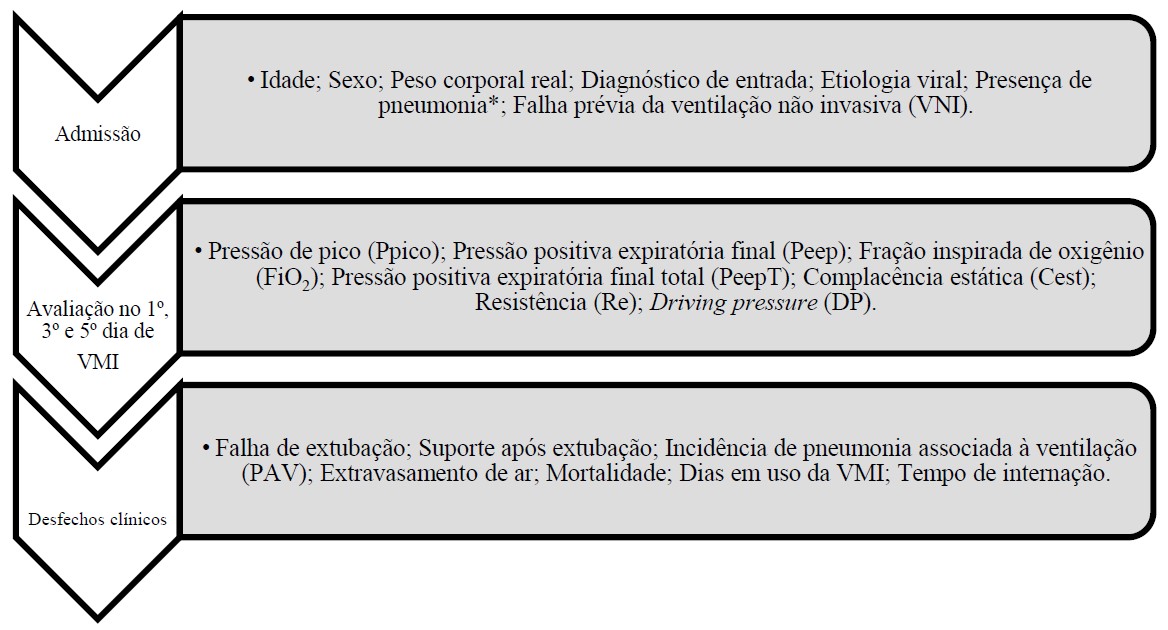
The retrospective data from the physical therapy protocol for daily monitoring lung mechanics is shown in Figure 1.
Institutional management of mechanical ventilation for bronchiolitis
The hospital unit in this study has Servo i® , Servo s® , Servo u® , Carescape R860® , and Babylog VN500 ® mechanical ventilators. All can perform inspiratory and expiratory pauses to measure lung mechanics values. These parameters were evaluated in a protocol manner during the first physical therapy daily evaluation. For this, the patients were either sedated and delivered to ventilation with RASS-5 or were to neuromuscular blockade. Patients weaning from ventilation or breathing spontaneously underwent no such measurements.
Also in protocol, infants with bronchiolitis were admitted to pressure regulated volume control (PRVC) IMV to actively control tidal volume (TV) via a protective ventilatory strategy targeting 6-8 ml/kg. Weaning occurred when peak pressure =<20cm/H2O. Patients were then administered controlled-pressure ventilation or pressure support ventilation as they regained consciousness (ultimately targeting extubation). The management of IMV in all infants began with passive humidification of ventilation; only cases of hypercapnia, increased airway resistance, and/or the presence of viscous tracheal secretion with the formation of stoppers or plugs required active humidification. The permissive hypercapnia strategy is also used in the unit considering a minimum 7.25 mmHg pH. All patients used intravenous corticosteroids 48 hours prior to extubation. All patients underwent it, only being extubated after positive spontaneous breathing trial results - only under sufficient inspiratory pressure: 6-8ml/kg TV, a 5-7 Peep, and a 40% FiO2 for at least 30 minutes. Extubation failure required the consideration of the need for IMV restitution within 72 hours.
Statistical analysis
Descriptive data analysis was performed on Epi info®, version 7.2, describing the variables as means and standard deviations for normally distributed continuous variables or as medians and minimum and maximum values for abnormally distributed continuous variables. Categorical variables were described as absolute frequencies and percentages. The unpaired t- and Mann-Whitney tests were used to compare the two groups. Significant p-values were considered if below 0.05.
RESULTS
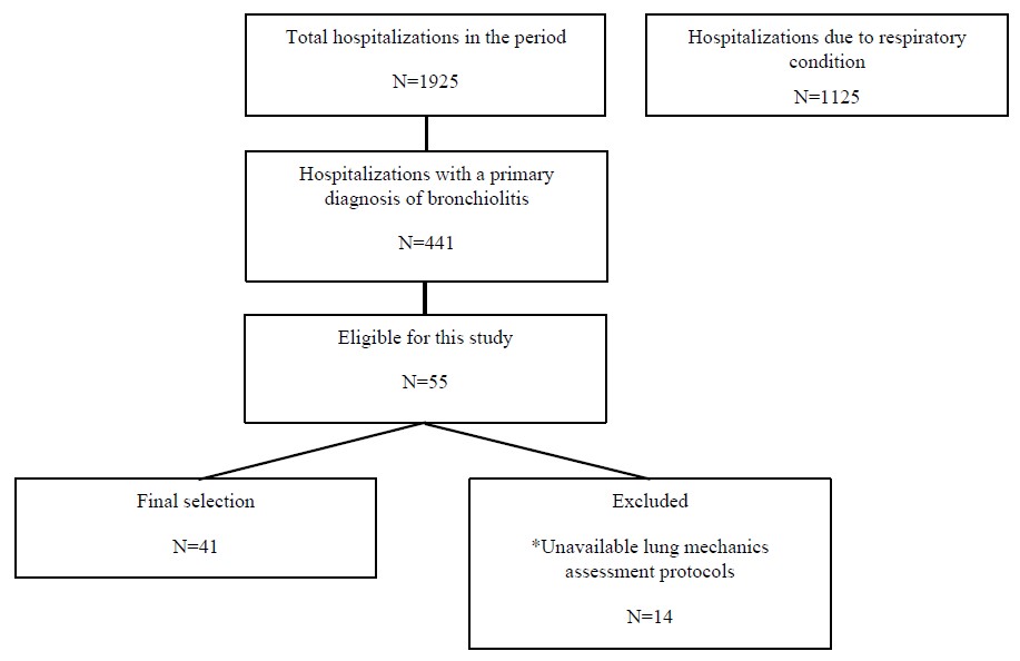
During the study period, the chosen unit admitted 441 infants with bronchiolitis, of whom 55 required invasive mechanical ventilation, a 12.4% AVB intubation rate. Figure 2 describes the study selection steps.
Of the final sample of 41 IMV events, 51.3% were boys with a 7.6-month median age and a 7.8-kg median weight. This study found a high incidence of bronchiolitis-associated pneumonia in its population (73.1%) but no statistically significant difference in clinical-demographic profiles when it stratified subgroups with and without associated pneumonia. Table 1 characterizes the study population.
The highest ventilatory parameters, with worse static compliance, resistance, and total Peep, occurred from the third to fifth days of IMV. The ml/kg ratio remained below 10ml/kg, with an 8.3ml/kg maximum mean. No ventilatory mechanic variables showed a statistically significant difference when comparing IMV admission and the other days. Table 2 describes the ventilatory mechanics and parameters.
This study found that 74% of extubation events used facilitative and/or rescue non-invasive ventilation. This research only had two cases of extubation failure, both in the same girl due to high obstruction. This research found no VAP cases despite its high rate of need for active humidification in its study population (74%). The rate of cases of air leakage and deaths in the population remained below 5%.
DISCUSSION
AVB manifests itself with the association of the cytopathological effect of the virus to the detriment of hosts’ inflammatory response, generating the signs and symptoms of the disease, which can vary according to the severity9),(10. Due to the variability of its clinical presentation, the need for ventilatory support and the population profile may be diverse in AVB, highlighting its higher prevalence in boys and the high incidence of RSV, as in this and other studies11-14. Acute respiratory infections configure an important cause of morbidity and mortality in children aged under five years worldwide. In total, two-thirds and 90% of these deaths occur in children aged under one year and in developing countries, respectively11. Estimates suggest that 90% of them are related to viral agents, the most common being RSV, influenza types A and B, parainfluenza, adenovirus, and metapneumovirus15. RSV manifestations can vary, associating complications and important sequelae for childhood and adulthood (wheezing, asthma, and airway hyperreactivity)11. In this study, the rate of co-infections (screening for two simultaneous viruses) totaled 40% in girls, corroborating the severity of the disease in the population that progressed to IMV.
The literature in AVB has no clearly defined criteria regarding the ideal time to institute an artificial airway, generally indicating it in cases of apneas, worsened respiratory efforts, hypoxemia, and hypercapnia16-19. Thus, the initial ventilatory adjustments in these patients should consider the predominantly obstructive pathophysiological characteristic of the disease. In its acute phase, reducing ventilatory work and improving gas exchange often require elevated parameters and intermittent neuromuscular blockade20. Thus, the continuous evaluation of lungs mechanics is essential to illustrate respiratory impairment in real time and to individualize mechanical ventilation adjustments and strategies.
This study observed the need for higher parameters from the third to the fifth day of IMV, with worse airway resistance and low compliance but without extrapolating the protective ventilatory strategy regarding TV levels, plateau pressure, and driving pressure21. According to the literature, although high airway resistance is the greatest characteristic of AVB, compliance may also change - in this case, decrease - due to air trapping, pulmonary hyperinflation, and parenchymal impairment due to consolidations, atelectasis, and possible progression to pediatric acute respiratory distress syndrome. The interpretation of these data can help to adapt mechanical ventilation parameters according to the pathophysiology and the predominant component in each phase of the disease8),(22. The high incidence of associated pneumonia may help explain the observed component of low pulmonary compliance, in addition to the obvious factor of ventilatory restriction due to hyperinflation.
In the evaluated patients, PRVC followed protocol, using TV as feedback to continuously adjust the pressure limit provided to patients. As main advantages, it can actively control the minute volume, self-adjusting the peak inspiratory pressure following variations in respiratory mechanics23. Despite the lack of robust studies in pediatrics and neonatology, positive reports exist on the use of PRVC in other conditions, such as after cardiac surgeries24 and in intubations due to acute respiratory syndrome associated with COVID-1925),(26. On the other hand, while some studies found no significant differences in hospital admission outcomes when comparing controlled-pressure ventilation with PRVC27, others have suggested that the latter tended to perform worse in severe asthma due to its relative risk of hypoventilation28. This study reinforces the importance of new and larger studies on the effectiveness of this ventilatory mode across pediatric conditions since the current guidelines agree that protective ventilation is to guide the management of ventilatory assistance in any condition21.
Regarding possible complications associated with IMV, VAP continues to be a major challenge in intensive care, which requires assessing its factors and risks of development. By definition, VAP occurs 48 hours after patients’ intubation, increasing hospital stay length and hospital costs, and may result in a high mortality rate (20-70%)29. Moreover, higher mortality rates are related to high-risk pathogens or situations in which initial antibiotic therapy is inappropriate for the causative agent30. Despite its high rate of active humidification, this study included no cases of VAP (and only two of its cases developed pneumothorax). Finally, the mortality in this study can be considered low in relation to the literature, the highest rates of which in this population total 30-50%18, which may support the importance of well-defined protocols, bundles, and good ventilatory practices as factors for reducing harm and improving final outcomes related to mechanical ventilation.
FINAL CONSIDERATIONS
Severe cases of AVB requiring IMV represent a small part of all cases. The high incidence of pneumonia and viral infection may worsen patients’ initial clinical picture. Regarding the mechanics of ventilation, we observed the need for moderate to high parameters in the worst phase of the disease (without breaking down the barriers to protective ventilatory strategies), high airway resistance, and low pulmonary compliance, corroborating the obstructive pattern that typifies the disease and adds a restrictive component. Moreover, PRVC and the institutional protocol to daily manage pulmonary mechanics actively monitored and controlled non-harmful pulmonary distension pressure levels, which may help to explain some of the observed positive outcomes, such as the low rate of cases of air leakage and deaths in the population.
The limitations of this retrospective descriptive study with data from an institutional protocol include its small sample (considering the total number of hospitalizations), the absence of randomization for comparisons with other ventilatory modes, the absence of the oxygenation index in the analyzed data, and the statistical impossibility of associating the variables with hospital admission outcomes as this is an observational study design. Finally, further studies should describe and compare PRVC with other ventilatory modes in AVB and other pediatric lung conditions to corroborate the advantages and limitations of this ventilatory strategy.
ACKNOWLEDGMENTS
We would like to thank the multidisciplinary team of the pediatric units of the Rios D’Or Hospital, especially physical therapy; the D’Or Institute for Research and Education; and to all study participants and their families.
Data Availability Statement:
The complete dataset supporting the findings of this study is available within the article.
REFERENCES
-
1 Dalziel SR, Haskell L, O’Brien S, Borland ML, Plint AC, et al. Bronchiolitis. Lancet. 2022;400(10349):392-406. doi: 10.1016/S0140-6736(22)01016-9
» https://doi.org/10.1016/S0140-6736(22)01016-9 -
2 Fujiogi M, Goto T, Yasunaga H, Fujishiro J, Mansbach JM, et al. Trends in bronchiolitis hospitalizations in the United States, 2000-2016. Pediatrics. 2019;144(6):e20192614. doi: 10.1542/peds.2019-2614
» https://doi.org/10.1542/peds.2019-2614 -
3 Tahamtan A, Samadizadeh S, Rastegar M, Nakstad B, Salimi V. Respiratory syncytial virus infection: why does disease severity vary among individuals? Expert Rev Respir Med. 2020;14(4):415-23. doi: 10.1080/17476348.2020.1724095
» https://doi.org/10.1080/17476348.2020.1724095 -
4 Arruvito L, Raiden S, Geffner J. Host response to respiratory syncytial virus infection. Curr Opin Infect Dis. 2015;28(3):259-66. doi: 10.1097/QCO.0000000000000159
» https://doi.org/10.1097/QCO.0000000000000159 - 5 Leung AKC, Kellner JD, Davies HD. Respiratory syncytial virus bronchiolitis. J Natl Med Assoc. 2005;97(12):1708-13.
-
6 Cruces P, González-Dambrauskas S, Quilodrán J, Valenzuela J, Martínez J, et al. Respiratory mechanics in infants with severe bronchiolitis on controlled mechanical ventilation. BMC Pulm Med. 2017;17(1):129. doi: 10.1186/s12890-017-0475-6
» https://doi.org/10.1186/s12890-017-0475-6 -
7 Ferlini R, Pinheiro FO, Andreolio C, Carvalho PRA, Piva JP. Características e evolução de crianças com bronquiolite viral aguda submetidas à ventilação mecânica. Rev Bras Ter Intensiva. 2016;28(1):55-61. doi: 10.5935/0103-507X.20160003
» https://doi.org/10.5935/0103-507X.20160003 -
8 Marlow RK, Brouillette S, Williams V, Lenihan A, Nemec N, et al. Risk factors associated with mechanical ventilation in critical bronchiolitis. Children (Basel). 2021;8(11):1035. doi: 10.3390/children8111035
» https://doi.org/10.3390/children8111035 -
9 Florin TA, Plint AC, Zorc JJ. Viral bronchiolitis. Lancet. 2017;389(10065):211-24. doi: 10.1016/S0140-6736(16)30951-5
» https://doi.org/10.1016/S0140-6736(16)30951-5 -
10 Wang EEL, Milner RA, Navas L, Maj H. Observer agreement for respiratory signs and oximetry in infants hospitalized with lower respiratory infections. Am Rev Respir Dis. 1992;145(1):106-9. doi: 10.1164/ajrccm/145.1.106
» https://doi.org/10.1164/ajrccm/145.1.106 -
11 Postiaux G, Zwaenepoel B, Louis J. Chest physical therapy in acute viral bronchiolitis: an updated review. Respir Care. 2013;58(9):1541-5. doi: 10.4187/respcare.01890
» https://doi.org/10.4187/respcare.01890 -
12 Simoes EAF. Environmental and demographic risk factors for respiratory syncytial virus lower respiratory tract disease. J Pediatr. 2003;143(5 Suppl):118-26. doi: 10.1067/S0022-3476(03)00511-0
» https://doi.org/10.1067/S0022-3476(03)00511-0 -
13 Meissner HC. Viral bronchiolitis in children. N Engl J Med. 2016;374(1):62-72. doi:10.1056/NEJMra1413456
» https://doi.org/0.1056/NEJMra1413456 -
14 Postiaux G, Louis J, Labasse HC, Gerroldt J, Kotik AC, et al. Evaluation of an alternative chest physiotherapy method in infants with respiratory syncytial virus bronchiolitis. Respir Care. 2011;56(7):989-94. doi: 10.4187/respcare.00721
» https://doi.org/10.4187/respcare.00721 - 15 World Health Organization. The World Health Report 2004: changing history. Geneva: WHO; 2004.
- 16 Frankel LR, Derish MT. Respiratory syncytial virus-induced respiratory failure in the pediatric patient. New Horizons. 1999;7(3):335-46.
-
17 Chevret L, Mbieleu B, Essouri S, Durand P, Chevret S, et al. Bronchiolites en unité de réanimation pédiatrique: facteurs pronostiques et devenir respiratoire des nourrissons ventilés. Arch Pediatr. 2005;12(4):385-90. doi: 10.1016/j.arcped.2005.01.008
» https://doi.org/10.1016/j.arcped.2005.01.008 -
18 Bueno FU, Piva JP, Garcia PCR, Lago PM, Einloft PR. Outcome and characteristics of infants with acute viral bronchiolitis submitted to mechanical ventilation in a Brazilian pediatric intensive care unit. Rev Bras Ter Intensiva. 2009;21(2):174-82. doi: 10.1590/S0103-507X2009000200010
» https://doi.org/10.1590/S0103-507X2009000200010 -
19 Tasker RC, Gordon I, Kiff K. Time course of severe respiratory syncytial virus infection in mechanically ventilated infants. Acta Paediatr. 2000;89(8):938-41. doi: 10.1111/j.1651-2227.2000.tb00412.x
» https://doi.org/10.1111/j.1651-2227.2000.tb00412.x -
20 Leclerc F, Scalfaro P, Noizet O, Thumerelle C, Dorkenoo A, et al. Mechanical ventilatory support in infants with respiratory syncytial virus infection. Pediatr Crit Care Med. 2001;2(3):197-204. doi: 10.1097/00130478-200107000-00002
» https://doi.org/10.1097/00130478-200107000-00002 -
21 Rodríguez Núñez A, Martinón Torres F, Martinón Sánchez JM. Ventilación mecánica en la bronquiolitis. An Pediatr (Barc). 2003;59(4):363-6. doi: 10.1016/S1695-4033(03)78195-8
» https://doi.org/10.1016/S1695-4033(03)78195-8 -
22 Kneyber MCJ, de Luca D, Calderini E, Jarreau PH, Javouhey E, et al. Recommendations for mechanical ventilation of critically ill children from the Paediatric Mechanical Ventilation Consensus Conference (PEMVECC). Intensive Care Med. 2017;43:1764-80. doi: 10.1007/s00134-017-4920-z
» https://doi.org/10.1007/s00134-017-4920-z - 23 Carvalho WB, Hirschheimer MR, Proença Filho JO, Freddi NA, Troster EJ. Ventilação com pressão regulada volume controlado e ventilação assistida proporcional. In: Carvalho WB, Hirschheimer MR, Proença FilhoJO, Freddi NA, Troster EJ, editors. Ventilação pulmonar mecânica em pediatria e neonatologia. São Paulo: Atheneu; 2005.
-
24 Kocis KC, Dekeon MK, Rosen HK, Bandy KP, Crowley DC, et al. Pressure-regulated volume control versus volume control ventilation in infants after surgery for congenital heart disease. Pediatr Cardiol. 2001;22(3):233-7. doi: 10.1007/s002460010210
» https://doi.org/10.1007/s002460010210 - 25 Beeching NJ, Fletcher TE, Fowler R. Doença do coronavírus 2019 (COVID-19). BMJ Best Pract. 2024.
-
26 Kneyber MCJ, Medina A, Modesto AV, Blokpoel R, Brierley J, Chidini G, et al. Practice recommendations for managing children with proven or suspected COVID-19. Geneva: European Society of Paediatric and Neonatal Intensive Care; 2020. Available from: https://www.espnic.eu/practice-recommendations-for-managing-children-with-proven-or-suspected-covid-19/
» https://www.espnic.eu/practice-recommendations-for-managing-children-with-proven-or-suspected-covid-19/ -
27 Kıhtır HS, Akçay N, Şevketoğlu E. Pressure-regulated volume control and pressure-control ventilation modes in pediatric acute respiratory failure. Turk J Anaesthesiol Reanim. 2022;50(1):18-23. doi: 10.1002/ppul.25730
» https://doi.org/10.1002/ppul.25730 -
28 Medina A, Modesto-Alapont V, Del Villar Guerra P, Redal MR, Cambra AM, et al. Pressure-regulated volume control versus volume control ventilation in severely obstructed patients. Med Intensiva. 2016; 40(4):250-2. doi: 10.1016/j.medin.2015.07.010
» https://doi.org/10.1016/j.medin.2015.07.010 - 29 Carvalho IB, Delvaux G, Silva MFO, Mateus SRM, Sousa RE, Meira DAB. Prevalência da mortalidade em pacientes com pneumonia associada à ventilação mecânica em UTI universitária. Anais Científicos. 2021.
-
30 Hunter JD. Ventilator associated pneumonia. Postgrad Med J. 2006;82(965):172-8. doi: 10.1136/pgmj.2005.036905
» https://doi.org/10.1136/pgmj.2005.036905
Edited by
-
Responsible editor:
Sônia LP Pacheco de Toledo

 IMV - invasive mechanical ventilation; *In this case, the presence of pneumonia at hospital admission is considered as a secondary diagnosis associated with bronchiolitis based on clinical and/or radiological criteria.