Abstracts
We report the case of a 61-year-old male patient who underwent surgical excision of a lung mass for anatomopathological study. The patient had previously presented with fever, dry cough, and chest pain, together with lung masses detected by chest X-ray, and had undergone thoracotomy for diagnostic investigation on two occasions (1976 and 1981), although a conclusive diagnosis had not been made. A CT scan of the chest revealed large masses with areas of calcification in both lung fields. The anatomopathological study was consistent with pulmonary hyalinizing granuloma. In the postoperative period, the patient experienced several episodes of bronchospasm, which was reversible with the use of symptomatic medication. At this writing, the patient was receiving maintenance therapy with prednisone (40 mg/day) and had shown clinical improvement.
Glucocorticoids; Mass chest X-ray; Granuloma
Relatamos o caso de um paciente de 61 anos, masculino, internado com objetivo de exérese de massa pulmonar para estudo anatomopatológico. O paciente apresentara anteriormente um quadro de febre, tosse seca e dor torácica, associado à presença de massas pulmonares detectadas por radiografia de tórax, tendo sido submetido em duas ocasiões (1976 e 1981) a toracotomia para a investigação diagnóstica, sem diagnóstico anatomopatológico conclusivo. A TC de tórax revelou volumosas massas com áreas de calcificação em ambos os campos pulmonares. O material do estudo anatomopatológico foi compatível com granuloma hialinizante de pulmão. No pós-operatório, o paciente apresentou vários episódios de broncoespasmo que foram revertidos com medicação sintomática. Foi mantido com prednisona na dose de 40 mg/dia com boa evolução clínica até o envio deste relato.
Glucocorticoides; Radiografia pulmonar de massa; Granuloma
CASE REPORT
Recurrent pulmonary hyalinizing granuloma*
Guilherme D'Andréa Saba ArrudaI; Paulo César Ribeiro de CarvalhoII; Mara Patrícia Guilhermino de AndradeIII; Maurício Campos CusmanichIV; Gustavo BandeiraV; Felipe Shigueo Passos TozakiVI
IResident in General Clinical Medicine. Dr. José de Carvalho Florence Municipal Hospital, Associação Paulista para o Desenvolvimento da Medicina/Universidade Federal de São Paulo - SPDM-UNIFESP, São Paulo State Association for the Development of Medicine/Federal University of São Paulo - São José dos Campos, Brazil
IIPulmonologist. Dr. José de Carvalho Florence Municipal Hospital, Associação Paulista para o Desenvolvimento da Medicina/Universidade Federal de São Paulo - SPDM-UNIFESP, São Paulo State Association for the Development of Medicine/Federal University of São Paulo - São José dos Campos, Brazil
IIIPathologist. Acta Laboratory, Taubaté, Brazil
IVThoracic Surgeon. Dr. José de Carvalho Florence Municipal Hospital, Associação Paulista para o Desenvolvimento da Medicina/Universidade Federal de São Paulo - SPDM-UNIFESP, São Paulo State Association for the Development of Medicine/Federal University of São Paulo - São José dos Campos, Brazil
VThoracic Surgeon. Dr. José de Carvalho Florence Municipal Hospital, Associação Paulista para o Desenvolvimento da Medicina/Universidade Federal de São Paulo - SPDM-UNIFESP, São Paulo State Association for the Development of Medicine/Federal University of São Paulo - São José dos Campos, Brazil
VIResident in General Surgery. Dr. José de Carvalho Florence Municipal Hospital, Associação Paulista para o Desenvolvimento da Medicina/Universidade Federal de São Paulo - SPDM-UNIFESP, São Paulo State Association for the Development of Medicine/Federal University of São Paulo - São José dos Campos, Brazil
Correspondence to
ABSTRACT
We report the case of a 61-year-old male patient who underwent surgical excision of a lung mass for anatomopathological study. The patient had previously presented with fever, dry cough, and chest pain, together with lung masses detected by chest X-ray, and had undergone thoracotomy for diagnostic investigation on two occasions (1976 and 1981), although a conclusive diagnosis had not been made. A CT scan of the chest revealed large masses with areas of calcification in both lung fields. The anatomopathological study was consistent with pulmonary hyalinizing granuloma. In the postoperative period, the patient experienced several episodes of bronchospasm, which was reversible with the use of symptomatic medication. At this writing, the patient was receiving maintenance therapy with prednisone (40 mg/day) and had shown clinical improvement.
Keywords: Glucocorticoids; Mass chest X-ray; Granuloma.
Introduction
Pulmonary hyalinizing granuloma is a rare benign lesion, the etiology and pathogenesis of which have yet to be well defined.(1) Initially described in 1977, pulmonary hyalinizing granuloma presents as multiple, mostly bilateral, recurrent pulmonary nodules that affect patients of both genders with equal frequency.(1) Clinically, pulmonary hyalinizing granuloma can be asymptomatic (25% of the patients) or can manifest as dry cough, chest pain, fever, dyspnea, and hemoptysis. Extrapulmonary involvement occurs in some cases.(2) The diagnosis is made through anatomopathological studies, which reveal collagen fibril deposition supplanting the lung parenchyma, together with a chronic inflammatory reaction in the periphery of the lung and formation of lymphoid aggregates. (2) Although the treatment of pulmonary hyalinizing granuloma has yet to be well established, there have been reports of patients who responded well to corticosteroids.(2)
Case report
We report the case of a 61-year-old Mulatto male who was hospitalized in February of 2009 in order to undergo surgical excision of a lung mass in the right upper lobe for anatomopathological study due to suspicion of pulmonary metastases.
The patient had been hospitalized three times, for the investigation of lung masses, at another facility. The first hospitalization occurred in August of 1976, when the patient was 28 years old, and was due to fever, dry cough, and chest pain, together with a left lung mass on chest X-ray. The patient underwent thoracotomy for surgical excision of the mass, and the anatomopathological study revealed the presence of a nonspecific chronic inflammatory process with calcifications.
In 1981, at 33 years of age, the patient was hospitalized with the same symptoms. At the time, a chest X-ray revealed a lung mass, this time in the right lung. Thoracotomy was again performed in order to remove the mass, and the anatomopathological results were similar to those obtained during the first hospitalization. One year after the second intervention, the patient noticed the formation of painless subcutaneous nodules. The nodules were mobile and fibroelastic, located on the posterior aspect of the right arm. A biopsy was performed in August of 1996, and the biopsy sample was defined as fibrous tissue with dystrophic calcification and areas suggestive of an amyloid deposit. At the time, the patient presented with recurrence of the symptoms. A CT scan of the chest showed two large lung masses, in the middle and upper fields of the left lung (the larger of the two presenting lobulated borders and measuring 9.0 cm along its longest axis), and a nodule of 1.5 cm in diameter, the appearance of which was homogeneous, in the middle field of the right lung (Figure 1).
The patient had systemic arterial hypertension, which was satisfactorily controlled with pharmacological treatment. He described himself as a nonsmoker and nonalcoholic with no history of exposure to smoke from woodburning stoves. The patient had no occupational history of prolonged exposure to toxic agents. However, he did have a 4-year occupational history of exposure to organic dust (having worked for a grain storage company).
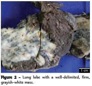
Bronchoscopy with biopsy provided no information that was relevant to the diagnosis. The patient again underwent thoracotomy for surgical excision of the mass. Macroscopic examination of the excised lobe revealed a welldelineated, firm, grayish-white mass, presenting calcified areas and measuring 10.0 × 4.0 × 3.5 cm (Figure 2). Microscopic examination showed that the lung parenchyma presented extensive hyaline fibrosis, areas of calcification, and foci of bone metaplasia (Figure 3). In the periphery of the lesion, moderate lymphoplasmacytic inflammatory infiltrate with formation of lymphoid aggregates, some foreign body granulomas, and foci of bronchiolitis obliterans organizing pneumonia were observed. Screening for birefringent material under polarized light, fungi, AFB, and amyloid yielded negative results. Theanatomopathologicalfindingswereconsistent with hyalinizing granuloma. In the postoperative period, the patient experienced several episodes of bronchospasm, which was reversible with the use of symptomatic medication. At this writing, the patient was receiving maintenance therapy with prednisone (40 mg/day) and had shown clinical improvement.
Discussion
Pulmonary hyalinizing granuloma was first reported in 1977.(1) Since then, few cases have been reported in the medical literature (fewer than 100 published cases).(2)
Pulmonary hyalinizing granuloma is a benign condition that can be recurrent.(3) In most cases, it presents as multiple nodules. The importance of pulmonary hyalinizing granuloma is that it is included in the differential diagnosis of diseases that are much more common, such as tuberculosis and histoplasmosis. Other possible differential diagnoses are inflammatory pseudotumor and solitary fibrous tumor, which have similar clinical and radiological characteristics.(3-5)
Although the etiology of pulmonary hyalinizing granuloma remains unclear, the disease has been associated with an abnormal reaction to antigens (fungi or the tuberculosis bacillus). Hyalinizing granuloma has also been related to certain immunological diseases, such as rheumatoid arthritis, sclerosing mediastinitis, retroperitoneal fibrosis, and uveitis, as well as to infectious diseases, such as tuberculosis, histoplasmosis, and aspergillosis.(6-8)
Patients with pulmonary hyalinizing granuloma can be asymptomatic, in which case suspicion of the disease is raised by radiological findings, or can present with dry cough, dyspnea, nonspecific chest pain, fatigue, and hemoptysis.(9) The diagnosis is based on anatomopathological examination, the microscopic aspects of which indicate thick deposits of collagen (keloid-like lesions) arranged concentrically or irregularly distributed, presenting central hyalinization and supplanting the lung parenchyma. At the interface between the lesion and the adjacent parenchyma, there is an increase in cellularity, together with the presence of lymphoplasmacytic inflammatory cells; in addition, there can be formation of lymphoid aggregates and foreign body granulomas.(2,8) There have been reports of extrapulmonary manifestations affecting the kidneys, larynx, and skin.(9,10) In the case reported here, the patient presented with skin involvement, characterized by a subcutaneous nodule. The nodule was investigated through biopsy and diagnosed as osteoma cutis, apparently unrelated to the pulmonary profile. Regarding the therapeutic approach, no specific drug is recommended; however, there have been reports of patients who responded well to corticosteroid therapy at initial doses of 40-60 mg/day.(10)
Acknowledgments
We would like to thank Dr. Ester Nei Aparecida Martins Coletta and Dr. Clarice Guimarães Freitas.
References
- 1. Engleman P, Liebow AA, Gmelich J, Friedman PJ. Pulmonary hyalinizing granuloma. Am Rev Respir Dis. 1977;115(6):997-1008.
- 2. Na KJ, Song SY, Kim JH, Kim YC. Subpleural pulmonary hyalinizing granuloma presenting as a solitary pulmonary nodule. J Thorac Oncol. 2007;2(8):777-9.
- 3. Fidan A, Ocal Z, Caglayan B, Dogusoy I, Gumrukcu G. An unusual cause of pulmonary nodules: Pulmonary hyalinizing granuloma with recurrence. Respir Med Extra. 2006;2(4):112-5.
- 4. Chalaoui J, Grégoire P, Sylvestre J, Lefebvre R, Amyot R. Pulmonary hyalinizing granuloma: a cause of pulmonary nodules. Radiology. 1984;152(1):23-6.
- 5. Esme H, Ermis SS, Fidan F, Unlu M, Dilek FH. A case of pulmonary hyalinizing granuloma associated with posterior uveitis. Tohoku J Exp Med. 2004;204(1):93-7.
- 6. Shibata Y, Kobayashi T, Hattori Y, Matsui O, Gabata T, Tamori S, et al. High-resolution CT findings in pulmonary hyalinizing granuloma. J Thorac Imaging. 2007;22(4):374-7.
- 7. O'Reilly KM, Boscia JA, Kaplan KL, Sime PJ. A case of steroid responsive pulmonary hyalinising granuloma: complicated by deep venous thrombosis. Eur Respir J. 2004;23(6):954-6.
- 8. Preuss J, Woenckhaus C, Thierauf A, Strehler M, Madea. B. Non-diagnosed pulmonary hyalinizing granuloma (PHG) as a cause of sudden unexpected death. Forensic Sci Int. 2008;179(2-3):e51-5.
- 9. Patel Y, Ishikawa S, MacDonnell KF. Pulmonary hyalinizing granuloma presenting as multiple cavitary calcified nodules. Chest. 1991;100(6):1720-1.
- 10. Shinohara T, Kaneko T, Miyazawa N, Nakatani Y, Nishiyama H, Shoji A, et al. Pulmonary hyalinizing granuloma with laryngeal and subcutaneous involvement: report of a case successfully treated with glucocorticoids. Intern Med. 2004;43(1):69-73.
Publication Dates
-
Publication in this collection
27 Oct 2010 -
Date of issue
Oct 2010
History
-
Received
05 Feb 2010 -
Accepted
02 June 2010


