 Inclusion body disease associated with salmonellosis in a Boa constrictor in the state of Ceará, northeastern Brazil
ABSTRACT:
Inclusion body disease (IBD) is a global viral infectious disease that affects snakes, mainly from the Boidae and Pythonidae families. The appearance of secondary infections is common in cases of IBD. The aim of this study was to report a case of inclusion body disease (IBD) associated with secondary infection in the respiratory system by Salmonella spp. in a captive Boa constrictor in the state of Ceará, northeastern Brazil. A male captive snake from a private captivity was sent for necropsy after presenting apathy and anorexia. It was sent for necropsy to the “Laboratório de Anatomia e Fisiologia Animal” at the “Universidade Federal do Cariri.” Macroscopically, there was a discrete focal brownish catarrhal content in the tracheal lumen. The lungs, especially the right one, were reddish, with multifocal to coalescing yellowish areas. On the cut surface, these areas extended to the lung parenchyma. They were soft, yellow and had a pasty texture. A brownish catarrhal exudate was also observed in the lung cavity. Microbiological examination of the tracheal swab and lungs resulted in the isolation of Salmonella spp. as well as Achromobacter denitrificans in the trachea. Fragments from all organs were collected, fixed in 10% buffered formalin solution and routinely processed to make histological slides stained with hematoxylin and eosin (HE). Microscopically, there was lymphoplasmacytic and heterophilic tracheitis, with hyperplasia of the mucosal epithelium associated with intracytoplasmic eosinophilic inclusion bodies. In the lungs, there was granulomatous pneumonia. The faveolar epithelium showed degeneration and necrosis associated with intracytoplasmic eosinophilic inclusion bodies. In the intestine, there was necrotic enteritis. In the liver there was random necrotic hepatitis. In the kidneys, there was degeneration and necrosis of the tubular epithelium and interstitial nephritis. Round to oval, intracellular eosinophilic to amphophilic inclusion bodies were also seen in the pancreas, liver, bile ducts, kidneys, and brain. Paraffin blocks of lung and intestine fragments were sent to the Veterinary Pathology Department at “Universidade Federal do Rio Grande do Sul” for immunohistochemistry with anti-Salmonella antibody, with positive immunostaining. The findings of inclusion bodies were consistent with infection by a virus of the genus Reptarenavirus. This appears to be the first reported case of IBD in a Boa constrictor from a private zoo in the state of Ceará, northeastern Brazil. In the present report, the disease was associated with secondary infection in the respiratory system by Salmonella sp. Additionally, this seems to be the first report of isolation of A. denitrificans from the microbiome of a respiratory sample in a snake in Brazil. Although Salmonella spp. infections are common in snakes, the isolation of A. denitrificans requires further studies on infection routes, pathogenicity and antimicrobial sensitivity.
INDEX TERMS:
Reptarenavirus; secondary infections; salmonellosis; immunohistochemistry
RESUMO:
A doença do corpúsculo de inclusão (“Inclusion Body Disease” - IBD) é uma doença infecciosa viral e de caráter global que acomete serpentes, principalmente das famílias Boidae e Pythonidae. O surgimento de infecções secundárias é comum em casos de IBD. Objetivou-se com este trabalho, relatar um caso da doença de corpúsculos de inclusão (IBD) associada a infecção secundária do sistema respiratório por Salmonella spp. em uma jiboia (Boa constrictor) de cativeiro, no estado do Ceará, nordeste do Brasil. Uma serpente, macho, proveniente de cativeiro particular foi encontrada morta em seu recinto após apresentar apatia e anorexia, e encaminhada para necropsia no Laboratório de Anatomia e Fisiologia Animal na Universidade Federal do Cariri. Macroscopicamente, no lúmen da traqueia havia conteúdo catarral acastanhado focal discreto. Os pulmões, principalmente o direito, estava avermelhado, com áreas multifocais a coalescentes amareladas. Ao corte, estendiam-se ao parênquima pulmonar, eram macias, amarelas e de consistência pastosa e na cavidade pulmonar havia exsudato catarral acastanhado. O exame microbiológico foi feito a partir de swab traqueal e pulmão resultou em isolamento de Salmonella spp., além de Achromobacter denitrificans em traqueia. Foram colhidos fragmentos de todos os órgãos, fixados em solução de formalina tamponada a 10% e, em seguida, processados rotineiramente para fazer confecção de lâminas histológicas coradas com hematoxilina e eosina (HE). Microscopicamente, havia traqueíte linfoplasmocitária e heterofílica com hiperplasia do epitélio da mucosa associada a corpúsculos de inclusão intracitoplasmáticos. No pulmão, havia pneumonia granulomatosa. No epitélio faveolar foi visto degeneração e necrose associada a corpúsculos de inclusão eosinofílicos intracitoplasmáticos. No intestino havia enterite necrótica. No fígado havia hepatite necrótica, aleatória. Nos rins, havia degeneração, necrose do epitélio tubular e nefrite intersticial linfoplasmocitária. Corpúsculos de inclusão eosinofílicos a anfofílicos, redondos a ovais, intracitoplasmáticos foram vistos ainda em pâncreas, fígado, ductos biliares, rins e encéfalo. Blocos de parafina de fragmentos de pulmão e intestino foram encaminhados ao Setor de Patologia Veterinária da Universidade Federal do Rio Grande do Sul para realização de imuno-histoquímica com anticorpo anti-Salmonella, com imunomarcação positiva. Os achados de corpúsculos de inclusões foram compatíveis com infecção por vírus do gênero Reptarenavirus. Este parece ser o primeiro relato de caso de doença do corpúsculo de inclusão em uma jiboia de um zoológico particular no estado do Ceará, Nordeste do Brasil. No presente relato, a doença foi associada à infecção secundária no sistema respiratório por Salmonella sp. Além disso, este parece ser o primeiro relato de isolamento de A. denitrificans do microbioma de uma amostra respiratória em uma serpente no Brasil. Embora infecções por Salmonella spp. sejam comuns em serpentes, o isolamento de A. denitrificans requer mais estudos sobre as vias de infecção, patogenicidade e sensibilidade antimicrobiana.
TERMOS DE INDEXAÇÃO:
Reptarenavirus; infecções secundárias; salmonelose; imuno-histoquímica
Introduction
Inclusion body disease (IBD) is a global viral infectious disease that affects snakes, mainly from the Boidae and Pythonidae families (Ossiborf 2018). Recently, it was identified that the virus responsible for IBD belongs to the Arenaviridae family and to the genus Reptarenavirus (Hetzel et al. 2013). Snakes with IBD may show nonspecific clinical signs such as anorexia, engorgement and cachexia. Additionally, neurological signs might be observed, including opisthotonus, imbalance, flaccid paralysis and inability to return to the original position when placed in a supine stance (Jacobson 2007).
In Brazil, Turchetti et al. (2013) reported the first case of IBD in a snake of the Boidae family, Corallus hortulana. Later, other studies have shown the occurrence of Reptarenavirus infection in Boa constrictor (Hardt et al. 2017, Argenta et al. 2020) and Epicrates cenchria (Ferreira et al. 2024), mostly associated with secondary infections. There are no published studies on IBD in Northeast Brazil, making this the first report of IBD infection in snakes in the state of Ceará. Studies reporting the occurrence of this disease are important for understanding the distribution of cases in the country, since many snakes kept in captivity come from different regions.
The development of secondary infections is common in cases of IBD and usually indicates an immunosuppressive effect. In snakes with IBD, most cell types in the haemolymphatic tissues are permissive for the causative reptarenavirus, expressing viral nucleoprotein and also bearing the typical cytoplasmic IB (Dervas et al. 2025). The aim of this study was to report a case of IBD associated with secondary infection in the respiratory system by Salmonella spp. in a captive Boa constrictor in the state of Ceará, Northeast Brazil.
Materials and Methods
Ethical approval. The results of this manuscript are part of the project “Anatomopathological findings of snakes kept in captivity in Cariri, Ceará”, submitted to the Ethics Committee for the Use of Animals (CEUA) of the “Universidade Federal do Cariri” (UFCA), under protocol 005/2025. The person responsible assigned the Donation Term and the Consent Term for performing the necropsy to the “Universidade Federal do Cariri”. The data are available upon request from the corresponding author.
A 101-cm-long captive Boa constrictor, of unknown age, weighing 460 g, from a private zoo in Juazeiro do Norte, Ceará, was found dead after showing anorexia and lack of appetite. The snake shared an enclosure with three other snakes of the same species, two of which had died approximately one year previously, with anatomopathological findings compatible with fibrinonecrotic colitis due to Salmonella sp. The breeder reported that the deaths occurred after the previous introduction of a ball python (Python regius) that presented with neurological signs and died after entering the zoo; however, this snake was not sent for necropsy. After this episode, he noticed that some snakes in the herd showed apathy and anorexia. In the approximately one-year interval between June and August 2023, four deaths occurred: the two boas mentioned above and two albino pythons. After approximately 11 months, another death occurred, this case being reported here, from May 2024, corresponding to a total of five deaths.
In this case, the corpse was refrigerated and sent to the “Laboratório de Anatomia e Fisiologia Animal” at the UFCA for necropsy examination. Samples from the trachea and right lung secretion were collected using Stuart’s swab and sent to a private laboratory for microbiological examination by automated seeding and identification using the VITEK® system. Fragments from all organs were collected, fixed in 10% buffered formalin solution, and routinely processed to make histological slides stained with hematoxylin and eosin (HE).
Paraffin blocks with sections of intestine and lung were sent and selected for immunohistochemistry (IHC) with anti-Salmonella antibody to the Veterinary Pathology Department at “Universidade Federal do Rio Grande do Sul” (UFRGS). To block endogenous peroxidase, the slides were incubated in 3% hydrogen peroxide for 15 minutes. Antigen recovery was carried out with protease XIV (Sigma®) for 15 minutes. The slides were incubated overnight with polyclonal anti-Salmonella primary antibody (Bio-Rad) diluted 1:1000. They were stained with AEC chromogen (Biocare Medical) and then counterstained with Mayer’s hematoxylin. Previously confirmed cases were used as positive controls.
Results
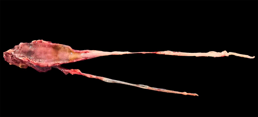
During necropsy, a regular body condition was observed. The trachea had a brownish catarrhal content in the lumen. The lungs, especially the right one, were reddish, with multifocal to coalescing yellowish areas. On the cut surface, these areas extended to the lung parenchyma. They were soft, yellow and had a pasty texture. A brownish catarrhal exudate was also observed in the lung cavity.
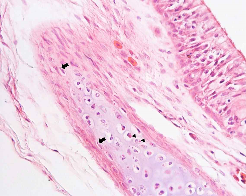
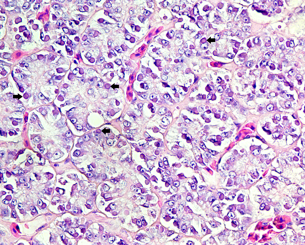
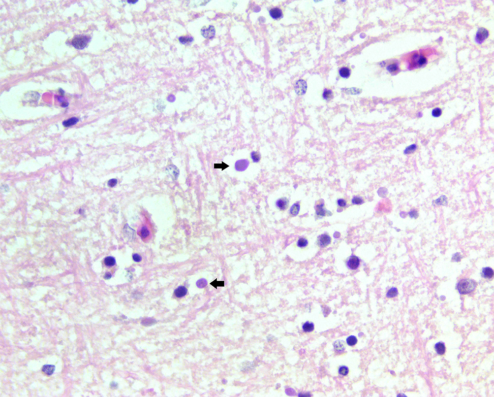
Microscopically, eosinophilic to amphophilic, round to oval, intracytoplasmic inclusion bodies of varying sizes were seen in several organs, including the lining epithelium of the pre-cardiac and post-cardiac esophagus, in the chondroblasts and chondrocytes of the hyaline cartilage of the trachea (Fig. 1), in the faveolar epithelium of the lungs, in the mucosal lining epithelium of the stomach and gastric glands, in the serous acini (Fig. 2) and ducts of the exocrine portion of the pancreas, gallbladder, in the lining epithelium of the mucosa of the small intestine, colon, cloaca (urodeum), renal tubular epithelium, seminiferous tubules of the testicle, sometimes associated with the inclusions there was vacuolar degeneration of the cells and sometimes transcytosis of inflammatory cells. In the liver, they were seen in hepatocytes, Kupffer cells and bile ducts. In the brain, they were observed in neurons, oligodendrocytes (Fig. 3), ependymal cells (Fig. 4) and meningothelial cells (Fig. 5), as well as macrophages and lymphoid cells from different tissues. The inclusion bodies varied in size from 27 to 74 micrometers, single or multiple in epithelial and parenchymal cells, with the largest ones predominating in the pancreas, kidneys and brain.
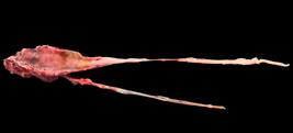
Lung, opened, with multifocal to coalescing yellowish areas associated with a discreet deposition of brown mucoid material.

Trachea, eosinophilic intracytoplasmic inclusions are seen in chondroblasts (arrow) and chondrocytes (arrowhead) of the hyaline cartilage. HE, obj. 20x.
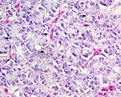
Pancreas, exocrine portion, amphophilic intracytoplasmic inclusion can be seen in the serous acini (arrows). HE, obj. 20x.
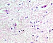
Brain, white matter, amphophilic intracytoplasmic inclusion in oligodendrocytes (arrows). HE, obj. 40x.
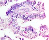
Brain, multiple eosinophilic intracytoplasmic inclusions are seen in the ependymal cells of the ependyma (arrows). HE, obj. 40x.
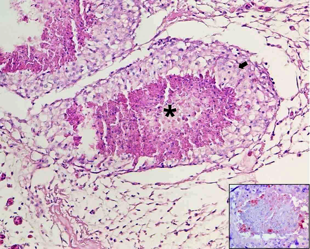
In the trachea, there was a focal area with hyperplasia of the epithelium and an inflammatory infiltrate composed of heterophils, lymphocytes and plasma cells in the lamina propria, multifocal and discrete. In the right lung, there were multifocal granulomas, characterized by central areas of caseous necrosis, surrounded by an infiltrate of epithelioid macrophages (Fig. 6), multinucleated giant cells, marked proliferation of fibrous connective tissue, and, in the periphery, an infiltrate of lymphocytes, plasma cells and heterophils. In the adjacent lung parenchyma, there was moderate interstitial and perivascular edema, heterophils and fibrin, as well as degeneration and necrosis of the faveolar epithelium, associated with intracytoplasmic inclusion bodies. In the left lung, there was slight degeneration of the faveolar epithelium associated with intracytoplasmic inclusion bodies, a slight infiltrate of heterophils in the lamina propria, moderate congestion and edema.
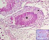
Lung, multifocal granulomas, characterized by a caseous necrotic center (asterisk), surrounded by numerous epithelioid macrophages (arrow). HE, obj. 20x. Inset: Lung, positive anti-Salmonella immunostaining at the center of the granuloma. IHC, obj. 40x.
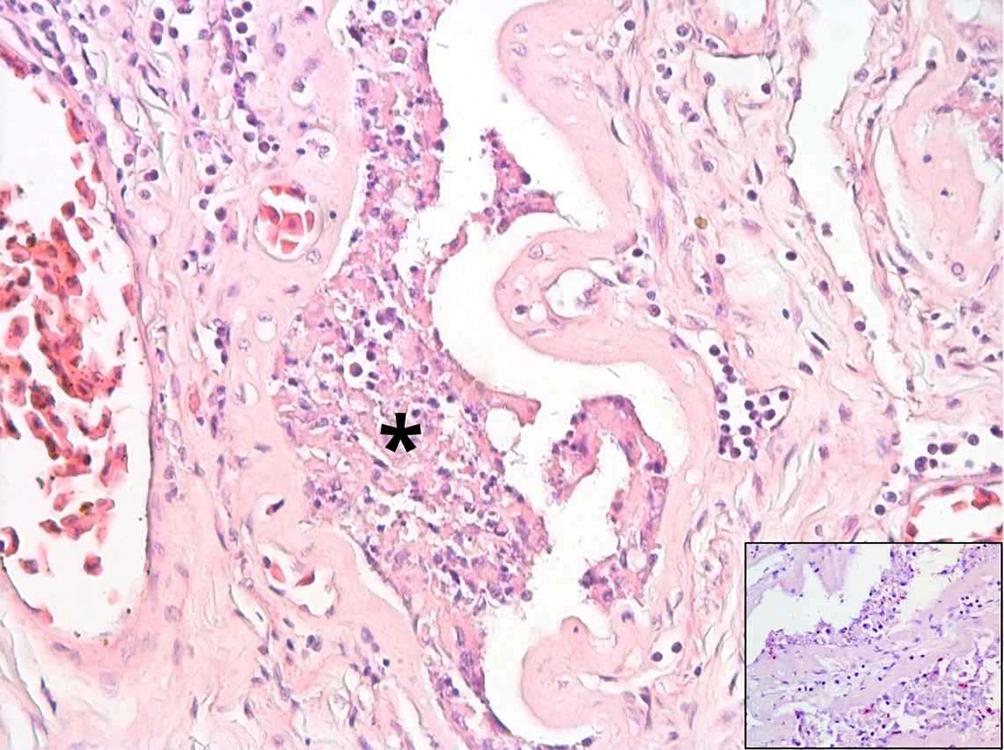
In the small and large intestines, in the duodenum and caecum segments respectively, there were multifocal areas of necrosis of the epithelium, with denudation of the mucosa and cell debris (Fig. 7). In the middle of the areas of necrosis, basophilic bacilli could be seen. In the caecum, there was also a discrete, mainly mononuclear infiltrate in the lamina propria, composed of lymphocytes, plasma cells, macrophages, and rarely heterophils. In the serous vessels of the intestine, there was also mixed vasculitis and perivasculitis, with lymphocytes, plasma cells and heterophils.
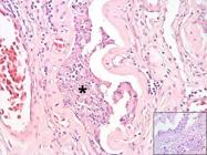
Small intestine, moderate necrotic enteritis (asterisk). HE, obj. 10x. Inset: Intestine, positive anti-Salmonella spp. immunostain in areas of mucosal necrosis and free in the lumen. IHC, obj. 40x.
In the liver, there was multifocal, random, discrete necrosis. In the kidneys, there was degeneration and necrosis of the tubular cells, associated with inclusion bodies, and a discrete focal inflammatory infiltrate, predominantly mononuclear, and some heterophils, characterizing a discrete, multifocal, mononuclear interstitial nephritis.
Microbiological analysis revealed the isolation of Salmonella spp. (from the trachea and lung samples) and Achromobacter denitrificans, only from the trachea. In the anti-Salmonella immunohistochemistry, there was positive immunolabeling of free bacillary bacteria in the lung, predominantly in the bronchial lumen and in the areas of necrosis (Fig. 5 inset), and in the intestine, free in the intestinal lumen and on the mucosal surface (Fig. 6 inset).
Discussion
The diagnosis was based on the clinical-pathological findings. It was possible to confirm IBD associated with secondary infection by Salmonella spp. in the respiratory tract, confirmed by microbiological and/or immunohistochemical tests, in a captive Boa constrictor from a private zoo in Northeast Brazil.
Similar to what was described by Turchetti et al. (2013), in this case, the clinical picture was characterized by nonspecific symptoms, such as lack of appetite and anorexia. Additionally, Ossiborf (2018) reported neurological symptoms such as head tremors and loss of proprioception, which were not observed in this report. The failure to describe neurological signs in this case may be associated with the absence of inflammation, which may or may not be present in the nervous system associated with inclusion bodies (Chang & Jacobson 2010) or the failure of breeders to observe the signs.
The diagnosis of IBD was confirmed by histopathological examination, which showed eosinophilic to amphophilic intracytoplasmic inclusions in various organs, a typical finding of IBD, as described by Jacobson (2007) and Hardt et al. (2017). The distribution and size of these inclusions were variable, present in tissues and organs of the respiratory, digestive and attached glands, lymphoid, urinary, reproductive and nervous systems, predominantly in the exocrine pancreas, renal epithelium, gastrointestinal tract and nervous system, as in the reports by Turchetti et al. (2013) and Hardt et al. (2017). Additionally, in this study, inclusion bodies were found in chondroblasts and chondrocytes of the tracheal cartilage, a finding that has not yet been described.
The occurrence of IBD in this case, coupled with its history of being kept in a captive environment and the entry of snakes from other farms, highlights the importance of effective biosecurity and health surveillance strategies in wildlife keeping institutions, including quarantine and monitoring through routine examinations, such as blood smears of peripheral blood stained with HE (Chang & Jacobson 2010) in asymptomatic animals and pathology in snakes that die (Hardt et al. 2017). Other tests are available at the research level, such as immunohistochemistry for detection of reptarenaviral nucleoprotein and reverse transcription polymerase chain reaction (RT-PCR) (Chang et al. 2016) and metatranscriptomic analysis (Argenta et al. 2020).
Chang & Jacobson (2010) conclude that the ease with which snakes can be transported around the world is probably responsible for the spread of IBD. In the present study, the occurrence of mortality in snakes from the Boidae and Pythonidae families over approximately one year, and the death of a member of the Python family with neurological symptoms, suggest the entry and/or circulation of the virus in the herd. However, in previous cases, due to the autolysis of the samples, confirmation was not possible. It is important to note that many cases of IBD are subclinical and snakes can remain asymptomatic for weeks to months, which makes the disease difficult to detect and control in collections of snakes kept in captivity (Ossiborf 2018).
When a case is detected in a herd, isolation or euthanasia is recommended for confirmed cases of IBD due to the risk of spreading both the reptarenavirus and the associated bacterial pathogens (Origgi 2019). In the Brazilian context, this case reinforces the urgent need for comprehensive epidemiological studies, because, as observed by Hardt et al. (2017), IBD may be underdiagnosed in our country.
Various bacterial infectious processes associated with IBD have been described, such as stomatitis, coelomitis, hepatitis, colitis, interstitial pneumonia and granulomatous spondylitis (Hardt et al. 2017, Simard et al. 2020). Bacteria of the genus Salmonella spp. are normal components of the intestinal microbiota, present in 90% or more of reptiles (Warwick et al. 2001). Among the most commonly described bacteria in snakes, Salmonella spp. has already been reported as a pathogen of secondary bacterial infection in cases of IBD (Hardt et al. 2017).
Salmonellosis is an important cause of death in snakes (Bertolini et al. 2021) and, in the present study, was associated with IBD. Despite the positive Salmonella staining in the lumen and mucosal surface, the association with necrotic enteritis suggests possible intestinal involvement in the present study, which we believe to be a finding. Studies demonstrating intestinal involvement show lesions characterized by fibrinonecrotic and granulomatous enteritis (Bertolini et al. 2021) and fibrinonecrotic enteritis associated with Salmonella spp. (Pereira et al. 2024).
Salmonella spp. infection in this case probably occurred due to the immunosuppression caused by IBD. Additionally, in this report, the Gram-negative bacteria Achromobacter denitrificans was isolated in a tracheal secretion sample. In humans, this agent has been implicated as an emerging cause of infection in immunosuppressed and immunocompetent populations (Awadh et al. 2017). In a study by Shek et al. (2009), A. denitrificans was isolated in the oropharynx of 10 of 32 Chinese snakes (Naja atra) as belonging to the oral bacterial microbiome. In our study, it was not possible to associate tracheitis with this agent due to infection by Salmonella spp. Additionally, this is the first report of isolation of A. denitrificans from the microbiome of a respiratory sample in a snake in Brazil.
Conclusion
This report seems to be the first recorded case of inclusion body disease (IBD) in a Boa constrictor from a private zoo in the state of Ceará, Northeast Brazil, in which anatomopathological findings reveal fundamental aspects of this disease, making it a differential diagnosis for snake diseases, especially with nonspecific clinical signs. In the present report, the disease was associated with secondary infection in the respiratory system by Salmonella sp. Although Salmonella spp. infections are common in snakes, the isolation of Achromobacter denitrificans requires further studies on infection routes, pathogenicity and antimicrobial sensitivity.
Acknowledgments
To the Animal Pathology Laboratory of “Universidade Federal de Campina Grande” (UFCG) and the Veterinary Pathology Sector of “Universidade Federal do Rio Grande do Sul” (UFRGS) for their diagnostic support.
References
-
Argenta FF, Hepojoki J, Smura T, Szirovicza L, Hammerschmitt ME, Driemeier D, Kipar A, Hetzel U. Identification of reptarenaviruses, hartmaniviruses, and a novel chuvirus in captive native Brazilian Boa Constrictors with boid inclusion body disease. J Virol 2020; https://doi.org/10.1128/jvi.00001-20
» https://doi.org/10.1128/jvi.00001-20 -
Awadh H, Mansour M, Aqtash O, Shweihat Y. Pneumonia due to a rare pathogen: Achromobacter xylosoxidans, subspecies denitrificans Case Rep Infect Dis 2017; https://doi.org/10.1155/2017/3969682
» https://doi.org/10.1155/2017/3969682 -
Bertolini M, Schwertz CI, Vielmo A, Piva MM, Bilhalva LC, Parvarini SP, Driemeier D, Sonne L. Pathological and microbiological findings in fatal cases of salmonellosis in captive bothrops snakes in Southern Brazil. J Comp Pathol 2021; https://doi.org/10.1016/j.jcpa.2021.04.011
» https://doi.org/10.1016/j.jcpa.2021.04.011 -
Chang L, Fu D, Stenglein MD, Hernandez JA, DeRisi JL, Jacobson ER. Detection and prevalence of boid inclusion body disease in collections of boas and pythons using immunological assays. Vet J 2016; https://doi.org/10.1016/j.tvjl.2016.10.006
» https://doi.org/10.1016/j.tvjl.2016.10.006 -
Chang L-W, Jacobson ER. Inclusion body disease, a worldwide infectious disease of boid snakes: a review. J Exotic Pet Med 2010; https://doi.org/10.1053/j.jepm.2010.07.014
» https://doi.org/10.1053/j.jepm.2010.07.014 -
Dervas E, Michalopoulou E, Hepojoki J, Thiele T, Baggio F, Hetzel U, Kipar A. Haemolymphatic tissues of captive boa constrictor (Boa constrictor): Morphological features in healthy individuals and with boid inclusion body disease. Dev Comp Immunol 2025; https://doi.org/10.1016/j.dci.2024.105302
» https://doi.org/10.1016/j.dci.2024.105302 -
Ferreira AB, Fonteque JH, Withoeft JA, Casagrande RA, Costa UM, Imkamp F, Göller P, Baggio F, Hepojoki J, Hetzel U, Kipar A. Multifocal cutaneous neoplastic vascular proliferations in a rainbow boa (Epicrates cenchria) collection with boid inclusion body disease. PLoS One 2024; https://doi.org/10.1371/journal.pone.0311015
» https://doi.org/10.1371/journal.pone.0311015 -
Hardt I, Gava MG, Paz JS, Silva ELF, Souza TD, Jabour FF, Leite FLG, Flecher MC. Doença do corpúsculo de inclusão e espondilite por Salmonella sp. em uma Boa constrictor constrictor Pesq Vet Bras 2017; https://doi.org/10.1590/S0100-736X2017000900014
» https://doi.org/10.1590/S0100-736X2017000900014 -
Hetzel U, Sironen T, Laurinmaki P, Liljeroos L, Patjas A, Henttonen H, Vaheri A, Artelt A, Kipar A, Butcher SJ, Vapalahti O, Hepojoki J. Isolation, identification, and characterization of novel arenaviruses, the etiological agents of boid inclusion body disease. J Virol 2013; https://doi.org/10.1128/jvi.01123-13
» https://doi.org/10.1128/jvi.01123-13 - Jacobson ER. Viruses and viral diseases of reptiles. In: Jacobson ER. Infectious Disease and Pathology of Reptiles: color atlas and text. Florida: CRC Press; 2007.
-
Origgi FC. Inclusion body disease (Reptarenavirus). In: Divers SJ, Stahl SJ. Mader’s Reptile and Amphibian Medicine and Surgery. 2019; https://doi.org/10.1016/B978-0-323-48253-0.00157-4
» https://doi.org/10.1016/B978-0-323-48253-0.00157-4 -
Ossiborf RJ. Serpentes. In: Terio KA, McAloose D, Leger JS. Pathology of Wildlife and Zoo Animals. 2018; https://doi.org/10.1016/B978-0-12-805306-5.00037-7
» https://doi.org/10.1016/B978-0-12-805306-5.00037-7 -
Pereira RMF, Oliveira Filho HS, Duarte JLC, Júnior FRP, Lima TS, Santos KMOL, Pereira ES, Silva NJ, Oliveira CJB, Araújo JL, Mendonça FS. Fibrinonecrotic enteritis and orchitis associated with Salmonella enterica subsp houtenae infection in a short-tailed boa (Boa constrictor amarali). J Comp Pathol 2024; https://doi.org/10.1016/j.jcpa.2024.01.002
» https://doi.org/10.1016/j.jcpa.2024.01.002 -
Shek KC, Tsui KL, Lam KK, Crow P, Ng KHL, Ades G, Yip KT, Grioni A, Tan KS, Lung DC, Lam TSK, Fung HT, Que TL, Kam CW. Oral bacterial flora of the Chinese cobra (Naja atra) and bamboo pit viper (Trimeresurus albolabris) in Hong Kong SAR, China. Hong Kong Med J 2009; https://www.hkmj.org/system/files/hkm0906p183.pdf
» https://www.hkmj.org/system/files/hkm0906p183.pdf -
Simard J, Marschang RE, Leineweber C, Hellebuyck T. Prevalence of inclusion body disease and associated comorbidity in captive collections of boid and pythonid snakes in Belgium. PLoS One 2020; https://doi.org/10.1371/journal.pone.0229667
» https://doi.org/10.1371/journal.pone.0229667 -
Turchetti AP, Tinoco HP, Malta MCC, Costa MELT, Pessanha AT, Soave SA, Paixão TA, Santos RL. Inclusion body disease in a Corallus hortulanus Braz J Vet Pathol 2013; https://shre.ink/oIvI
» https://shre.ink/oIvI -
Warwick C, Lambiris AJL, Westwood D, Steedman C. Reptile-related salmonellosis. J Royal Soc Med 2001; https://doi.org/10.1177/014107680109400306
» https://doi.org/10.1177/014107680109400306
-
Data availability statement
The data used in this study are available and can be accessed upon request to the corresponding author.
Edited by
-
Editor-in-Chief
Fabiano José Ferreira de Sant’Ana.
The data used in this study are available and can be accessed upon request to the corresponding author.