 Serological survey of brucellosis in camels from the Aegean region of Turkey
ABSTRACT:
Brucella melitensis and Brucella abortus are known to cause brucellosis in camels. It is especially seen in camels raised together with ruminants in regions where brucellosis is endemic. However, since this disease causes only a few clinical signs, unlike cattle, it is challenging to diagnose camel brucellosis. While camels bred in Asia and Africa are used for milk, meat, and transportation purposes, camel breeding in Turkey consists of small male camel populations raised under special conditions for cultural purposes. The aim of this study was to investigate the seroprevalence of brucellosis in camels in the Aegean region of Turkey using three serological tests - Rose Bengal plate test (RBPT), serum agglutination test (SAT), and complement fixation test (CFT) - and to compare their diagnostic performance. A total of 76 camel sera (72 Camelus dromedarius and four Camelus bactrianus) were collected from three cities (İzmir, Aydın, and Denizli) via random sampling. All sera were tested simultaneously using RBPT, SAT, and CFT. A positive control serum from a camel vaccinated with conjunctival B. abortus S19 and a negative control serum from a brucellosis-free newborn camel calf. It was determined that all 76 camel sera screened, except the positive control serum, gave negative results in RBP, SA, and CF tests. This comprehensive serological survey represents the first documented evidence of a brucellosis-free status in the camel population in Turkey. The findings suggest that the isolated and special conditions in which the camels are raised, the lack of contact with ruminants, and the predominantly male camel population contribute to the low risk of brucellosis in this region. These results highlight the importance of region-specific management practices in controlling camel brucellosis.
INDEX TERMS:
Camel; brucellosis; serosurvey; Turkey
RESUMO:
Brucella melitensis e Brucella abortus são conhecidas por causar brucelose em camelos. É especialmente observada em camelos criados em conjunto com ruminantes em regiões onde a brucelose é endêmica. No entanto, como essa doença causa apenas alguns sinais clínicos, ao contrário do que acontece com o gado bovino, o diagnóstico da brucelose em camelos é desafiador. Enquanto a criação de camelos na Ásia e na África é principalmente para produção de leite, carne e transporte, a criação de camelos na Turquia consiste em pequenas populações de camelos machos criados em condições especiais para fins culturais. O objetivo deste estudo foi investigar a soroprevalência da brucelose em camelos na região do Mar Egeu, na Turquia, utilizando três testes sorológicos - teste de placa rosa bengala (TPRB), teste de aglutinação sorológica (TAS) e teste de fixação de complemento (TFC) - e comparar seus desempenhos diagnósticos. Soros de 76 camelos (72 Camelus dromedarius e quatro Camelus bactrianus) foram coletados em três cidades (Izmir, Aydın e Denizli) por meio de amostragem aleatória. Todos os soros foram testados simultaneamente com RBPT, SAT e CFT. Foram incluídos um soro de controle positivo de um camelo vacinado com B. abortus S19 conjuntival e um soro de controle negativo de um camelo recém-nascido livre de brucelose. Foi determinado que todos os 76 soros de camelo examinados, exceto o soro de controle positivo, apresentaram resultados negativos nos testes PRB, AS e FC. Este abrangente levantamento sorológico representa a primeira evidência documentada de um status livre de brucelose na população de camelos na Turquia. Os resultados sugerem que as condições isoladas e especiais em que os camelos são criados, a falta de contato com ruminantes e a população predominantemente masculina de camelos contribuem para o baixo risco de brucelose nesta região. Esses resultados destacam a importância de práticas de manejo específicas para cada região no controle da brucelose em camelos.
TERMOS DE INDEXAÇÃO:
Camelo; brucelose; inquérito sorológico; Turquia
Introduction
According to data from the Food and Agriculture Organization of the United Nations (FAO), the number of camels in the world is reported to be more than 35 million. Of this, 25.89 million are dromedary camels, and 80% are found in Africa (Faye 2020). Approximately one million camels are reported in Ethiopia, 7.1 million in Somalia, and approximately five million in Sudan (Kena 2022). According to the 2023 data of the Turkish Statistical Institute, the total number of camels is 1,197. The distribution by city is as follows: 432 in Aydın, 27 in Denizli, 172 in İzmir, 62 in Muğla, 30 in Manisa, 163 in Antalya, 44 in Nevşehir, 216 in Çanakkale, and 51 in other cities (TÜİK 2023).
According to the World Health Organization (WHO), brucellosis is one of the most common zoonoses transmitted from animals, and human brucellosis causes serious public health problems in endemic areas (Berhanu & Pal 2020). Camel brucellosis is caused by Brucella abortus and Brucella melitensis (Wernery 2014). As in other farm animals, brucellosis can be transmitted to camels through contact with aborted fetal material or through the spillage of fetal amniotic fluid from aborted camels (Sprague et al. 2012). The possibility of infection also increases in farms where infected sheep, goats, or cattle are kept together (Ahad et al. 2024). Camels reach sexual maturity when females are three to four years old and males are four to five years old. Since camels mate seasonally, this type of infection is rare (Faye et al. 2023). Compared to other farm animals, the disease progresses more insidiously in camels without showing any signs. In female camels, there are not many signs other than abortion and premature birth. However, mastitis, metritis, and infertility may occur (Wernery 2014). In males, orchitis and arthritis are the most common signs (Khalafalla & Hussein 2021). When it infects large herds, it can lead to significant productivity losses through delayed sexual maturity, longer calving intervals, and lower milk yields (Mohammed et al. 2023). Since brucellosis is not reliably present in camels, the most definitive diagnostic method is culture and polymerase chain reaction (PCR) from aborted fetal tissues. Screening can be done on a herd basis. The disease can be diagnosed with serological tests, such as the complement fixation test (CFT), the Rose Bengal plate test (RBPT), the serum agglutination test (SAT), and the enzyme-linked immunosorbent test (ELISA), which have different levels of sensitivity and specificity (Adel 2022). The disease is reported to be seen in African Countries (Egypt, Ethiopia, Libya, Kenya, Nigeria, Sudan), Arabian Peninsula countries (Saudi Arabia, Jordan, Kuwait) and Asian countries (Iran, Pakistan, Mongolia, Azerbaijan) that breed and trade camels, but brucellosis has not been reported in the wild camel population in Australia (Teshome et al. 2003, Al-Majali et al. 2008, Musa et al. 2008, Gwida et al. 2012, Fatima et al. 2016, Adel 2022). Brucellosis has been reported in sheep and cattle in Turkey (Demeli & Fındık 2021, Akar et al. 2024), but there is no data about camel brucellosis.
This study aimed to determine the prevalence of brucellosis in camels in the Aegean region of Turkey, where unique husbandry and management practices were used, using three different serological tests.
Materials and Methods
Ethical approval. The Ethical Committee of the Faculty of Veterinary Medicine, Selcuk University, Turkey (No. 2024/175) approved this study.
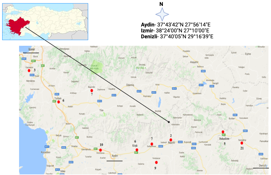
Sample collection. For this study, the number of animals required was determined based on official data and camel farms in the Aegean region of Turkey, where sampling was conducted. The number of samples to be taken was calculated based on the official number of 1,197 camels in Turkey, with a 10% margin of error, an estimated 50% prevalence, and a 90% confidence interval. The minimum number of samples to be taken was found to be 65 (Fig. 1). Various criteria were taken into consideration when collecting samples from camels. Camels were selected to reflect the diverse management systems of the farms (small-scale cultural rearing vs. larger farms). The study deliberately included a wide age range to assess exposure across life stages. Both sexes, males and females, were sampled to account for possible differences in disease transmission (e.g., reproductive risks in females). Samples were collected predominantly from Camelus dromedarius and small-scale Camelus bactrianus to select camels with no observable clinical signs (e.g., lameness, abortion, orchitis, mastitis, weight loss, or infertility in the last two years) to assess rare breed susceptibility.
The ten districts in three cities where the samples were collected and the number of samples.
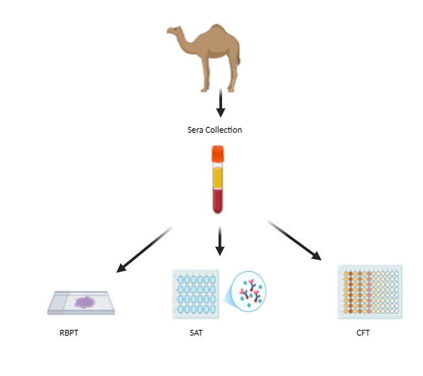
Serological tests. Blood collection from camels was done sterilely via the vena subcutanea abdominis. The blood samples taken were brought to the laboratory in a cold chain at +4 °C. For serum isolation, blood tubes were centrifuged at 5000 rpm for 5 min. For all serological tests, camel serums were inactivated at 60 °C for 30 min. In order to compare the specificity and sensitivity of serological tests used in the diagnosis of brucellosis in camel sera, RBP, SA, and CF tests were performed simultaneously according to the rules specified in the World Organisation for Animal Health (WOAH) regulations (Fig. 2) (MAF 2022).

The study hypothesis was that camel sera would be tested simultaneously, rather than sequentially, using three different serological methods - Rose Bengal plate test (RBPT), serum agglutination test (SAT), and complement fixation test (CFT) - to assess the presence of brucellosis antibodies.
For RBPT (RBPT, Vetal A.Ş.), 25 µl of test antigen was mixed with an equal amount of camel sera on a slide, which was rotated for four minutes. According to WOAH regulations, the Brucella abortus S99 antigen is used to diagnose B. abortus and Brucella melitensis infections because they have common antigenic regions (WOAH 2022).
A fixed amount of SAT antigen (SAT Antigen, Vetal A.Ş.) and diluted suspicious camel sera were added to the wells of 96-well U-bottom plates at 1/10 dilution to 1/1280 dilution and left to incubate overnight at 37 °C. The wells with antigen-antibody agglutinations were examined (WOAH 2022).
For CFT, diluted sera were distributed to the wells of the plates. A fixed amount of antigen (SN 437, IDEXX) and veronal buffer (KM0030, Virion-Serion GmbH) were added to the wells. Antigen-camel sera mixture and control wells were incubated at 37 °C for 30 min. Then, a complement (KM0038, Virion-Serion GmbH) was added to each well and incubated at 37 °C for 30 min. After this step, amboceptor and sheep red blood cells (SRBC) were also distributed to each well. Plates were incubated at 37 °C for 30 min and then at 4 °C for 2 h to allow the non-lysed cells to settle, after which the results were evaluated (Legesse et al. 2023). In the study, blood sera obtained from a female camel vaccinated (prime and boost) with the conjunctival B. abortus S19 vaccine were used as a positive control in the tests. Serum obtained by taking blood from a newborn camel calf before it ingested colostrum was used as a negative control serum.
Statistical analysis. The number of samples to be taken was calculated based on the official number of camels in Turkey, with a 10% margin of error, an estimated 50% prevalence, and a 90% confidence interval. The data were evaluated using the statistical package program IBM SPSS Statistics Standard Concurrent User V 265. Descriptive statistics were given as number of units (n), percentage (%), mean ± standard deviation (x ̅±ss), median (M), minimum (min) and maximum (max) values. Chi-square test was used to compare categorical variables with each other. A value of P< 0.05 was considered statistically significant.
Results
A total of 76 camels were included in the study, including 16 from İzmir, 39 from Aydın, and 21 from Denizli. The study included 49 males and 27 females, with average ages of 10.24 and 14.18 years, respectively. The oldest camel was a 28-year-old male, while the youngest was a 10-month-old male calf. The overall mean age of the sampled camels was 11.64 years. Among them, four were male Bactrian camels, averaging 12.25 years in age (Table 1).
All 76 samples were found to be brucellosis-negative except the positive control (Table 2). Therefore, no difference in sensitivity or specificity could be detected between the tests performed. However, all RBP, SA, and CF tests performed gave reactions in the positive control camel serum (Table 3). There were no reports of abortion or early delivery in any of the female camel farms.
Discussion
Camel brucellosis was first reported in 1931 (Dadar et al. 2022). Brucellosis is observed in camels in countries neighboring Turkey, such as Iran, Iraq, Syria, and other African countries (Gwida et al. 2012). However, there has been no report on camel brucellosis in Turkey. According to sero-survey results in countries where camel brucellosis is seen, positivity rates were reported as follows: Egypt (1-20%), Libya (1.4-4%), Sudan (0-84.2%), Ethiopia (5.7%), Nigeria (1-11.4%), Somalia (0.3-3.9%), Kenya (6.4%), Jordan (12.1-14.2%), Saudi Arabia (1.4-8%), Kuwait (14.8%), Pakistan (0-8%), Republic of Yemen (0%) (Moghney 2004, Alshaikh et al. 2007, Al-Mali et al. 2008, Musa et al. 2008, El-Boshy et al. 2009, Ghanem et al. 2009, Gwida et al. 2012, Adel 2022, Ahmed et al. 2024). It was reported that 9.21% of the 76 camels were diagnosed with brucellosis through a serological study conducted in Kirkuk, Iraq, using the SAT method (Yawoz et al. 2021). In a survey conducted with an RBP test on 66 camels in a similar region, the number of brucellosis-positive camels was 3.03% (Yawoz et al. 2012). A brucellosis identification study conducted in the northern part of Iran by Zowghi & Ebadi (1989) using RBPT, SAT, and CFT on 953 camels reported that 8% were serologically positive for brucellosis. Brucella melitensis was reported to be isolated from 1% of the camels in the culture of lymph nodes (Zowghi & Ebadi 1989). Khadjeh et al. (1999) reported that 1.9% of the 258 camels in the southern part of Iran were found to be brucellosis-positive in a survey using RBPT and SAT (Khadjeh et al. 1999).
In a study conducted by Gul et al. (2014) using RBPT, SAT, and ELISA on 100 camels in Punjab, Pakistan, all camels were found to be negative for brucellosis (Gul et al. 2014). El-Ansary et al. (2001) conducted a serosurvey on ruminants and camels in the eastern Sudan provinces and found that all 64 camel sera were negative for brucellosis (El-Ansary et al. 2001). In a study conducted in Yemen by Al-Shamahy (1999), 105 camels were screened for brucellosis using ELISA; all camels in the sample were reported to be negative for brucellosis (Al-Shamahy 1999). In the aforementioned serosurvey studies on camel brucellosis conducted in Pakistan, Sudan, and Yemen, respectively, it was observed that the camels were brucellosis-negative, similar to the findings of this study.
Researchers have argued that the variation in the prevalence of brucellosis among animal species may stem from factors such as regional disease prevalence, diagnostic methods, close contact with infected domestic and wild animals, population density, mixed-species housing, or the type of animal husbandry system applied (Rossetti et al. 2022).
Brucellosis eradication regulations aim to prevent false-positive and false-negative results by combining the RBP with the SA and CF tests (Bányász et al. 2023). It was determined that the positive control serum gave compatible results in all three tests used in screening (Table 3). However, since all camels screened in the study were serologically brucellosis-negative, it is not possible to comment on the effectiveness of the tests on each other in the diagnosis of camel brucellosis. Because all camels in this study were negative, the relationship between infection and age could not be established. Additionally, no abortion or premature birth data were reported in any of the female camel farms. This was found to be compatible with the results obtained.
Conclusions
Although brucellosis is prevalent in ruminants in Turkey and has been reported in camels in neighboring countries, the absence of brucellosis in the tested camel sera from the Aegean region is likely due to the unique cultural breeding practices of camels in Turkey. This study appears to be the first to report the absence of brucellosis in camels from the Aegean region, likely due to the unique breeding practices, where camels are raised individually or in controlled environments, minimizing exposure to ruminants.
Additionally, vaccination programs may help control the disease. However, brucellosis remains a risk in Turkey, and camel farmers should continue regular screenings and molecular tests, particularly in case of abortion or reproductive issues, to maintain brucellosis-free herds. These results demonstrate how unique rearing systems can create practical barriers against zoonotic transmission.
Acknowledgments
This study was completed without support from any institution and has not been presented as a presentation in Congress or published. We would like to thank Camel Breeders Federation of Turkey (DEGUF) for their effort and support of this study.
References
-
Adel M. Brucella transmission from domestic and wild animals to dromedary camel: diagnostic methods and zoonotic threats - a review. Open Vet Sci 2022; https://doi.org/10.1515/ovs-2022-0113
» https://doi.org/10.1515/ovs-2022-0113 -
Ahad AA, Megersa B, Edao BM. Brucellosis in camel, small ruminants, and Somali pastoralists in Eastern Ethiopia: a One Health approach. Front Vet Sci 2024; https://doi.org/10.3389/fvets.2024.1276275
» https://doi.org/10.3389/fvets.2024.1276275 -
Ahmed MIK, Shigidi M, El Sanousi E, Ahmed S, Salih S. The seroprevalence and geographic distribution of camel brucellosis in Kordofan states, western Sudan. Vet Ital 2024; https://doi.org/10.12834/VetIt.3009.31185.2
» https://doi.org/10.12834/VetIt.3009.31185.2 -
Akar K, Brangsch H, Jamil T, Öz GY, Baklan EA, Eroğlu B, Atıl E, Gürbilek SE, Keskin O, Tel OY, Yücetepe AG, Sandalakis V, Boukouvala E, Psaroulaki A, El Tawab AAA, Melzer F, Pletz MW, Neubauer H, Wareth G. Genomic analysis of Brucella isolates from animals and humans, Türkiye, 2010 to 2020. Eurosurveillance 2024; https://doi.org/10.2807/1560-7917.ES.2024.29.38.2400105
» https://doi.org/10.2807/1560-7917.ES.2024.29.38.2400105 -
Al-Majali AM, Al-Qudah KM, Al-Tarazi YH, Al-Rawashdeh OF. Risk factors associated with camel brucellosis in Jordan. Trop Anim Health Prod 2008; https://doi.org/10.1007/s11250-007-9080-7
» https://doi.org/10.1007/s11250-007-9080-7 - Alshaikh M, Al-Haidary A, Aljumaah R, Al-Korashi M, El-Nabi A, Hussein M. Camel brucellosis in Riyadh Region, Saudi Arabia. J Camel Pract Res 2007;14(2):113-117.
-
Al-Shamahy HA. Seropositivity for brucellosis in a sample of animals in the Republic of Yemen. East Mediterr Health J 1999; https://doi.org/10.26719/1999.5.5.1042
» https://doi.org/10.26719/1999.5.5.1042 -
Bányász B, Antal J, Dénes B. False positives in brucellosis serology: wrong bait and wrong pond? Trop Med Infect Dis 2023; https://doi.org/10.3390/tropicalmed8050274
» https://doi.org/10.3390/tropicalmed8050274 -
Berhanu G, Pal M. Brucellosis: a highly infectious zoonosis of public health and economic importance. J Emerg Environm Technol Health Protect 2020; https://telegeco.gr/3_1.pdf
» https://telegeco.gr/3_1.pdf -
Dadar M, Omar SS, Shahali Y, Fakhri Y, Godfroid J, Khaneghah AM. The prevalence of camel brucellosis and associated risk factors: a global meta-epidemiological study. Qual Assur Saf Crops Foods 2022; https://doi.org/10.15586/qas.v14i3.1088
» https://doi.org/10.15586/qas.v14i3.1088 -
Demeli A, Fındık M. Assessment of cattle and sheep Brucellosis in Turkey. Veteriner Hekimler Derneği Dergisi 2021; https://doi.org/10.33188/vetheder.678481
» https://doi.org/10.33188/vetheder.678481 -
El-Ansary EH, Mohammed BA, Hamad A-RA, Karom A-GO. Brucellosis among animals and human contacts in eastern Sudan. Saudi Med J 2001; https://smj.org.sa/content/smj/22/7/577.full.pdf
» https://smj.org.sa/content/smj/22/7/577.full.pdf -
El-Boshy M, Abbas H, El-Khodery S, Osman S. Cytokine response and clinicopathological findings in Brucella infected camels (Camelus dromedarius). Vet Med 2009; https://doi.org/10.17221/3044-VETMED
» https://doi.org/10.17221/3044-VETMED -
Fatima S, Khan I, Nasir A, Younus M, Saqib M, Melzer F, Neubauer H, El-Adawy H. Serological, molecular detection and potential risk factors associated with camel brucellosis in Pakistan. Trop Anim Health Prod 2016; https://doi.org/10.1007/s11250-016-1148-9
» https://doi.org/10.1007/s11250-016-1148-9 -
Faye B. How many large camelids in the world? A synthetic analysis of the world camel demographic changes. Pastoralism 2020; https://doi.org/10.1186/s13570-020-00176-z
» https://doi.org/10.1186/s13570-020-00176-z -
Faye B, Konuspayeva G, Magnan C. Large Camel Farming: a care-management guide from breeding to camel products. 2023; https://doi.org/10.1007/978-94-024-2237-5
» https://doi.org/10.1007/978-94-024-2237-5 -
Ghanem YM, El-Khodery SA, Saad AA, Abdelkader AH, Heybe A, Musse YA. Seroprevalence of camel brucellosis (Camelus dromedarius) in Somaliland. Trop. Anim Health Prod 2009; https://doi.org/10.1007/s11250-009-9377-9
» https://doi.org/10.1007/s11250-009-9377-9 -
Gul ST, Ahrar Khan AK, Farzana Rizvi FR, Iftikhar Hussain IH. Sero-prevalence of brucellosis in food animals in the Punjab, Pakistan. Pakistan Vet J 2014; https://www.pvj.com.pk/pdf-files/34_4/454-458.pdf
» https://www.pvj.com.pk/pdf-files/34_4/454-458.pdf -
Gwida M, El-Gohary A, Melzer F, Khan I, Rösler U, Neubauer H. Brucellosis in camels. Res Vet Sci 2012; https://doi.org/10.1016/j.rvsc.2011.05.002
» https://doi.org/10.1016/j.rvsc.2011.05.002 -
Kena D. Review on camel production and marketing status in Ethiopia. Pastoralism 2022; https://doi.org/10.1186/s13570-022-00248-2
» https://doi.org/10.1186/s13570-022-00248-2 -
Khadjeh G, Zowghi E, Zarif-Fard MR. Incidence of brucellosis in one-humped camels of Boushehr, Iran. Arch Razi Inst 1999; https://archrazi.areeo.ac.ir/article_109183_bffc2db977a10b907db34ff57ba7103d.pdf
» https://archrazi.areeo.ac.ir/article_109183_bffc2db977a10b907db34ff57ba7103d.pdf -
Khalafalla AI, Hussein MF. Infectious Diseases of Dromedary Camels: a concise guide. 2021; https://doi.org/10.1007/978-3-030-79389-0
» https://doi.org/10.1007/978-3-030-79389-0 -
Legesse A, Mekuriaw A, Gelaye E, Abayneh T, Getachew B, Weldemedhin W, Tesgera T, Deresse G, Birhanu K. Comparative evaluation of RBPT, I-ELISA, and CFT for the diagnosis of brucellosis and PCR detection of Brucella species from Ethiopian sheep, goats, and cattle sera. BMC Microbiol 2023; https://doi.org/10.1186/s12866-023-02962-2
» https://doi.org/10.1186/s12866-023-02962-2 -
MAF. Regulation on Control and Eradication of Brucella with Conjunctival Vaccine. Ministry of Agriculture and Forestry. 2022. Accessed on May 15, 2023. https://www.tarimorman.gov.tr/Konu/2103/Brusella_Conjuctival_Vaccine_control_eradication
» https://www.tarimorman.gov.tr/Konu/2103/Brusella_Conjuctival_Vaccine_control_eradication -
Moghney ARFA. A preliminary study on brucellosis on camels at Behira province. Ass Univ Bull Environ Res 2004; https://doi.org/10.21608/auber.2004.150592
» https://doi.org/10.21608/auber.2004.150592 -
Mohammed A, Ahmed M, Ahmed AO, Yousof S, Hamad S, Shuaib Y, Ibrahim N. Seroprevalence and risk factors of brucellosis in dromedary camels (Camelus dromedarius) in Sudan from 1980 to 2020: a systematic review and meta-analysis. Vet Q 2023; https://doi.org/10.1080/01652176.2023.2248233
» https://doi.org/10.1080/01652176.2023.2248233 -
Musa MT, Eisa MZM, El Sanousi EM, Wahab MBA, Perrett L. Brucellosis in camels (Camelus dromedarius) in Darfur, western Sudan. J Comp Pathol 2008; https://doi.org/10.1016/j.jcpa.2007.10.005
» https://doi.org/10.1016/j.jcpa.2007.10.005 -
Rossetti CA, Maurizio E, Rossi UA. Comparative review of brucellosis in small domestic ruminants. Front Vet Sci 2022; https://doi.org/10.3389/fvets.2022.887671
» https://doi.org/10.3389/fvets.2022.887671 -
Sprague LD, Al-Dahouk S, Neubauer H. A review on camel brucellosis: a zoonosis sustained by ignorance and indifference. Pathog Glomunbal Health 2012; https://doi.org/10.1179/2047773212Y.0000000020
» https://doi.org/10.1179/2047773212Y.0000000020 -
Teshome H, Molla B, Tibbo M. A seroprevalence study of camel brucellosis in three camel-rearing regions of Ethiopia. Trop Anim Health Prod 2003; https://doi.org/10.1023/A:1025874310261
» https://doi.org/10.1023/A:1025874310261 -
TÜİK. Türkiye’s Annual Camel Wealth Number 2023. Türkiye İstatistik Kurumu. 2023. Accessed on Apr 27, 2024. https://biruni.tuik.gov.tr/medas/?kn=101&locale=tr
» https://biruni.tuik.gov.tr/medas/?kn=101&locale=tr -
Wernery U. Camelid brucellosis: a review. Rev Sci Tech 2014; https://doi.org/10.20506/rst.33.3.2322
» https://doi.org/10.20506/rst.33.3.2322 -
WOAH. Brucellosis (Brucella abortus, B. melitensis and B. suis) (infection with B. abortus, B. melitensis and B. suis). World Organisation for Animal Health. 2022. Accessed on Mar 10, 2024. http://www.oie.int/en/international-standard-setting/terrestrial-manual/access-online/
» http://www.oie.int/en/international-standard-setting/terrestrial-manual/access-online/ -
Yawoz M, Jaafar SE, Alali F, Babur C. Seroprevalence of camels listeriosis, brucellosis and toxoplasmosis from Kirkuk province - Iraq. Pakistan Vet J 2021 https://doi.org/10.29261/pakvetj/2021.030
» https://doi.org/10.29261/pakvetj/2021.030 -
Yawoz M, Jaafar SE, Salih AI, Abdullah MH. A serological study of brucellosis in camels south of Kirkuk, Iraq. Iraqi J Vet Sci 2012; https://doi.org/10.33899/ijvs.2012.67484
» https://doi.org/10.33899/ijvs.2012.67484 -
Zowghi E, Ebadi A. Brucellosis in camels in Iran. Arch Razi Inst 1989;40(1):111-115. https://doi.org/10.20506/rst.7.2.355
» https://doi.org/10.20506/rst.7.2.355
The data supporting the findings of this study are available within the article. Additional or derived data are available from the corresponding author upon reasonable request.