 Anatomopathological findings and etiological characterization of swine organs in Brazilian abattoirs
ABSTRACT:
Brazil stands out as one of the leading pork exporters, driven by advances in pig farming and the adoption of strict sanitary standards. However, non-compliance with these regulations still significantly contributes to the occurrence of infectious and non-infectious diseases, which are among the main causes of pig mortality and carcass condemnation at slaughter. Identifying recurring issues in slaughterhouses can help develop better regulations and improve pig farming production. The present study identifies and describes the main diseases, injuries, and etiologies found in samples of slaughtered pigs submitted for analysis at the Veterinary Pathology Sector of the Federal University of Rio Grande do Sul (SPV-UFRGS) over the 21 years studied. For this, 1,206 samples from Brazil were analyzed, obtained through a retrospective and prospective study, with greater representation from the South region. Among these samples, 1,100 received a conclusive diagnosis (91.2%). Of the diagnosed diseases, 77.5% were of infectious origin, with a focus on respiratory diseases, lymphadenitis caused by Mycobacterium sp., and infections associated with swine circovirus, which together accounted for nearly 80% of the cases. Non-infectious lesions accounted for 22.5% of diagnoses and were classified as neoplastic and non-neoplastic. In the non-neoplastic group, integumentary changes were the most frequent, with umbilical hernias being the main manifestation. On the other hand, in the neoplastic group, lymphoma was the most frequent, with 35.5% of the samples having more than one organ affected in the same animal. Thus, this study aims to demonstrate the importance of continuous surveillance and adherence to sanitary guidelines to enhance swine farming further.
INDEX TERMS:
Swine; infectious diseases; neoplasia; abattoir; condemnation
RESUMO:
O Brasil se destaca como um dos principais exportadores de carne suína, impulsionado pelos avanços na suinocultura e pela adoção de rígidos padrões sanitários. No entanto, o não cumprimento destas regulamentações ainda contribui significativamente para a ocorrência de doenças infecciosas e não infecciosas, que estão entre as principais causas de mortalidade de suínos e de condenação de carcaças no abate. A identificação de problemas recorrentes nos abatedouros pode ajudar a desenvolver melhores regulamentações e melhorar a produção suinícola. O presente trabalho identifica e descreve as principais doenças, lesões e etiologias encontradas em amostras de suínos abatidos submetidas para análise no Setor de Patologia Veterinária da Universidade Federal do Rio Grande do Sul (SPV-UFRGS) ao longo dos 21 anos estudados. Para isso, foram analisadas 1.206 amostras do Brasil, obtidas através de estudo retrospectivo e prospectivo, com maior representatividade da região Sul. Dessas amostras, 1.100 receberam diagnóstico conclusivo (91,2%). Das doenças diagnosticadas, 77,5% eram de origem infecciosa, com destaque para doenças respiratórias, linfadenites causadas por Mycobacterium sp. e infecções associadas ao circovírus suíno, que juntas representaram quase 80% dos casos. As lesões não infecciosas representaram 22,5% dos diagnósticos e foram classificadas em neoplásicas e não neoplásicas. No grupo não neoplásico, as alterações tegumentares foram as mais frequentes, sendo as hérnias umbilicais a principal manifestação. Por outro lado, no grupo neoplásico, o linfoma foi o mais frequente, com 35.5% das amostras apresentando mais de um órgão afetado no mesmo animal. Portanto, este estudo tem como objetivo demonstrar a importância da vigilância contínua e do cumprimento das diretrizes sanitárias para melhorar ainda mais a suinocultura.
TERMOS DE INDEXAÇÃO:
Suíno; doenças infecciosas; neoplasia; abatedouro; condenação
Introduction
Brazil ranks fourth in global pork production and exports, generating 5.305 million tons in 2024, primarily from the southern region (ABPA 2025). This success is attributed to advancements in pig farming techniques and integrated systems, along with cooperatives and inspection services at various levels that play a vital role in ensuring food safety and in sustaining the industry’s growth and Brazil’s global economic prominence (Brasil 2020). Failure to meet these criteria leads to declines in zootechnical standards and economic losses (Harley et al. 2012, Brum et al. 2013, Johnson et al. 2019, Bruns & Stalder 2019, Konradt et al. 2020, Piva et al. 2020, Morés et al. 2022, Souza et al. 2022).
Diseases affecting finishing pigs include infectious diseases, mainly respiratory, and non-infectious conditions, such as gastric ulcers, mesenteric torsions, and cardiomyopathies (Piva et al. 2020). Some of these conditions can reach the slaughterhouse, where the destination needs to be recognized and determined (Brasil 2020). Recognizing lesions of sanitary importance in the swine slaughter line is extremely important for both animal and human (Brasil 2020, Rodarte et al. 2023). For this, guidelines and protocols of conduct were created and imposed to help slaughterhouse workers, farm workers, and veterinarians act in suspected cases of diseases of health importance (Brasil 2020).
Diseases such as pneumonia, enteritis, arthritis, and some neoplasms, for example, are easily recognized in both ante mortem and post mortem examination, and the affected parts are appropriately destined (Brasil 2020, Morey-Matamalas et al. 2021). However, due to the brief time in which the carcasses and viscera are examined on the inspection line, the lack of preparation of employees, technical managers, and, sometimes, veterinarians, some diseases end up not being identified correctly because they are subtle and confusing with other diseases and, consequently, receive a faulty or incomplete diagnosis (Morey-Matamalas et al. 2021). Comprehensive studies detailing the most common findings in slaughterhouses are crucial for filling out the current knowledge gap. They will provide a foundational reference for future research, as no such compilation currently exists. Thus, this study aimed to identify and characterize pig slaughterhouse lesions, with morphological and etiological diagnoses, to better outline areas for improvement in Brazilian pig farming.
Materials and Methods
Ethical approval. Samples were submitted as part of routine clinical diagnostic testing following institutional and national guidelines, not requiring ethical approval.
Retrospective and prospective study. A retrospective and prospective study of cases from swine slaughterhouses was conducted along with the “Setor de Patologia Veterinária” of the “Universidade Federal do Rio Grande do Sul” (SPV-UFRGS) report archive, dated from January 2001 to December 2020, along with received material and in-locus collection from January 2021 to December 2022. The data collected during the review of reports included epidemiological and anatomopathological findings. Epidemiological aspects included gender, lineage, age, and provided history.
In the macroscopic evaluation of the received samples, not only was the pathologist’s description recorded in the report, but, whenever possible, the macroscopic pattern was reassessed based on the photographic file. In some cases, when considered necessary, the formalin-fixed paraffin-embedded tissue samples present in the SPV-UFRGS file were retrieved, and histological sections of 3-5 µm were made and stained with hematoxylin and eosin (HE) for reassessment.
Supplementary exams. After the first histological analyses, when necessary, complementary exams were performed. Bacteriology (Quinn et al. 2011), polymerase chain reaction (PCR) (Renzhammer et al. 2023), and immunohistochemistry (IHC) (Stafford et al. 2021) were performed using the internal protocols present at the SPV-UFRGS and used in research projects, and of the laboratories with which the SPV-UFRGS has a partnership. Special histochemical stains, such as Ziehl-Neelsen (ZN), Warthin-Starry (WS), and Grocott (GR), were performed at SPV-UFRGS (Arrington 1992, Gaffney 1992, Huerta et al. 2003).
Results
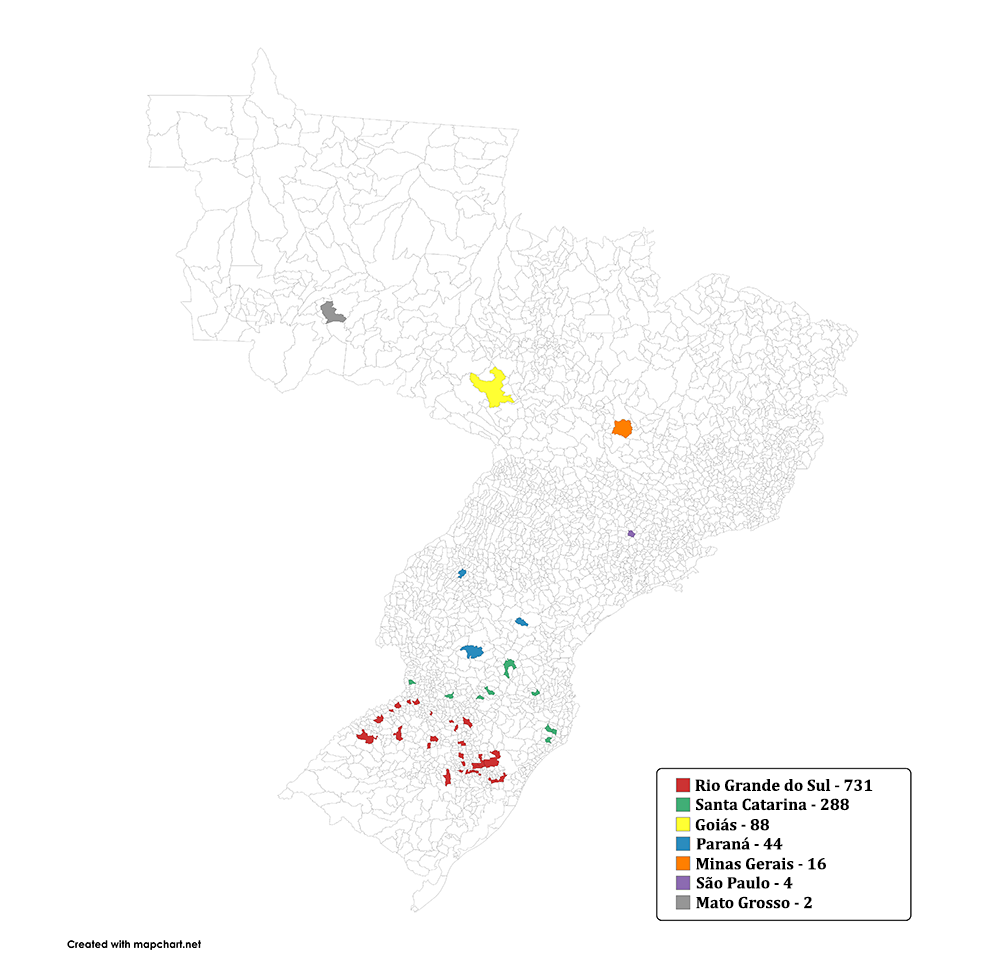
From January 2001 to December 2022, SPV-UFRGS received 16,087 swine samples. Of these, 1,206 cases originated from slaughterhouse samples, sent by veterinary companies or through research project collections (1,206/16,087; 7.5%). There was information about the pigs’ age in 492 reports (492/1,206; 40.8%), with an average of 178.8 days (standard deviation 72.55 days). Most samples came from large slaughterhouses located in Rio Grande do Sul (60.6%), followed by Santa Catarina (23.9%), Goiás (7.3%), Paraná (3.7%), Minas Gerais (1.3%), São Paulo (0.3%), and Mato Grosso (0.2%), the rest being without state information (33/1206) (Fig. 1). Of the available sample data, 54.4% were female (118/217), 45.6% were male (99/217), being all composed of commercial lineage.
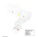
Swine origin distribution of samples received by the SPV-UFRGS from 2001 to 2022 (n = 1100). Source: mapchart.net
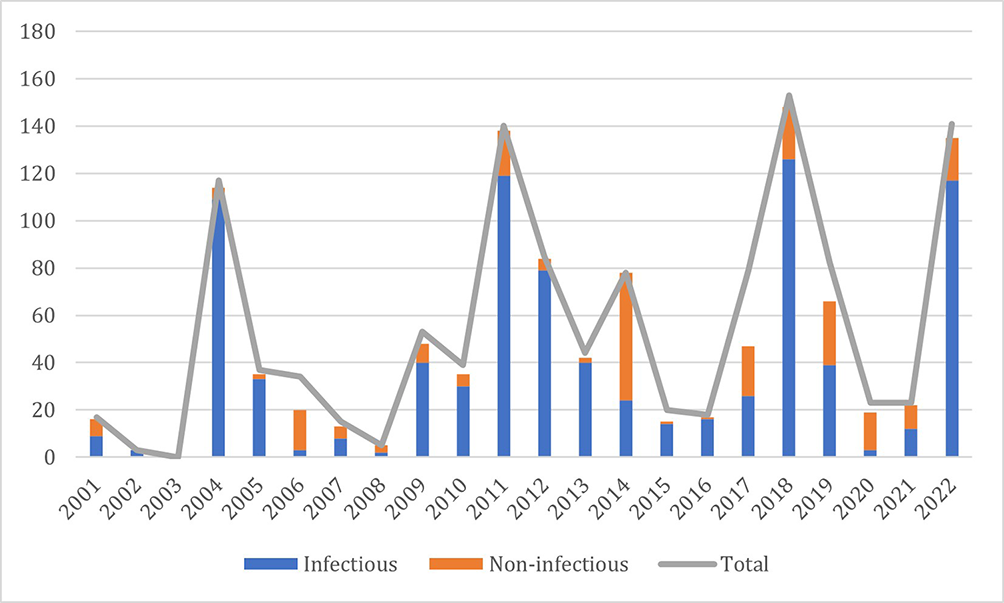
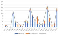
Of the 1,206 selected reports, 1,100 contained conclusive diagnoses (91.2%) while 106 reports were inconclusive (8.8%). The diagnoses were divided into two categories for a better presentation of the data: infectious and non-infectious. The distribution of cases per year is shown in Figure 2.
Infectious causes
The infectious causes accounted for 77.5% (852/1100) of the conclusive samples, represented by bacterial, viral, parasitic, and fungal causes, further grouped and divided into lymphadenitis, respiratory diseases, and other infectious diseases. The quantification of the identified etiologies and their percentages is detailed in Table 1.
Lymphadenitis
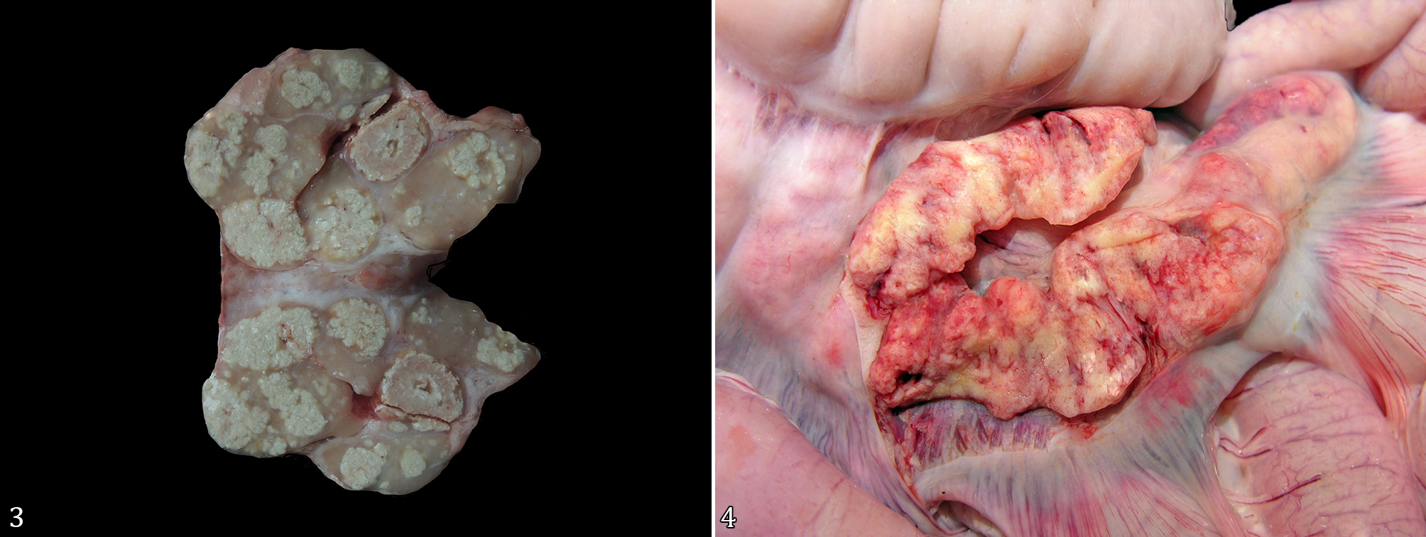
A total of 340 cases of bacterial and viral lymphadenitis (340/852, 39.9%) were diagnosed, many of which came from experiments (119/340, 35%). Most lymphadenitis were bacterial, the majority being granulomatous lymphadenitis with multifocal to coalescing white and friable areas that histologically showed fibrous tissue proliferation encircling caseous necrosis and an accentuated inflammatory infiltrate of epithelioid macrophages and neutrophils, caused by Mycobacterium sp. (Fig. 3), followed by granulomatous and bacterial lymphadenitis with no determined etiology. Of the diagnoses suggestive of Mycobacterium spp., 51% showed positive acid-fast bacilli (AFB) via ZN special staining. Due to their chronicity, the remaining 58 cases were given as suggestive of mycobacterial lymphadenitis in regression. Viral lymphadenitis was represented entirely by porcine circovirus type 2 (PCV2) infections characterized by enlarged to atrophied lymph nodes (Fig. 4), with granulomatous lymphadenitis and lymphoid depletion and, in cases that included the kidneys and skin, fibrinossupurative glomerulonephritis, dermatitis, and dermal vasculitis, respectively, and were included in the post-weaning multisystemic wasting syndrome (PMWS) and porcine dermatitis and nephropathy syndrome (PDNS). PCV-2 was identified by IHC in 15.4% of cases; the remaining cases were diagnosed through histopathological lesions such as lymphoid depletion, inflammatory infiltration of multinucleated giant cells, and fibrous tissue proliferation. The majority of samples (121/130, 93.1%) were from slaughterhouses in Goiás and Santa Catarina states from 2004 to 2005, displaying PDNS concomitant with bacterial infections.
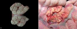
Lymphadenitis in slaughterhouse swine received by the SPV-UFRGS from 2001 to 2022. (3) Mycobacteriosis. Lymph node on cut surface with multifocal to coalescing white and friable areas. (4) Porcine Circovirus. Markedly large, with multifocal to coalescing areas of hemorrhage.
Respiratory diseases
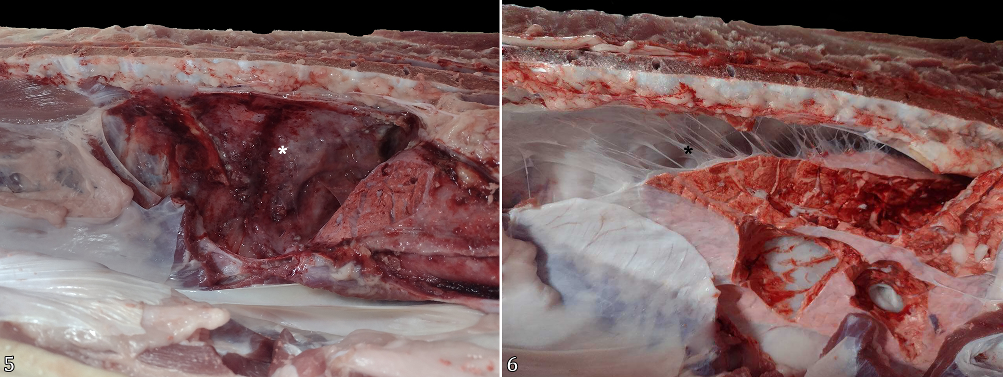
A total of 322 samples were from the lower respiratory tract, specifically lungs (322/852, 37.8%), where 210 had a diagnosis of bacterial or viral pneumonia (65.2%), and 112 of pleuropneumonia (34.8%) (Fig. 5). The latter having 43 cases only presenting as chronic pleuritis, characterized by fibrosis of the visceral pleura (Fig. 6). It was possible to identify the etiological agent causing the disease and coinfections in 253 cases (78.6%), being confirmed through PCR in 78 samples, 56 by bacterial isolation, and one case by special ZN staining. The remaining 118 etiologies were suggested through their morphological and histological patterns, such as fibrinonecrotic or purulent bronchopneumonia, fibrinous pleuropneumonia, and necrotic bronchiolitis. The most frequently observed etiologies were type A swine influenza, Mycoplasma hyopneumoniae, Pasteurella multocida, and Actinobacillus pleuropneumoniae. Respiratory diseases with no identified etiology totaled 21.4% of cases.
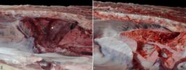
Respiratory diseases in slaughterhouse swine received by the SPV-UFRGS from 2001 to 2022. (5) Markedly diffuse fibrinous pleuropneumonia (white asterisk). (6) Chronic pleuritis highlighting fibrous projections between parietal and visceral pleura (black asterisk).
Pathogens in 140 cases were associated with one or more other pathogens (43.5%), a few examples being: swine influenza virus with M. hyopneumoniae, 25 of these also coinfected with P. multocida; swine influenza virus with P. multocida; M. hyopneumoniae with P. multocida, among others, in a smaller number or with unidentified secondary infections. Cases without coinfections accounted for 56.5% of samples. P. multocida was seen more as a secondary cause of respiratory infection in this study, with 53.9% of cases being such.
Other infectious diseases
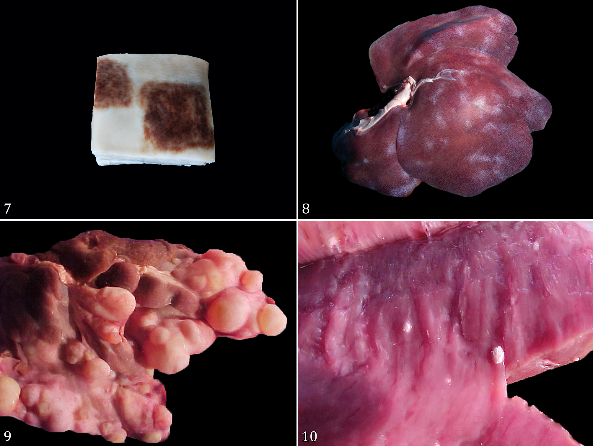
There were also 190 infectious diseases (190/852, 22.3%) not included in the previous categories. This category was mainly composed of bacterial dermatitis due to Erysipelothrix rhusiopathiae (Fig. 7), bacterial polyserositis, and hepatitis, in which the latter included bacterial, parasitic (Ascaris suum and Echinococcus granulosus) (Fig. 8 and 9) and fungal etiologies. Arthritis due to Mycoplasma hyosynoviae accounted for only a small portion of this study. Also, in the photographic archive of the SPV-UFRGS, a picture of a calcified cyst of Taenia solium in the myocardium was recovered and included to illustrate this section (Fig. 10). It was possible to confirm the etiological agents in 27 cases with PCR, 15 with bacteriology, and four with WS and one with GR silver stains, respectively.
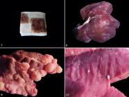
Other infectious diseases in slaughterhouse swine received by the SPV-UFRGS from 2001 to 2022. (7) Erysipelosis. Dark red multifocal geometric lesions, compatible with dermal vasculitis by Erysipelothrix rhusiopathiae. (8) Ascariasis. Multifocal to coalescent whitish areas of fibrosis compatible with parasitic migration of Ascaris suum (“milk-spots”). (9) Taeniasis. Multifocal to coalescent whitish nodules compatible with calcified Taenia solium cysts. (10) Echinococcosis. Multifocal to coalescent cysts compatible with Echinococcus granulosus.
Non-infectious
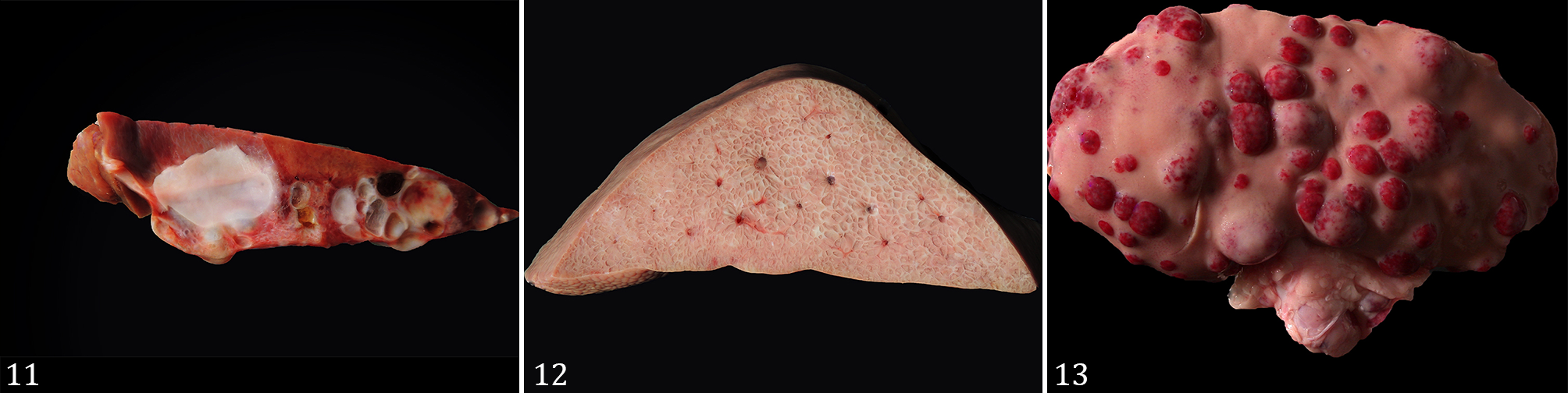
Non-infectious origin accounted for 22.5% of the diagnoses (248/1106). It was further divided into neoplastic and non-neoplastic origins, with the latter including degenerative, metabolic/nutritional, toxic, traumatic causes and malformations, represented in Table 2. The non-neoplastic section is mainly represented by umbilical hernias, lymphoid hyperplasia, and hepatic, lymphoid, and renal cysts (Fig. 11). The neoplastic group was mostly composed of lymphoma (Fig. 12 and 13), nephroblastoma, and hepatocellular carcinoma. Lymphoma was the most common neoplasia, reported in 11 cases in which more than one organ of the same animal was affected, denominated “multicentric”. Of these, the liver was present in 9/11 cases in this subcategory, followed by lymph nodes (8/11), spleen (6/11), kidneys (3/11), stomach, and heart (1/11 each).
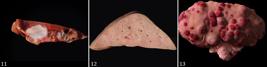
Non-infectious lesions in slaughterhouse swine received by the SPV-UFRGS from 2001 to 2022. (11) Polycystic liver with multifocal to coalescent cysts. (12) Hepatic lymphoma. Parenchyma is markedly white with a marked lobular pattern. (13) Renal lymphoma. Multifocal to coalescent whitish to reddish nodules protruding from the renal capsule.
Discussion
Infectious diseases were the main cause of condemnation in the study, corroborating the most common causes of diversion and condemnation of carcasses in slaughterhouses, and with retrospective studies or field studies already carried out (Brum et al. 2013, Konradt et al. 2020, Piva et al. 2020, Filippini & Freitas 2021, Costa et al. 2022). Histological diagnosis was essential to identify conditions in which isolation or identification by complementary exams was not possible, due to sample storage in 10% formalin (Melo et al. 2010, Hykin et al. 2015).
Lymphadenitides were the most diagnosed during the studied period. They were mainly composed of lymphadenitis by Mycobacterium spp., confirmed via characteristic lesions and ZN staining. The species most commonly affecting pigs is the Mycobacterium avium complex, with the subspecies hominissuis as the most prevalent (Stromerova & Faldyna 2018). However, infection with Mycobacterium bovis and Mycobacterium tuberculosis may also occur via wild boars and humans (Lara et al. 2011, Lopes et al. 2021). The species of Mycobacterium involved in this study was not determined because the sample was fixed in 10% formalin solution, which made it impossible to perform the isolation (Melo et al. 2010). The M. avium complex is usually restricted to mesenteric and gastro-hepatic lymph nodes, while M. tuberculosis and M. bovis are more disseminated to the lungs, liver, and spleen (Agdestein et al. 2012, Barandiaran et al. 2015, Stromerova & Faldyna 2018). Although the majority of the samples in this study were from mesenteric lymph nodes, the lack of other lymph nodes may create a diagnostic bias. The diagnosis of this agent signals a break in the sanitary barriers on farms, as it indicates contact with other infected animal species or humans (Lipiec et al. 2019, Kern et al. 2021). Viral lymphadenitis’ were represented by both clinical syndromes of circovirus infection. This was of great importance in the first years of the comprehended study, as the disease was entering the country, causing peaks in cases until 2004, which, as also shown in this study, subsequently reduced the number of cases, due to vaccination (Brum et al. 2013, Konradt et al. 2020). Since this disease immunocompromises the animal, secondary bacterial infections are a common finding.
Respiratory diseases formed a significant group in this study, indicating that diseases commonly observed during the growing-finishing phase may also manifest in the slaughterhouse (Konradt et al. 2020, Piva et al. 2020). Recognizing these diseases as representative examples is imperative to establishing them as a basis for formulating additional regulations and laws governing slaughterhouse operations. M. hyopneumoniae was one of the etiological agents with the highest frequency, frequently associated with other infectious agents in the samples, showing the importance of exams such as histopathology, PCR, and IHC to confirm. In this study, type A swine influenza stands out, with 57 cases of coinfection, corroborating other studies that have shown this association (Morés et al. 2015, Rech et al. 2018). Pasteurella multocida was also a pathogen of immense importance in the study as a cause of respiratory disease. This bacterium is currently treated both as a primary and secondary pathogen (Takeuti et al. 2013, Paladino et al. 2017, Oliveira Filho et al. 2018), being predominantly secondary in this study, due to primary lesions caused by other pathogens such as M. hyopneumoniae and type A swine influenza virus (Liu et al. 2019, Konradt et al. 2020). A. pleuropneumoniae was found to be the most common etiology of pleuropneumonias in this study, showing the importance of this disease in swine farming. This bacterium can be found in the upper respiratory system; its infection is triggered by stressful situations or by other immunosuppressing infections, such as PCV-2 or swine influenza virus infections. Morés & Morés (2012) state that 8% of opportunistic bacteria in the porcine respiratory disease complex syndrome are Actinobacillus pleuropneumoniae, similar to what was found. It is possible that some cases of chronic pleuritis were caused by this pathogen, but because of the chronic nature of the lesions, we were unable to confirm this.
Other bacterial respiratory disease pathogens found in this study were Glaesserella parasuis and Mycobacterium spp. (Cezar et al. 2019, Dazzi et al. 2020). However, the lesions from G. parasuis were not related to any injury to the lungs, possibly due to the frequent chronicity and advanced age of finishing animals, in addition to the commensal nature of this bacterium in the lower respiratory system and the possibility of it being detected by highly sensitive techniques, such as PCR (Cezar et al. 2019, Wiencek et al. 2022). On the other hand, three respiratory conditions (two cases of pneumonia and one of pleuropneumonia) were diagnosed as mycobacteriosis: two through suggestive lesions, and one using ZN staining. Both cases of pneumonia had concomitant lymphadenitis by Mycobacterium spp. (Lara et al. 2011, Lipiec et al. 2019, Kern et al. 2021), What likely also happened in the pleuropneumonia case, which unfortunately only came with a lung sample.
Viral pneumonia was an important diagnosis in this study, which was almost entirely comprised of type A swine influenza virus (Piva et al. 2020), and was frequently associated with other pathogens. Morphologically, the remaining five cases could be suggestive of swine influenza virus, but due to the chronicity of the lesions, the secondary bacterial infection overriding the viral infection, and the lack of other complementary exams for further differential diagnosis, an accurate diagnosis of influenza could not be made. Given the absence of a diagnosis of porcine reproductive and respiratory syndrome virus in Brazil (Gava et al. 2022) and the need for complementary exams such as PCR and IHC for differential diagnosis, these cases were diagnosed as viral pneumonia.
Other infectious diseases, such as Erysipelothrix rhusiopathiae dermatitis, polyserositis by G. parasuis; parasitic, bacterial, and fungal hepatitis; and parasitic myositis, accounted for a smaller portion of this study but are nonetheless of great importance. Although some are currently uncommon diagnoses in Brazil, these pathogens are still strong indicators of sanitary barrier breakage (Ferreiro et al. 2007, Costa et al. 2022, Rodarte et al. 2023), along with important causes of economic losses (Cezar et al. 2019, Dazzi et al. 2020, Pereira et al. 2020, 2022).
Identifying zoonotic diseases is of utmost importance not just for the well-being of consumers who eventually buy meat or its byproducts, but also, most importantly, for slaughterhouse employees who may come into direct contact with these pathogens (Matos et al. 2021, Rodarte et al. 2023, Sattar et al. 2023). In this study, they accounted for 27.8% of all conclusive cases presenting one or more zoonotic pathogens. Of these, Mycobacterium spp. and type A swine influenza were those of most importance, with other bacteria such as E. rhusiopathiae and Streptococcus suis also occurring on a smaller scale (Roesel & Fries 2018, Andersen et al. 2022, Rodarte et al. 2023). Although some of these pathogens are not of great concern due to sanitary legislation, such as Ascaris suum, Taenia solium, and Echinococcus granulosus, all documented in this study, and the fact that, for a human to get infected by a few of them, they must be somewhat immunocompromised, these pathogens are still of utmost importance to public health (Rodarte et al. 2023). Today, Brazil is one of the top producers and exporters of pork in the world, mainly due to these guidelines and standards implemented to guarantee national and international food safety (Cavalheiro et al. 2022, ABPA 2025).
Non-infectious diseases were comprised of lymphoid and integumentary lesions, along with neoplasms, being mostly asymptomatic and easily identified on the slaughterhouse line and discarded rapidly, due to the repercussions on the carcass and repugnant appearance. This leads to greater economic losses for the slaughterhouse due to condemnation of the viscera and occasionally affected muscles (Brum et al. 2015, Brasil 2020, Morey-Matamalas et al. 2021). Some lymphoid conditions have macroscopic similarities between granulomatous lymphadenitis and lymphatic cysts, showing the importance of sending samples for a confirmatory diagnosis of what is causing these lesions. This study comprised hernias without intestines, most likely the acquired form, although we lack information regarding the pigs’ installations before arriving at the abattoirs to confirm (Souza et al. 2020, Hovmand-Hansen et al. 2021). Lymphoid neoplasms were the most common, affecting multiple organs (Brum et al. 2015, Morey-Matamalas et al. 2021). No additional testing was performed to determine the lymphoma cell type. However, those originating from B-cells are the most common in pigs (Ogihara et al. 2012, Brum et al. 2015, Valli et al. 2016). Nephroblastoma, hepatocellular carcinoma, and neurofibroma were also observed in this study, with pathological aspects already described in the literature (Brum et al. 2015, Meuten & Meuten 2017).
Although our study received samples from various Brazilian states and provided a basic panorama of national swine production and the situation in slaughterhouses, it may have some limitations, as most of the analyses were performed on samples sent to the laboratory, mostly in 10% formalin, which prevented many complementary examinations. Receiving samples from only one affected area of the animal, rather than many, also prevented us from understanding the entire situation in the slaughterhouse. The little-to-no in-locus sample collection also limits this study, potentially leading to an underestimation of the prevalence of some diseases. Thus, it is suggested that the veterinarians responsible for the inspection submit more samples to compile the archive and make it more trustworthy. Therefore, more studies are needed to assess the impact of these diseases on production.
Conclusion
Infectious diseases dominated the findings in this study, particularly lymphadenitis and respiratory conditions, underscoring their significant impact on swine health and the economic repercussions for the industry. The identification of zoonotic pathogens, such as Mycobacterium spp., Pasteurella multocida, and type A swine influenza, highlights the need for robust sanitary practices to protect both public health and industry workers. Non-infectious conditions, while less prevalent, also contributed to economic losses, emphasizing the importance of diagnosis to distinguish between similar presentations. Addressing these challenges through more systematic and comprehensive sample submissions would enhance the reliability of future analyses. Overall, the findings reinforce the importance of continuous surveillance, strict adherence to sanitary guidelines, and ongoing research to reduce the impact of swine diseases and ensure food safety.
Acknowledgments
This study was developed and financed together with the support of the “Coordenação de Aperfeiçoamento de Pessoal de Nível Superior” (CAPES), Brazil, Finance Code 001, under project number 41347. We thank all members of the Department of Veterinary Pathology of the “Universidade Federal do Rio Grande do Sul”.
References
-
ABPA. Relatório Anual 2025. Associação Brasileira de Proteína Animal; 2025. Accessed on June 2, 2025. https://abpa-br.org/wp-content/uploads/2025/04/ABPA.-Relatorio-Anual-2025.pdf
» https://abpa-br.org/wp-content/uploads/2025/04/ABPA.-Relatorio-Anual-2025.pdf -
Agdestein A, Johansen TB, Kolbjørnsen Ø, Jørgensen A, Djønne B, Olsen I. A comparative study of Mycobacterium avium subsp. avium and Mycobacterium avium subsp. hominissuis in experimentally infected pigs. BMC Vet Res 2012; https://doi.org/10.1186/1746-6148-8-11
» https://doi.org/10.1186/1746-6148-8-11 -
Andersen KM, Vestergaard LS, Nissen JN, George SJ, Ryt-Hansen P, Hjulsager CK, Krog JS, Skov MN, Alexandersen S, Larsen LE, Trebbien R. Severe human case of zoonotic infection with swine-origin Influenza A virus, Denmark, 2021. Emerg Infect Dis 2022; https://doi.org/10.3201/eid2812.220935
» https://doi.org/10.3201/eid2812.220935 - Arrington JB. Bacteria, fungi, and other microorganisms, p.203-234. In: Prophet EB, Mills B, Arrington JB, Sobin LH. Laboratory Methods in Histotechnology. Washington: American Registry of Pathology; 1992.
-
Barandiaran S, Pérez AM, Gioffré AK, Vivot MM, Cataldi AA, Zumárraga MJ. Tuberculosis in swine co-infected with Mycobacterium avium subsp. hominissuis and Mycobacterium bovis in a cluster from Argentina. Epidemiol Infect 2015; https://doi.org/10.1017/S095026881400332X
» https://doi.org/10.1017/S095026881400332X -
Brasil. Decreto nº 10.468, de 18 de agosto de 2020. Altera o Decreto nº 9.013, de 29 de março de 2017, que regulamenta a Lei nº 1.283, de 18 de dezembro de 1950, e a Lei nº 7.889, de 23 de novembro de 1989, que dispõem sobre o regulamento da inspeção industrial e sanitária de produtos de origem animal. Presidência da República, Secretaria-Geral, Subchefia para Assuntos Jurídicos, Diário Oficial da União, Brasília; 2020. Accessed on Jan 26, 2023. https://www.planalto.gov.br/ccivil_03/_ato2019-2022/2020/decreto/d10468.htm
» https://www.planalto.gov.br/ccivil_03/_ato2019-2022/2020/decreto/d10468.htm -
Brum JS, Konradt G, Bazzi T, Fighera RA, Kommers GD, Irigoyen LF, Barros CSL. Características e frequência das doenças de suínos na Região Central do Rio Grande do Sul. Pesq Vet Bras 2013; https://doi.org/10.1590/S0100-736X2013001000006
» https://doi.org/10.1590/S0100-736X2013001000006 -
Brum JS, Martins TB, Viélmo A, Hammerschmitt ME, Talini R, Minozzo CD, Barros CSL. Neoplasmas em suínos: 37 casos. Pesq Vet Bras 2015; https://doi.org/10.1590/S0100-736X2015000600009
» https://doi.org/10.1590/S0100-736X2015000600009 - Bruns CE, Stalder KJ. Genetics and health, p. 42-49. In: Zimmerman JJ, Karriker LA, Ramirez A, Schwartz KJ, Stevenson GW, Zhang J. Diseases of Swine. 11th ed. Hoboken: Wiley-Blackwell; 2019.
-
Cavalheiro LG, Gené LA, Coldebella A, Kich JD, Ruiz VLA. Microbiological quality of pig carcasses in a slaughterhouse under risk-based inspection system. Foods 2022; https://doi.org/10.3390/foods11243986
» https://doi.org/10.3390/foods11243986 -
Cezar GA, Barbosa CN, Morés N. Doença de Glässer: uma revisão. Medicina Veterinária (UFRPE) 2019; https://doi.org/10.26605/medvet-v13n1-2604
» https://doi.org/10.26605/medvet-v13n1-2604 -
Costa RA, Leite PAG, Barros CGG, Lopes GMB. Principais causas de condenações em vísceras comestíveis de suínos abatidos em um matadouro frigorífico sob inspeção estadual na região de Alagoinhas-BA. RVZ 2022; https://rvz.emnuvens.com.br/rvz/article/view/1009/558
» https://rvz.emnuvens.com.br/rvz/article/view/1009/558 -
Dazzi CC, Guizzo JA, Prigol SR, Kreutz LC, Driemeier D, Chaudhuri S, Schryvers AB, Frandoloso R. New pathological lesions developed in pigs by a non-virulent strain of Glaesserella parasuis Front Vet Sci 2020; https://doi.org/10.3389/fvets.2020.00098
» https://doi.org/10.3389/fvets.2020.00098 - Ferreiro L, Spanamberg A, Carregaro FB, Cavallini EMS. Principais micoses dos suínos. Anais do II Simpósio UFRGS sobre Produção, Reprodução e Sanidade Suína, Porto Alegre, 2007. Acta Scient Vet 2007;35(Supl.):S113-S120.
-
Filippini V, Freitas ES. Frequência de condenação de vísceras e carcaças de suínos abatidos no ano de 2018 em um frigorífico localizado na região oeste do Paraná. Arq Bras Med Vet FAG 2021; https://themaetscientia.fag.edu.br/index.php/ABMVFAG/article/view/389/483
» https://themaetscientia.fag.edu.br/index.php/ABMVFAG/article/view/389/483 - Gaffney E. Carbohydrates, p.151-174. In: Prophet EB, Mills B, Arrington JB, Sobin LH. Laboratory Methods in Histotechnology. Washington: American Registry of Pathology; 1992.
-
Gava D, Caron L, Schaefer R, Silva VS, Weiblen R, Flores EF, Lima M, Takeda GZ, Ciacci-Zanella JR. A retrospective study of porcine reproductive and respiratory syndrome virus infection in Brazilian pigs from 2008 to 2020. Transbound Emerg Dis 2022; https://doi.org/10.1111/tbed.14036
» https://doi.org/10.1111/tbed.14036 -
Harley S, More S, Boyle L, O’Connell N, Hanlon A. Good animal welfare makes economic sense: potential of pig abattoir meat inspection as a welfare surveillance tool. Ir Vet J 2012; https://doi.org/10.1186/2046-0481-65-11
» https://doi.org/10.1186/2046-0481-65-11 -
Hovmand-Hansen T, Nielsen SS, Jensen TB, Vestergaard K, Nielsen MBF, Jensen HE. Survival of pigs with different characteristics of umbilical outpouching in a prospective cohort study of Danish pigs. Prev Vet Med 2021; https://doi.org/10.1016/j.prevetmed.2021.105343
» https://doi.org/10.1016/j.prevetmed.2021.105343 -
Huerta B, Arenas A, Carrasco L, Maldonado A, Tarradas C, Carbonero A, Perea A. Comparison of diagnostic techniques for porcine proliferative enteropathy (Lawsonia intracellularis infection). J Comp Path 2003; https://doi.org/10.1016/S0021-9975(03)00030-6
» https://doi.org/10.1016/S0021-9975(03)00030-6 -
Hykin SM, Bi K, McGuire JA. Fixing formalin: a method to recover genomic-scale DNA sequence data from formalin-fixed museum specimens using high-throughput sequencing. PLoS One 2015; https://doi.org/10.1371/journal.pone.0141579
» https://doi.org/10.1371/journal.pone.0141579 - Johnson AK, Colpoys JD, Edwards-Callaway LN, Calvo-Lorenzo M, McGlone JJ, Millman ST, Phillips CE, Ritter MJ, Sutherland MA, Anita L, Tucker AL, Webb AR. Behavior and welfare, p.17-41. In: Zimmerman JJ, Karriker LA, Ramirez A, Schwartz KJ, Stevenson GW, Zhang J. Diseases of Swine. 11th ed. Hoboken: Wiley-Blackwell; 2019.
-
Kern PL, Pinto AT, Schmidt V. Mycobacterium bovis detection in slaughtered pigs in the state of Rio Grande do Sul, Brazil. J Biosci 2021; https://doi.org/10.14393/BJ-v37n0a2021-41762
» https://doi.org/10.14393/BJ-v37n0a2021-41762 -
Konradt G, Bassuino DM, Siqueira LC, Siqueira MV, Sonne L, Driemeier D, Pavarini SP. Infectious diseases dynamics in growing/finishing pigs in Southern Brazil (2005-2016). Pesq Vet Bras 2020; https://doi.org/10.1590/1678-5150-PVB-6510
» https://doi.org/10.1590/1678-5150-PVB-6510 -
Lara GHB, Ribeiro MG, Leite CQF, Paes AC, Guazzelli A, Silva AV, Santos ACB, Listoni FJP. Occurrence of Mycobacterium spp. and other pathogens in lymph nodes of slaughtered swine and wild boars (Sus scrofa). Res Vet Sci 2011; https://doi.org/10.1016/j.rvsc.2010.06.009
» https://doi.org/10.1016/j.rvsc.2010.06.009 -
Lipiec M, Radulski Ł, Szulowski K. A case of bovine tuberculosis in pigs in Poland - a country free from the disease. Ann Agric Environ Med 2019; https://doi.org/10.26444/aaem/90979
» https://doi.org/10.26444/aaem/90979 -
Liu W, Zhou D, Yuan F, Liu Z, Duan Z, Yang K, Guo R, Li M, Li S, Fang L, Xiao S, Tian Y. Surface proteins mhp390 (P68) contributes to cilium adherence and mediates inflammation and apoptosis in Mycoplasma hyopneumoniae Microb Pathog 2019; https://doi.org/10.1016/j.micpath.2018.10.035
» https://doi.org/10.1016/j.micpath.2018.10.035 -
Lopes BC, Vidaletti MR, Soares DH, Argenta FF, Mello LS, Pavarini SP, Mayer FQ, Driemeier D. Tuberculosis outbreak in intensive swine farming from southern Brazil. Ciência Rural 2021; https://doi.org/10.1590/0103-8478cr20200991
» https://doi.org/10.1590/0103-8478cr20200991 -
Matos FBP, Farias LABG, Dias CN, Mendes LP, Bessa PPN. Meningitis due to Streptococcus suis in two patients with occupational exposure from Northeastern Brazil. Case Rep Infect Dis 2021; https://doi.org/10.1155/2021/5512303
» https://doi.org/10.1155/2021/5512303 -
Melo MR, Martins AR, Barbosa IV, Romano P, Shcolnik W. Coleta, transporte e armazenamento de amostras para diagnóstico molecular. J Bras de Patol Med Lab 2010; https://doi.org/10.1590/S1676-24442010000500006
» https://doi.org/10.1590/S1676-24442010000500006 -
Meuten DJ, Meuten TLK. Tumors of the urinary system, p.632-688. In: Meuten DJ. Tumors in Domestic Animals. 2017; https://doi.org/10.1002/9781119181200.ch15
» https://doi.org/10.1002/9781119181200.ch15 -
Morés MAZ, Oliveira Filho JX, Rebelatto R, Klein CS, Barcellos DEN, Coldebella A, Morés N. Aspectos patológicos e microbiológicos das doenças respiratórias em suínos de terminação no Brasil. Pesq Vet Bras 2015; https://doi.org/10.1590/S0100-736X2015000800004
» https://doi.org/10.1590/S0100-736X2015000800004 - Morés N, Barcellos DEN, Takeuti KL. Variáveis de estresse, manejo, ambiente e pressão de infecção relacionadas a ocorrência de doenças, p. 915-928. In: Barcellos D, Guedes R. Doenças dos Suínos. 3ª ed. Porto Alegre: Professor David Barcellos; 2022.
- Morés N, Morés MAZ. O Vírus Influenza No Complexo de Doença Respiratória dos Suínos e Formas de Controle. V Simpósio Brasil Sul de Suinocultura, Chapecó, Santa Catarina; 2012.
-
Morey-Matamalas A, Vidal E, Martínez J, Alomar J, Ramis A, Marco A, Domingo M, Segalés J. Neoplastic lesions in domestic pigs detected at slaughter: literature review and a 20-year review (1998-2018) of carcass inspection in Catalonia. Porc Health Manag 2021; https://doi.org/10.1186/s40813-021-00207-0
» https://doi.org/10.1186/s40813-021-00207-0 -
Ogihara K, Ohba T, Takai H, Ishikawa Y, Kadota K. Lymphoid neoplasms in swine. J Vet Med Sci 2012; https://doi.org/10.1292/jvms.11-0277
» https://doi.org/10.1292/jvms.11-0277 -
Oliveira Filho JX, Morés MAZ, Rebelatto R, Kich JD, Cantão ME, Klein CS, Guedes RMC, Coldebella A, Barcellos DESN, Morés N. Pathogenic variability among Pasteurella multocida type A isolates from Brazilian pig farms. BMC Vet Res 2018; https://doi.org/10.1186/s12917-018-1565-2
» https://doi.org/10.1186/s12917-018-1565-2 -
Paladino ES, Gabardo MP, Lunardi PN, Morés N, Guedes RMC. Anatomopathological pneumonic aspects associated with highly pathogenic Pasteurella multocida in finishing pigs. Pesq Vet Bras 2017; https://doi.org/10.1590/s0100-736x2017001000009
» https://doi.org/10.1590/s0100-736x2017001000009 -
Pereira PR, Bianchi RM, Hammerschmitt ME, Cruz RAS, Hesse KL, Sonne L, Pavarini SP, Driemeier D. Primary skin diseases and cutaneous manifestations of systemic diseases in swine. Pesq Vet Bras 2020; https://doi.org/10.1590/1678-5150-PVB-6704
» https://doi.org/10.1590/1678-5150-PVB-6704 -
Pereira PR, De Lorenzo C, Lopes BC, Sonne L, Pavarini SP, Driemeier D. Anatomopathological aspects and the use of immunohistochemistry in slaughter pigs with cutaneous lesions of erysipelas. Pesq Vet Bras 2022; https://doi.org/10.1590/1678-5150-PVB-6997
» https://doi.org/10.1590/1678-5150-PVB-6997 -
Piva MM, Schwertz CI, Bianchi RM, Kemper RT, Henker LC, Nagae RY, Cê TRM, Barcellos DESN, Driemeier D, Pavarini SP. Causes of death in growing-finishing pigs in two technified farms in southern Brazil. Pesq Vet Bras 2020; https://doi.org/10.1590/1678-5150-PVB-6708
» https://doi.org/10.1590/1678-5150-PVB-6708 - Quinn PJ, Markey BK, Leonard FC, Fitzpatrick ES, Fanning S, Hartigen PJ. Veterinary Microbiology and Microbial Disease. 2nd ed. Ames: Wiley-Blackwell; 2011.
-
Rech RR, Gava D, Silva MC, Fernandes LT, Haach V, Ciacci-Zanella JR, Schaefer R. Porcine respiratory disease complex after the introduction of H1N1/2009 influenza virus in Brazil. Zoonoses Public Health 2018; https://doi.org/10.1111/zph.12424
» https://doi.org/10.1111/zph.12424 -
Renzhammer R, Auer A, Loncaric I, Entenfellner A, Dimmel K, Walk K, Rümenapf T, Spergser J, Ladinig A. Retrospective analysis of the detection of pathogens associated with the porcine respiratory disease complex in routine diagnostic samples from Austrian swine stocks. Vet Sci 2023; https://doi.org/10.3390/vetsci10100601
» https://doi.org/10.3390/vetsci10100601 -
Rodarte KA, Fair JM, Bett BK, Kerfua SD, Fasina FO, Bartlow AW. A scoping review of zoonotic parasites and pathogens associated with abattoirs in Eastern Africa and recommendations for abattoirs as disease surveillance sites. Front Public Health 2023; https://doi.org/10.3389/fpubh.2023.1194964
» https://doi.org/10.3389/fpubh.2023.1194964 -
Roesel K, Fries R. Occupational disease risks for handlers of pigs and pork. 2018. Accessed on Dec 30, 2023. https://www.pig333.com/articles/occupational-disease-risks-for-handlers-of-pigs-and-pork_13618/
» https://www.pig333.com/articles/occupational-disease-risks-for-handlers-of-pigs-and-pork_13618/ -
Sattar MA, Ijaz M, Mahmood M, Yar MK, Ashraf J, Younas MA, Ilyas S, Ullah S. Diagnosis and identification of zoonotic diseases associated with cattle at abattoirs: current trends and future prospectus. In: Kükürt A, Gelen V, Payan-Carreira R. Cattle Diseases - Molecular and Biochemical Approach. 2023; https://doi.org/10.5772/intechopen.109984
» https://doi.org/10.5772/intechopen.109984 -
Souza MR, Ibelli AMG, Savoldi IR, Cantão ME, Peixoto JO, Mores MAZ, Lopes JS, Coutinho LL, Ledur MC. Transcriptome analysis identifies genes involved with the development of umbilical hernias in pigs. PloS One 2020; https://doi.org/10.1371/journal.pone.0232542
» https://doi.org/10.1371/journal.pone.0232542 - Souza SG, Simão GMR, Linhares DCL. Biossegurança, p. 893-914. In: Barcellos D, Guedes R. Doenças dos Suínos. 3ª ed. Porto Alegre: Professor David Barcellos; 2022.
-
Stafford VV, Streltsova EB, Zaberezhny AD, Aliper TI, Gulyukin AM. Immunohistochemical method for detection PCV-2 antigen in pigs. IOP Conf Ser Earth Environ Sci 2021; https://doi.org/10.1088/1755-1315/666/5/052017
» https://doi.org/10.1088/1755-1315/666/5/052017 -
Stromerova HN, Faldyna M. Mycobacterium avium complex infection in pigs: a review. Comp Immunol Microbiol Infect Dis 2018; https://doi.org/10.1016/j.cimid.2018.06.005
» https://doi.org/10.1016/j.cimid.2018.06.005 -
Takeuti KL, Watanabe TTN, Castro LA, Driemeier D, Barcellos DESN. Caracterização histopatológica e imuno-histoquímica da pneumonia causada pela co-infecção por Pasteurella multocida e Mycoplasma hyopneumoniae em suínos. Acta Scient Vet 2013; https://www.redalyc.org/pdf/2890/289031817023.pdf
» https://www.redalyc.org/pdf/2890/289031817023.pdf -
Valli VE, Bienzle D, Meuten DJ. Tumors of the hemolymphatic system, p. 203-321. In: Meuten DJ. Tumors in Domestic Animals. 2016; https://doi.org/10.1002/9781119181200.ch7
» https://doi.org/10.1002/9781119181200.ch7 -
Wiencek I, Hartmann M, Merkel J, Trittmacher S, Kreienbrock L, Hennig-Pauka I. Temporal patterns of phenotypic antimicrobial resistance and coinfecting pathogens in Glaesserella parasuis strains isolated from diseased swine in Germany from 2006 to 2021. Pathogens 2022; https://doi.org/10.3390/pathogens11070721
» https://doi.org/10.3390/pathogens11070721
-
Data availability statement
The data presented are available in the report archives of the “Setor de Patologia” of the “Universidade Federal do Rio Grande do Sul” (SPV-UFRGS) and will be made available upon request.
Edited by
-
Editor-in-Chief
Fabiano José Ferreira de Sant’Ana
The data presented are available in the report archives of the “Setor de Patologia” of the “Universidade Federal do Rio Grande do Sul” (SPV-UFRGS) and will be made available upon request.