ABSTRACT
Objective
To analyze magnetic resonance imaging (MRI) of the elbow area to quantify the presence of the anconeus epitrochlearis muscle.
Methods
A total of 232 exams were analyzed; 218 were included, of which 141 were of men and 77, women.
Results
Presence of the muscle was observed in 29 cases (13.3%), demonstrating that the presence of this muscle on images does not have a statistical correlation with the gender or age of the individual.
Conclusion
The prevalence of the anconeus epitrochlearis muscle is variable, without a pattern of normality.
Keywords
Anatomical variation; Upper limb; Musculoskeletal system
RESUMO
Objetivo
Analisar imagens de ressonância magnética da região do cotovelo para quantificar a presença o músculo ancôneo epitroclear.
Métodos
Foram analisados 232 exames, foram incluídos 218, dos quais 141 eram homens e 77 mulheres.
Resultados
Observou-se a presença do músculo em 29 casos (13,3%), a presença desse músculo em imagens não apresentou correlação estatística com o gênero ou com a idade do indivíduo.
Conclusão
A prevalência do músculo ancôneo epitroclear é variável, sem a presença de um padrão de normalidade.
Palavras-chave
Variação anatômica; Membro superior; Sistema musculoesquelético
Introduction
Muscular anatomical variations are commonly observed; they may consist of the absence of a muscle in the majority of the population, accessory or surplus muscles, or deviation from the normal course, as well as presenting anomalous origin or insertion, or having a belly or supernumerary origin. Accessory muscles are anatomical variations that represent additional muscles, distinct from those found in most individuals.
With the technological advances in diagnostic imaging and the innovation of its equipment, especially regarding the quality of sectional images (such as computed tomography and magnetic resonance imaging), the analysis and study of muscular anatomical variations have become much simpler, due to the precision in distinguishing muscle tissue from other tissues. Furthermore, as it is an in vivo method, it is not necessary to dissect cadavers, which generates a significant increase in the population available for study.
The anconeus epitrochlearis muscle (AEM) is present in several animal species, such as reptiles, amphibians, and mainly mammals; in humans, it is considered an anomalous or accessory muscle.11 Galton JC. Note on the epitrochleo-anconeus or anconeus sextus (gruber). J Anat Physiol. 1874;9:168-75.–33 Dekelver I, Van Glabbeek F, Dijs H, Stassijns G. Bilateral ulnar nerve entrapment by the M. anconeus epitrochlearis. A case report and literature review. Clin Rheumatol. 2012;31(7):1139-42. It follows the same path as the fibrous retinaculum, which forms the roof of the cubital tunnel, or the Osborne's ligament,44 Capdarest-Arest N, Gonzalez JP, Türker T. Hypotheses for ongoing evolution of muscles of the upper extremity. Med Hypotheses. 2014;82(4):452-6. considered by Testut to be a fibrous remnant of this ligament.55 Testut L. Les anomalies musculaires chez l’homme. Expliquées par l’anatomie comparée. Leur importance en anthropologie. Paris: Masson; 1884. It originates in the inferior region of the medial epicondyle, inserting posteromedially on the olecranon.
Its prevalence has varied greatly among authors since it was first described in 1866 by Gruber,66 Gruber W. Über die muskel epitrochleo-anconeus des menschen und den Saugethieren. Mem Imp Acad Sci St Petersbourg. 1866;10(5):1-26. who termed it the sixth anconeus muscle: it ranges from 1% to 34%.22 Gessini L, Jandolo B, Pietrangeli A, Occhipinti E. Ulnar nerve entrapment at the elbow by persistent epitrochleoanconeus muscle. J Neurosurg. 1981;55(5):830-1.,77 Sookur PA, Naraghi AM, Bleakney RR, Jalan R, Chan O, White LM. Accessory muscles: anatomy, symptoms and radiologic evaluation. Radiographics. 2008;28(2):481-99.–1010 Li X, Dines JS, Gorman M, Limpisvasti O, Gambardella R, Yocum L. Anconeus epitrochlearis as a source of medial elbow pain in baseball pitchers. Orthopedics. 2012;35(7):1129-32. When compared to its constant presence in most primates, it is considered an evolutionary remnant in humans.11 Galton JC. Note on the epitrochleo-anconeus or anconeus sextus (gruber). J Anat Physiol. 1874;9:168-75.,44 Capdarest-Arest N, Gonzalez JP, Türker T. Hypotheses for ongoing evolution of muscles of the upper extremity. Med Hypotheses. 2014;82(4):452-6.,1111 Aversi-Ferreira TA, Maior RS, Carneiro-e-Silva FO, Aversi-Ferreira RA, Tavares MC, Nishijo H, et al. Comparative anatomical analyses of the forearm muscles of Cebus libidinosus (Rylands et al., 2000): manipulatory behavior and tool use. PLoS ONE. 2011;6(7):1-8.
The clinical importance of this accessory muscle is justified when its presence is symptomatic and is associated with ulnar tunnel syndrome, compressive neuritis of the ulnar nerve, and other painful elbow syndromes. In most cases, the AEM causes compression of the ulnar nerve in its passage through the groove of the ulnar nerve in the humerus, medially to the trochlea.22 Gessini L, Jandolo B, Pietrangeli A, Occhipinti E. Ulnar nerve entrapment at the elbow by persistent epitrochleoanconeus muscle. J Neurosurg. 1981;55(5):830-1.,33 Dekelver I, Van Glabbeek F, Dijs H, Stassijns G. Bilateral ulnar nerve entrapment by the M. anconeus epitrochlearis. A case report and literature review. Clin Rheumatol. 2012;31(7):1139-42.,99 Husarik DB, Saupe N, Pfirrmann CWA, Jost B, Hodler J, Zanetti M. Elbow nerves: MR findings in 60 asymptomatic subjects – normal anatomy, variants ad pitfalls. Radiology. 2009;252(1):148-56.,1010 Li X, Dines JS, Gorman M, Limpisvasti O, Gambardella R, Yocum L. Anconeus epitrochlearis as a source of medial elbow pain in baseball pitchers. Orthopedics. 2012;35(7):1129-32.,1212 Lin TY, Teixeira MJ, Picarelli H, Okane SY, Romano MA, Benegas E, et al. Síndromes dolorosas dos membros superiores. Rev Med. 2001;80(2):317-34.,1313 Jeon IH, Kim PT, Park IH, Kyung HS, Ihn JC. Cubital tunnel syndrome due to the anconeus epitrochlearis in a amateur weight lifter. Sicot Case Rep. 2002:1-6. Available from: http://www.sicot.org/resources/File/IO_reports/07-2002/1-07-2002.pdf [accessed May 2014].
http://www.sicot.org/resources/File/IO_r...
Objective
To analyze magnetic resonance imaging of the elbow region and assess the presence of the AEM. To quantify the presence of this muscle in relation to gender and age.
Methods
A total of 232 magnetic resonance elbow exams were recorded on a PACS database, created in a Signa Horizon HDXT model using a 1.5 T magnetic field, from General Electric Medical Systems. The analyzed images were T1- and T2-weighted with fat saturation, without contrast, acquired in the axial planes – the machine was programmed perpendicular to the plane of the elbow joint, starting at 10 cm above the joint until the radius tuberosity. The exams were provided by the diagnostic center for images of the institution involved, according to the opinion of the Ethics Committee (No. 1.051.245); the study was registered at Plataforma Brasil (CAAE: 42869015.0.0000.0070). The exams were filed on CD-R media and analyzed using the Centricity software DICOM viewer® 3.0 (General Electric Medical Systems), provided automatically in the filing of the exams. The identity of the patients was confidential; only gender, age, and the studied side were recorded.
In the evaluation of the presence of the AEM, the following reference points were considered: in the axial plane, from proximal to distal, the cuts were observed from the beginning of the medial epicondyle (medial supracondylar ridge), passing through the entire olecranon until the ulnar tuberosity level. The presence of a muscular belly was observed in the region of ulnar nerve groove in the humerus, in a section in which the capitellum, the trochlea, both humeral epicondyles, and the olecranon can be visualized on a single cut, characterizing the presence of the AEM.
Exams from healthy adults and without any previous surgery or placement of osteosynthesis in the region examined were included; those in which there was evident muscle injury or presence of neoplasias that altered the normal anatomy of the region of interest were excluded. The exams were separately assessed for the presence or absence of the AEM by two observers, who have over seven years of experience in sectional human anatomy by magnetic resonance imaging.
After the quantification of the AEM, metric and volumetric analysis were performed using AnalyzePro 1.0 software from AnalyzeDirect. Measurements of length and volume were considered as the mean calculated from the measurements made by each of the observers separately, using the same version of the same software.
Results
Of the 232 exams evaluated, 218 met the inclusion criteria proposed in the present study's methods.
Of these, 141 were male (65%) and 77 female (35%); 127 exams were from right elbows (58%) and 91, left (42%). Of the elbows analyzed, 29 had the AEM (13.3%); Table 1 presents the distribution between genders and sides.
The mean age of the evaluated patients was 44 years; the youngest patient was 13 years old and the oldest, 83. Table 2 indicates that a higher prevalence of AEM was observed in young adults and adults (30–59 years).
Prevalence of the anconeus epitrochlearis muscle in relation to the age of the evaluated individuals.
Muscle length ranged from 8.31 mm to 26.2 mm (mean of 18.12 mm, ±5.42 mm), and its volume ranged from 295.05 mm3 to 1967.92 mm3 (mean of 882.94 mm3, ±295.05 mm3; Fig. 1).
Compilation of the images from the AnalyzePro volumetry software. (A) Coronal reconstruction of the axial sequence shown in (D); the AEM is highlighted from the cut-to-cut selection in an axial plane. (B) Volumetric selection in a frontal plane. (C) Volumetric selection in a lateral plane. (D) Axial T1-weighted image, highlighting the AEM.
The intra-observer agreement was moderate (Kappa test: 0.574); the disagreements were reassessed, first individually and then with the observation together, so that a consensus could be reached by the observers.
Discussion
The discrepancy regarding the AEM starts by its nomenclature. In 1866, it was termed the anconeus sextus muscle by Gruber66 Gruber W. Über die muskel epitrochleo-anconeus des menschen und den Saugethieren. Mem Imp Acad Sci St Petersbourg. 1866;10(5):1-26.; unlike the fourth and fifth anconeus muscles, which are treated as variations of the triceps brachii muscle, the AEM is considered a variation of the flexor carpi ulnaris. It is also called the anconeus internus, the epitrochleo-olecranonis, and the epitrochleocubital.1414 Morris H, McMurrich JP. Morris’s human anatomy – a complete systematic treatise by English and American authors. 4th ed. Philadelphia: Blakiston’s Son & Co; 1907. p. 392.,1515 Tubbs RS, Shoja MM, Loukas M, editors. Bergman’s comprehensive encyclopedia of human anatomic variation. New Jersey: Wiley Blackwell; 2016.
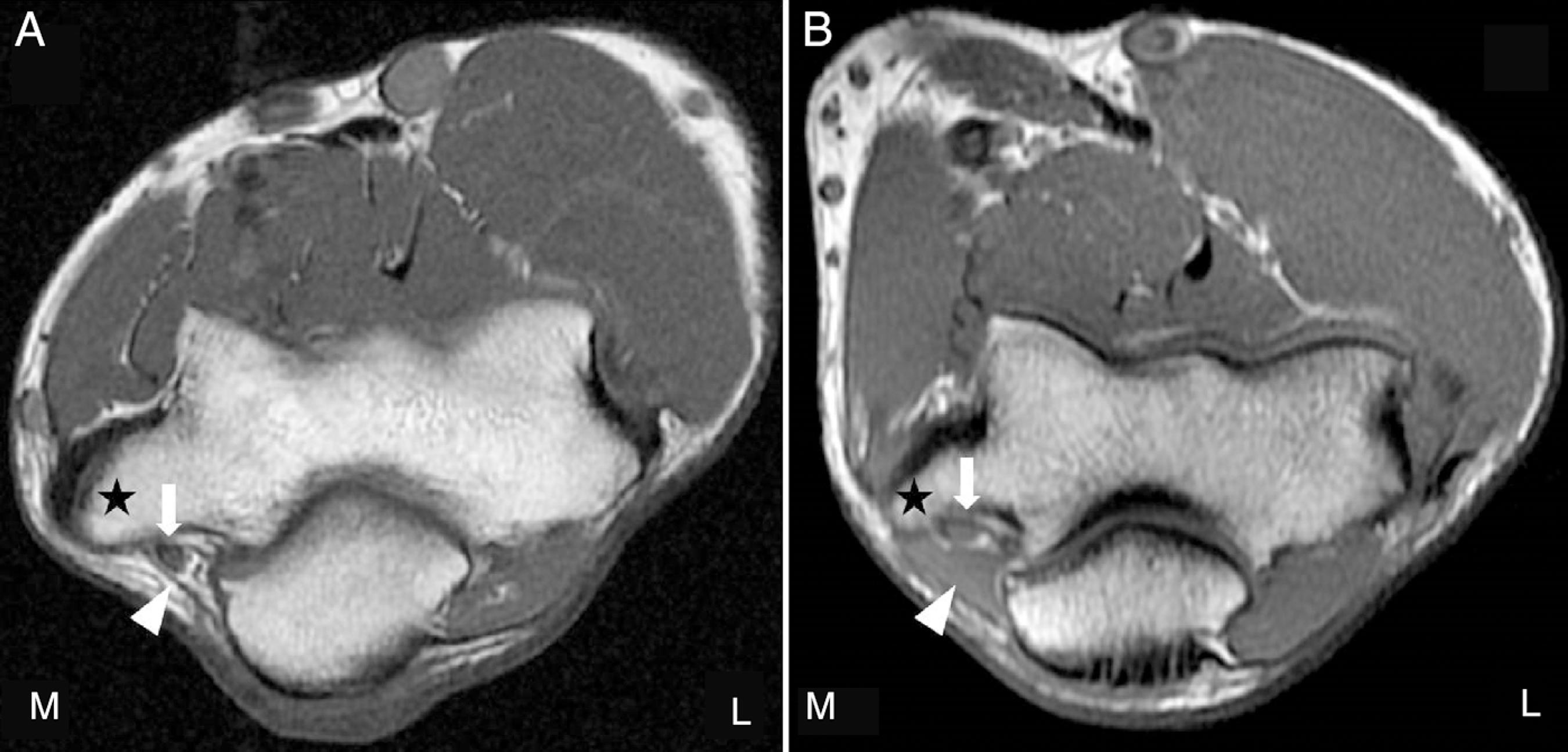
Since its first description, its prevalence is also quite divergent among the authors,66 Gruber W. Über die muskel epitrochleo-anconeus des menschen und den Saugethieren. Mem Imp Acad Sci St Petersbourg. 1866;10(5):1-26.,99 Husarik DB, Saupe N, Pfirrmann CWA, Jost B, Hodler J, Zanetti M. Elbow nerves: MR findings in 60 asymptomatic subjects – normal anatomy, variants ad pitfalls. Radiology. 2009;252(1):148-56.,1616 Testut L. Traité d’anatomie humaine. 4th ed. Paris: Octave Doin; 1899.–2222 Gervasio O, Zaccone C. Surgical approach to ulnar nerve compression at the elbow caused by the epitrochleoanconeus muscle and a prominent medial head of the triceps. Neurosurgery. 2008;62(3–1):186-92. ranging from 3% to 34%. In the present study, the AEM was observed in 29 elbows (Fig. 2), a result similar to previous studies.66 Gruber W. Über die muskel epitrochleo-anconeus des menschen und den Saugethieren. Mem Imp Acad Sci St Petersbourg. 1866;10(5):1-26.,99 Husarik DB, Saupe N, Pfirrmann CWA, Jost B, Hodler J, Zanetti M. Elbow nerves: MR findings in 60 asymptomatic subjects – normal anatomy, variants ad pitfalls. Radiology. 2009;252(1):148-56.,1717 Le-Double AF. Traite des variations du syteme musclulaire de l’homme et de leur significantion au pointe de vue de l’anthropologie zoologiques. Paris: Schleicher Freres; 1897.
Left elbow axial T1-weighted magnetic resonance imaging. (A) Absence of AEM. Star, medial epicondyle; arrow, ulnar nerve; arrowhead, fibrous retinaculum (Osborne's ligament). (B) Presence of AEM. Star, medial epicondyle; arrow, ulnar nerve; arrowhead, AEM; L, lateral; M, medial.
The inconstant and variable presence of this muscle is not an exclusive characteristic of the human species. Galton11 Galton JC. Note on the epitrochleo-anconeus or anconeus sextus (gruber). J Anat Physiol. 1874;9:168-75. reported the presence of AEM in other species of mammals, such as the Capuchin monkey, eastern quoll, Eurasian hare, giant armadillo, Philippine flying lemur, wombat, echidna, sloth, seal, anteater, lion, and grizzly bear. In a comparative study, Abdala and Diogo2323 Abdala V, Diogo R. Comparative anatomy, homologies and evolution of the pectoral and forelimb musculature of tetrapods with special attention to extant limbed amphibians and reptiles. J Anat. 2010;217(5):536-73. reported the presence of AEM in the salamander, sand frog, red-eared slider turtle, ocellated lizard, broad-snouted caiman, and even in the domestic cock. As in humans, the AEM is variable in primates; it appears only in certain species of orangutans, such as the Borneo orangutan, and in chimpanzees.2424 Diogo R, Potau JM, Pastor JF, de Paz FJ, Ferrero EM, Bello G, et al. Photographic and descriptive musculoskeletal atlas of chimpanzees. Boca Raton: CRC Press; 2013.,2525 Diogo R, Potau JM, Pastor JF, de Paz FJ, Ferrero EM, Bello G, et al. Photographic and descriptive musculoskeletal atlas of orangutans. Boca Raton: CRC Press; 2013. Unlike humans, primates and other mammals have the AEM classified as a tensor muscle of the forearm fascia,2424 Diogo R, Potau JM, Pastor JF, de Paz FJ, Ferrero EM, Bello G, et al. Photographic and descriptive musculoskeletal atlas of chimpanzees. Boca Raton: CRC Press; 2013. and as an ulnar flexor of the forearm in amphibians, reptiles, and birds.2323 Abdala V, Diogo R. Comparative anatomy, homologies and evolution of the pectoral and forelimb musculature of tetrapods with special attention to extant limbed amphibians and reptiles. J Anat. 2010;217(5):536-73.
AEM prevalence is higher in lower primates and lemurs, tending to disappear in arthropod monkeys,44 Capdarest-Arest N, Gonzalez JP, Türker T. Hypotheses for ongoing evolution of muscles of the upper extremity. Med Hypotheses. 2014;82(4):452-6. providing an evolutionary clue regarding this muscle.
In the present sample, no statistical correlation was observed between AEM presence and gender (chi-squared: 0.01) or age (chi-squared: 0.955).
The fact that more samples are available at the moment, due to the increasing advance of in vivo techniques allows a more accurate and assertive verification of the actual prevalence of this muscle.
Conclusion
The prevalence of the AEM in humans and other species is variable, without a pattern of normality. No statistical correlation was observed between the presence of this muscle and age or gender of the individuals.
-
☆
Study conducted at the Hospital Alemão Oswaldo Cruz, São Paulo, SP, Brazil.
REFERENCES
-
1Galton JC. Note on the epitrochleo-anconeus or anconeus sextus (gruber). J Anat Physiol. 1874;9:168-75.
-
2Gessini L, Jandolo B, Pietrangeli A, Occhipinti E. Ulnar nerve entrapment at the elbow by persistent epitrochleoanconeus muscle. J Neurosurg. 1981;55(5):830-1.
-
3Dekelver I, Van Glabbeek F, Dijs H, Stassijns G. Bilateral ulnar nerve entrapment by the M. anconeus epitrochlearis. A case report and literature review. Clin Rheumatol. 2012;31(7):1139-42.
-
4Capdarest-Arest N, Gonzalez JP, Türker T. Hypotheses for ongoing evolution of muscles of the upper extremity. Med Hypotheses. 2014;82(4):452-6.
-
5Testut L. Les anomalies musculaires chez l’homme. Expliquées par l’anatomie comparée. Leur importance en anthropologie. Paris: Masson; 1884.
-
6Gruber W. Über die muskel epitrochleo-anconeus des menschen und den Saugethieren. Mem Imp Acad Sci St Petersbourg. 1866;10(5):1-26.
-
7Sookur PA, Naraghi AM, Bleakney RR, Jalan R, Chan O, White LM. Accessory muscles: anatomy, symptoms and radiologic evaluation. Radiographics. 2008;28(2):481-99.
-
8Macalister A. Additional observations on muscular anomalies in human anatomy (third series), with a catalogue of the principal muscular variations hitherto published. Trans R Ir Acad Sci. 1872;25(1):134.
-
9Husarik DB, Saupe N, Pfirrmann CWA, Jost B, Hodler J, Zanetti M. Elbow nerves: MR findings in 60 asymptomatic subjects – normal anatomy, variants ad pitfalls. Radiology. 2009;252(1):148-56.
-
10Li X, Dines JS, Gorman M, Limpisvasti O, Gambardella R, Yocum L. Anconeus epitrochlearis as a source of medial elbow pain in baseball pitchers. Orthopedics. 2012;35(7):1129-32.
-
11Aversi-Ferreira TA, Maior RS, Carneiro-e-Silva FO, Aversi-Ferreira RA, Tavares MC, Nishijo H, et al. Comparative anatomical analyses of the forearm muscles of Cebus libidinosus (Rylands et al., 2000): manipulatory behavior and tool use. PLoS ONE. 2011;6(7):1-8.
-
12Lin TY, Teixeira MJ, Picarelli H, Okane SY, Romano MA, Benegas E, et al. Síndromes dolorosas dos membros superiores. Rev Med. 2001;80(2):317-34.
-
13Jeon IH, Kim PT, Park IH, Kyung HS, Ihn JC. Cubital tunnel syndrome due to the anconeus epitrochlearis in a amateur weight lifter. Sicot Case Rep. 2002:1-6. Available from: http://www.sicot.org/resources/File/IO_reports/07-2002/1-07-2002.pdf [accessed May 2014].
» http://www.sicot.org/resources/File/IO_reports/07-2002/1-07-2002.pdf -
14Morris H, McMurrich JP. Morris’s human anatomy – a complete systematic treatise by English and American authors. 4th ed. Philadelphia: Blakiston’s Son & Co; 1907. p. 392.
-
15Tubbs RS, Shoja MM, Loukas M, editors. Bergman’s comprehensive encyclopedia of human anatomic variation. New Jersey: Wiley Blackwell; 2016.
-
16Testut L. Traité d’anatomie humaine. 4th ed. Paris: Octave Doin; 1899.
-
17Le-Double AF. Traite des variations du syteme musclulaire de l’homme et de leur significantion au pointe de vue de l’anthropologie zoologiques. Paris: Schleicher Freres; 1897.
-
18Clemens HJ. Zur morphologie des ligamentum epitrochleo-anconeum. Anat Anz. 1957;104:343-4.
-
19Mori M. Statistics on the musculature of the Japanese. Okajimas Fol Anat. 1964;40:195-300.
-
20Dellon AL. Musculotendinous variations about the medial humeral epicondyle. J Hand Surg Eur. 1986;11(2):175-81.
-
21Okamoto M, Abe M, Shirai H, Ueda N. Diagnostic ultrasonography of the ulnar nerve in cubital tunnel syndrome. J Hand Surg Eur. 2000;25(5):499-502.
-
22Gervasio O, Zaccone C. Surgical approach to ulnar nerve compression at the elbow caused by the epitrochleoanconeus muscle and a prominent medial head of the triceps. Neurosurgery. 2008;62(3–1):186-92.
-
23Abdala V, Diogo R. Comparative anatomy, homologies and evolution of the pectoral and forelimb musculature of tetrapods with special attention to extant limbed amphibians and reptiles. J Anat. 2010;217(5):536-73.
-
24Diogo R, Potau JM, Pastor JF, de Paz FJ, Ferrero EM, Bello G, et al. Photographic and descriptive musculoskeletal atlas of chimpanzees. Boca Raton: CRC Press; 2013.
-
25Diogo R, Potau JM, Pastor JF, de Paz FJ, Ferrero EM, Bello G, et al. Photographic and descriptive musculoskeletal atlas of orangutans. Boca Raton: CRC Press; 2013.
Publication Dates
-
Publication in this collection
May-Jun 2018
History
-
Received
14 Jan 2017 -
Accepted
2 May 2017 -
Published
05 Apr 2018


