 Six new records of polypores (Agaricomycetes, Basidiomycota) from Southeast Brazil (Atlantic Forest) and perspectives in basic mycological research
Abstract
The present study aims to present six new records of polypores from Southeast Brazil (Atlantic Forest): Fibroporia gossypium, Fomitiporia subtilissima, Fuscoporia marquesiana, Microporellus terrestris, Neodictyopus atlanticae and Perenniporia brasiliensis, as well as to discuss perspectives in basic mycological research through contextual analysis of primary and secondary metadata from inventories, monographs and revisions of existing reference collections. Polypores are wood-inhabiting macrofungi with tubular hymenophores. They play a major role in decomposition within terrestrial ecosystems and hold significant importance for humans. The Atlantic Forest, a biodiversity hotspot with high levels of diversity and endemism, is an ideal source for discovering unknown fungi. Thus, our fungal inventory was built from collections made in the natural remnants of the São Paulo Green Belt Biosphere Reserve, which are strategic sites within the Brazilian Atlantic Forest, located in the municipality of Juquitiba, São Paulo state, Brazil. We provide full descriptions, photoplates, illustrations, remarks on geographic distribution, and additional comments for the newly recorded polypores.
Key words:
Hymenochaetales; Mycology; Polyporales; Taxonomy; Tropical America
Resumo
Este trabalho tem por objetivo apresentar seis novos registros de políporos do sudeste brasileiro (Floresta Atlântica): Fibroporia gossypium, Fomitiporia subtilissima, Fuscoporia marquesiana, Microporellus terrestris, Neodictyopus atlanticae e Perenniporia brasiliensis, assim como discutir perspectivas da pesquisa micológica básica através da análise contextual de metadados primários e secundários de inventários, monografias e coleções físicas existentes. Políporos são macrofungos lignícolas com himenóforos tubulares. Eles desempenham funções cruciais na decomposição dos ecossistemas terrestres e possuem uma importância significativa para os humanos. A Floresta Atlântica, hotspot da biodiversidade com níveis elevados de diversidade e endemismo, é uma fonte ideal para descobertas de fungos desconhecidos. Sendo assim, o inventário dos fungos foi elaborado a partir de coletas em remanescentes naturais da Reserva da Biosfera do Cinturão Verde de São Paulo, que são locais estratégicos da Floresta Atlântica brasileira, situados no município de Juquitiba, estado de São Paulo, Brasil. Além do mais, este artigo provê descrições completas, pranchas fotográficas, ilustrações, informações sobre distribuições geográficas e comentários adicionais para os seis novos registros de políporos.
Palavras-chave:
Hymenochaetales; Mycologia; Polyporales; Taxonomia; America Tropical
Introduction
Polypores are wood-inhabiting macrofungi with tubular hymenophores (Rajchenberg 2011). They play a major role in decomposition within forests (Dighton 2003), and hold significant biological, cultural, and economic importance for humans (Willis et al. 2018; Antonelli et al. 2020).
Global fungal diversity is estimated in millions of species but only less than 10% of them are scientifically known, i.e., ca. 148,000 spp. (Hawksworth & Lücking 2017; Willis et al. 2018). Unknown fungi are emerging primarily from: (1) tropical regions and biodiversity hotspots; (2) little-explored sites and habitats; (3) morphologically and ecologically cryptic species; (4) existing reference collections (Hawksworth & Lücking 2017).
Brazil is one of the richest countries in fungal diversity in Tropical America - ca. 6,320 known spp./ - 200,000 estimated spp. (Lewinsohn & Prado 2002; Maia et al. 2015; BFG 2021). Therefore, the number of undetected species shows that Brazilian fungal studies are still far from reaching its census (Maia et al. 2015; BFG 2021; Hawksworth & Lücking 2017).
The Atlantic Forest, a biodiversity hotspot with high levels of diversity and endemism, retains about 28% of its original cover (Myers et al. 2000; Rezende et al. 2018) and is the richest and most well-known biome harboring fungal species in Brazil (BFG 2021). Nevertheless, considering the estimated number of species above, it still has enormous potential for new discoveries (Maia et al. 2015; Pires et al. 2016; Palacio et al. 2017; Motato-Vasquez et al. 2020; Pagin-Cláudio et al. 2022; Westphalen et al. 2022).
The state of São Paulo (Southeast Brazil) has 32.6% of its area within Atlantic Forest conservation units (IF 2020), and is well-known for hosting the greatest known fungal diversity in the country - ca. 1,900 known spp. (BFG 2021). Still, new taxa have been described by means of inventories, monographs and revisions of existing reference collections in basic mycological research.
The present study aims to present six newly recorded polypore species from Southeast Brazil: Fibroporia gossypium (Speg.) Parmasto, Fomitiporia subtilissima Alves-Silva, Reck & Drechsler-Santos, Fuscoporia marquesiana Gibertoni & C.R.S. de Lira, Microporellus terrestris (Gibertoni & Ryvarden) Decock, Neodictyopus atlanticae Palacio, Robledo & Drechsler-Santos, and Perenniporia brasiliensis C.R.S. de Lira, A.M.S. Soares, Ryvarden & Gibertoni. It will also synthesize perspectives in basic mycological research using contextual analysis of primary and secondary metadata from inventories, monographs, and revisions of existing reference collections. We provide full descriptions, photoplates, illustrations, remarks on geographic distribution, and additional comments for the newly recorded polypores.
Materials and Methods
Fieldwork and inventory zone
Fieldwork was carried out in remnants (24°01’20.9”S, 47°04’06.7”W) of Dense Ombrophilous Montane Forest within the Atlantic Forest province (IBGE 2012), located in the municipality of Juquitiba, São Paulo state, Brazil (Fig. 1). Those natural remnants are managed by Instituto Terra Luminous (ITL 2023) and are strategic for conservation due to their circumscription in UNESCO-designated sites, such as Mata Atlântica Biosphere Reserve and São Paulo Green Belt Biosphere Reserve (RBRB 2023). In addition, they are in the environmental preservation area of Serra do Mar, the buffer zones of Parque Estadual da Serra do Mar and Parque Estadual do Jurupará, as well as in spring protection and recovery areas (ITL 2023).
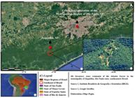
a-c. Research area - a. geographical disposition of the inventory zone in relation to the municipality of Juquitiba and the urban agglomeration of the São Paulo metropolitan region; b. an overview of the inventory zone in the municipality of Juquitiba, located in the state of São Paulo; c. an overview of the Southeast region of Brazil, situated in South America.
The inventory zone covers an area of approximately 270 ha., with a maximum altitude of nearly 760 m.a.s.l., mean temperatures of 18 °C, and the highest rainfall depth occurring in summer (PMGIRS Juquitiba 2009; ITL 2023).
Sample collection and preservation
Our fungal inventory was built from collections made through active sampling on trails and in forest interiors, conducted bimonthly from October 2021 to November 2022; the first author collected all specimens. Samples were collected and preserved using the traditional methods applied in the taxonomy of polypores (Fidalgo & Fidalgo 1989; Lodge et al. 2004). During the survey, basidiomata where photographed, collected with a knife and individually packed in paper bags. Samples were preserved by dehydration in an oven with air ventilation at room temperature and then frozen at -20 °C. Subsequently, vouchers were deposited as heritage in the SP-FUNGI collection (Maria Eneyda Pacheco Kauffmann Fidalgo), at Instituto de Pesquisas Ambientais da Secretaria de Meio Ambiente, Infraestrutura e Logística do Estado de São Paulo (IPA/SEMIL).
Morphologic analysis of basidiomata
Basidiomata were described using concepts and measurements applied in the taxonomy of polypores (Fidalgo & Fidalgo 1967; Vellinga 1988; Ryvarden 2004). Microscopic analysis was performed using freehand longitudinal and transversal cuts of the basidiomata. In order to hydrate the sections, a KOH solution was used (3-5%). Phloxine solution (1%) was used to stain the protoplasm, and Melzer’s reagent was adopted to verify for chemical reactions in microstructures (Lodge et al. 2004). A minimum of thirty measurements were taken of microstructures, when founded. The following abbreviations and codes were used for measurements: Lm × Wm = mean length and width, Q = range of length/width ratio, Qm = length/width mean, and n = x/y [x = number of measurements from a given number (y) of specimens].
Species taxonomy and their geographic distribution
Species taxonomy - current names, classifications, authors and synonyms - as well as geographic distribution were verified using metadata from specialized databases and relevant literature [Ryvarden & Gilbertson 1993; Ryvarden 2004, 2015, 2016; Maia et al. 2015; Li et al. 2016; Crous et al. 2017; Palacio et al. 2017; Rajchenberg et al. 2022, Flora do Brasil 2020 (continuously updated), Index Fungorum electronic database 2023 (continuously updated), among others].
Results
Species taxonomy and remarks
1. Fibroporia gossypium (Speg.) Parmasto, Conspectus Systematis Corticiacearum (Tartu): 207 (1968). Figs. 2a-c; 9a

a-c. Fibroporia gossypium. Macromorphology of the basidiomata - a. general view of the basidioma; b. margin showing rhizomorphs and pores in the regular portion of the hymenial surface; c. pores in the irregular portion of the hymenial surface. (a-c. Pagin et al. 528). Scale bars: a = 1 cm; b-c = 1 mm.
Basidiomata resupinate, seasonal, white to cream, in age or drying becoming sordid cream, adnate to effuse, soft when young turning wax-resinous and brittle when old, growing as radial meshes, up to 1 mm thick. Margin white, rhizomorphic to fimbriate, sterile, up to 1 mm of extension. Subiculum cottony, white, less than 1 mm thick. Tubes sordid cream to whitish cream, less than 1 mm deep. Hymenial surface poroid. Pores easily to hardly seen to the naked eye, circular to angular, decurrent in some parts, (3-)5-7 per mm. Dissepiments dentate to lacerated, pubescent. Hyphal system dimitic. Generative hyphae frequent in the trama of young parts and scarce in old parts, with unilateral clamps, thin-walled, hyaline, 2-3.5(-4) μm diam. Skeletal hyphae abundant in the subiculum, thick-walled to almost solid, sparingly branching, 3-4 μm diam. Cystidia absent. Cystidioles scarce to frequent, fusiform, hyaline, thin-walled. Basidia not found. Basidiospores frequent, slightly thick-walled, hyaline, cylindrical to oblong-ellipsoid in frontal view and allantoid to slightly obovoid in lateral view, (4.3-)4.5-5.7(-5.9) × 2-2.4(-2.5) μm, Lm × Wm = 5 × 2.3 μm; Q = 2-2.5(-2.6); Qm = 2.2 (n = 32/1).
Material examined: Juquitiba, São Paulo Green Belt Biosphere Reserve, Trilha da Onça Parda, 13.VII.2022, F. Pagin et al. FP 528, (SP 528856).
Xylophagous on gymnosperm wood and rarely on angiosperm wood, the species causes brown rot (Ryvarden & Gilbertson 1993; Ryvarden 2016).
This species has a circumpolar distribution into the coniferous forests of Europe, warm parts of temperate zone, Argentina, Brazil, North America and Asia (Rick 1960; Lombard 1990; Ryvarden & Gilbertson 1993; Spirin 2007; Yuan & Dai 2008). Previously recorded from the Brazilian Atlantic Forest of the state of Rio Grande do Sul (Rick 1960). This is the second record from Brazil and the first record from the Southeast of Brazil.
The type locality of F. gossypium is La Plata, Buenos Aires, Argentina (Spegazzini 1899, as Poria gossypium Speg.).
Fibroporia gossypium can be recognized in the field by its resupinate, seasonal, soft to wax-resinous, white to sordid cream basidiomata, rhizomorphic to fimbriate margin, cottony subiculum, as well as by its circular to angular pores. In addition, the dimitic hyphal system composed of clamped generative hyphae and of skeletal hyphae plus the cylindrical to broadly-ellipsoid, slightly thick-walled, (4.3-)4.5-5.7(-5.9) × 2-2.4(-2.5) μm, basidiospores are diagnostic.
Fibroporia vaillantii (DC.) Parmasto is a morphologically similar species, but differs by a softer, not wax-resinous to brittle basidiomata, as well as by larger, 5-7 × 3-4 μm, basidiospores (Ryvarden & Gilbertson 1993). Antrodia porothelioides (Cke) Ryvarden, another morphologically similar species, differs by host preference (angiosperms wood), has slightly smaller pores and slightly longer basidiospores (Ryvarden & Gilbertson 1993; Ryvarden 2015).
Fibroporia gossypium was first described from La Plata (Argentina, South Hemisphere) and later identified from countries of the Northern Hemisphere (Spegazzini 1899; Ryvarden & Gilbertson 1993); molecular metadata show conspecificity between specimens from both Hemispheres (Rajchenberg et al. 2006). Fibroporia gossypium presents an antitropical distribution with conifers being its main hosts in the Northern Hemisphere and with Nothofagus spp. being its main hosts in Patagonia (Argentina, South Hemisphere), where Fibroporia vaillantii colonizes conifers (Rajchenberg 2022).
Our record is the second for Fibroporia gossypium in Brazil, the first one was made by Rick (1960) from an unidentified host at Rio Grande do Sul, and this species was classified as native (Rick 1960; Maia & Carvalho 2010), agreeing with its antitropical distribution. In our fungal inventory, F. gossypium was recorded in a site near to the Tropic of Capricorn, deviating from its typical distribution. This suggests that F. gossypium is an opportunistic and rare species in this region where it is associated with coniferous hosts; in this case sampled from a dead stump of Pinus sp. The morphologically similar Fibroporia vaillantii had never been recorded in Brazil, and Antrodia porothelioides was recorded only once by Rick (1960) in Rio Grande do Sul, though with differences in morphology and substrate preference when compared to Fibroporia gossypium.
2. Fomitiporia subtilissima Alves-Silva, Reck & Drechsler-Santos, inLi et al., Fungal Diversity 78 (2016). Figs. 3a-c; 9b
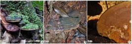
a-c. Fomitiporia subtilissima. Macromorphology of the basidiomata - a. young basidiomata covered with mosses; b. an aged basidioma, partially covered with dead plant material; c. hymenial surface. (a. Pagin et al. 504; b-c. Pagin et al. 548). Scale bars: a-c = 1 cm.
Basidiomata perennial, pileate, woody, sessile to pseudostipitate due to concrescence of layers, aplanate, flabelliform, dimidiate, convex to elongate, single or laterally connate and thus horizontally imbricate, gregarious, growing at the base and roots of trees, 4-22 × 4.5-15.5 cm, up to 1.5 cm thick. Abhymenial surface zonate, concentric zones slightly to strongly sulcate and narrow, crustose, dark brown to reddish-brown, glabrous, rimose, often with moss growing on. Margin obtuse, copper-brown, glabrous. Context homogeneous to slightly zonate, fibrous, with a resinous dark line near the abhymenial surface, golden-brown to slightly reddish-brown, up to 7 mm. Tubes unistratified to pluristratified, concolorous, up to 6 mm thick, old ones filled with white mycelium. Hymenial surface poroid, brownish-gray to copper gold. Pores invisible to the naked eye, round to slightly angular, (7-)8-9(-10) per mm. Dissepiments thick, tomentose, entire. Hyphal system dimitic. Generative hyphae more easily observed at the dissepiment, simple septate, hyaline, thin-walled, rarely with refringent content, 3-5(-6) μm diam. Skeletal hyphae dominant, moderately to strongly dextrinoid, thick-walled to solid, sulfurous-yellow to reddish-yellow, often aseptate to occasionally septate, sparingly branched, with or without refringent content, 3-5(-7) μm diam. Setae absent. Basidia not found. Basidiospores rare to frequent, subglobose to globose, slightly to moderately dextrinoid, hyaline, thick-walled, without content, (4.4-)4.5-5.2(-5.5) × (4.1-)4.2-5(-5.2) μm, Lm × Wm = 4.8 × 4.3 μm; Q = 1-1.18(-1.2), Qm = 1.1 (n = 24/2).
Material examined: Juquitiba, São Paulo Green Belt Biosphere Reserve, Trilha da Chama Violeta, 9.II.2022, F. Pagin et al. FP 504 (SP 528834); 14.VII.2022, F. Pagin et al. FP 548 (SP 527875).
Growing on angiosperm wood (Li et al. 2016).
The species was found in Brazil (Li et al. 2016). Previously recorded from the Brazilian Atlantic Forest in the state of Santa Catarina (Li et al. 2016). This is the first record from the Southeast of Brazil.
The type locality of Fomitiporia subtilissima is Blumenau, Santa Catarina, Brazil (Li et al. 2016).
Fomitiporia subtilissima can be recognized in the field by its perennial, gregarious, aplanate, sessile to pseudostipitate basidiomata, narrowly sulcate abhymenial surface, dark resinous line in the context, obtuse margin, brownish-gray to copper-gold hymenial surface, and by pores which are invisible to the naked eye as well. In addition other diagnostic features are its globose and dextrinoid basidiospores with 4-6 μm in diam. and not surpassing 6 μm in diam. (Alves-Silva et al. 2020). This set of morphological features used to identify F. subtilissima is quite unique within the genus.
In our fungal inventory, as well as in Li et al. (2016), basidiomata were collected from the base and roots of trees, both dead or alive, suggesting that this species may oscillate between being a parasite and a saprotroph.
3. Fuscoporia marquesiana Gibertoni & C.R.S. de Lira, inYuan et al., Fungal Diversity (2020). Figs. 4a-c; 9c-d

a-c. Fuscoporia marquesiana. Macromorphology of the basidiomata - a. close view of the basidiomata; b. basidiomata; c. hymenial surface. (a-c. Pagin et al. 508). Scale bars: a-c = 1 cm.
Basidiomata seasonal to perennial, pileate, aplanate to slightly reflexed on the base, coriaceous to sub-woody, flabelliform, semicircular, elongate to convex, gregarious, up to 1 cm deep, 1-7(-8) × 1-4 cm. Abhymenial surface zonate, zones slightly to moderately sulcate and numerous, glabrous to pubescent or tomentose, sometimes radially striate and rugose, brownish-yellow, yellowish-orange to faintly purplish-dark. Margin acute to subobtuse. Context zonate, fibrous-velutinate, gold-yellowish to yellowish-brown, 1-5 mm thick. Tubes unistratified to pluristratified, distinctly stratified, up to 8 mm deep. Hymenial surface poroid, brownish-purple, often with a grayish tint. Pores invisible to the naked eye, round, 7-9 per mm. Dissepiments entire, thick, pubescent. Hyphal system dimitic. Generative hyphae frequent, simple septate, hyaline to yellow, thin- to slightly thick-walled, straight, branches proximal, 2-3 μm diam. Skeletal hyphae dominant, thick-walled to solid, often aseptate, yellowish-red to reddish-brown, 3-4 μm diam. Setae hymenial, frequent, covering about 3/4 of the pores, apex acute to slight- or even moderately hook, lanceolate to subulate, brown, thick-walled, aseptate to simple septate close to the apex, (14-)15-23.8(-25) × (3.4)4.1-5.7(-6.7) μm, Lm × Wm = 20 × 4.8 μm, Q = (2.6-)3-5.4(-6.3), Qm = 4.2 (n = 26/1). Basidia not found. Basidiospores few, widely ellipsoid, hyaline, thin-walled, smooth, often with refringent content, 4-6.9(-7) × 2.5-4.5(-4.6) μm, Lm × Wm = 5.1 × 3.6 μm; Q = 1.2-1.6; Qm = 1.4 (n = 16/1).
Material examined: Juquitiba, São Paulo Green Belt Biosphere Reserve, Tenda Tipi, 9.II.2022, F. Pagin et al. FP 508 (SP 528838).
Xylophagous on angiosperm wood, where it may cause white rot like other species in the genus.
The species was found in Brazil (Yuan et al. 2020). Previously recorded from the Brazilian Atlantic Forest of the state of Bahia (Yuan et al. 2020). This is the second record for the species since its description and the first record from the Southeast of Brazil.
The type locality of Fuscoporia marquesiana is Santa Teresinha, Bahia, Brazil (Yuan et al. 2020).
Fuscoporia marquesiana can be recognized in the field by its aplanate, seasonal to perennial basidiomata, narrowly to densely sulcate abhymenial surface, often acute margin, brown to purple-brown hymenial surface, as well as by pores which are invisible to the naked eye. In addition, it’s both hooked and acute hymenial setae plus broadly ellipsoid basidiospores are diagnostic.
The material examined (SP 528838), has slightly larger basidiospores compared to the original description, but only few were found and the original description for F. marquesiana was based on a single specimen (URM 83094), which is insufficient to verify intraspecific variations (Yuan et al. 2020; present study). Fuscoporia wahlbergii (Fr.) T. Wagner & M. Fisch. is a morphologically similar species, but differs by the strongly hooked setae (vs. slightly to moderately hooked in F. marquesiana), besides the type of insertion, margin thickness, and coloration of the basidiomata (Yuan et al. 2020; present study).
4. Microporellus terrestris (Gibertoni & Ryvarden) Decock, Czech Mycol. 59(2): 161 (2007). Figs. 5a-c; 9e

a-c. Microporellus terrestris. Macromorphology of the basidiomata - a. basidiomata; b. inferior view of the basidiomata; c. pores. (a-c. Pagin et al. 494). Scale bars: a-b = 1 cm; c = 1 mm.
Basidiomata pileate, laterally to eccentrically stipitate, circular, coriaceous, up to 1 cm high, up to 1 cm in diameter, up to 2 mm thick. Abhymenial surface radially striate, faintly zonate, up to 2 zones, infundibuliform at the center, dark reddish-brown, glabrous. Margin obtuse to sub-truncate. Stipe darkish-brown, up to 0.5 mm high, up to 0.3 mm in diam., rugose, glabrous to pubescent in some parts. Context fibrous, pale brown, up to 1 mm thick. Tubes concolorous, up to 2 mm deep. Hymenial surface poroid, grayish-brown to pale brown. Pores visible to the naked eye, irregular, 2-3 per mm. Dissepiments entire, glabrous, moderately thick. Hyphal system dimitic. Generative hyphae with unilateral clamps, hyaline, simple septate, thin-walled, with refringent content, 3-4 μm diam. Skeletal-binding hyphae aseptate, thick-walled to solid, hyaline to brownish, variably dextrinoid, 1-3 μm diam. Cystidia absent. Basidia not found. Basidiospores frequent to abundant, hyaline, cylindrical to navicular, thin-walled to slightly thick-walled, often with ferruginous content, (7-)7.1-10(-11.5) × 3-4(-4.4) μm, Lm × Wm = 8.4 × 3.5 μm; Q = 1.9-2.9(-3.6), Qm = 2.4, (n = 77/1).
Material examined: Juquitiba, São Paulo Green Belt Biosphere Reserve, Zé da Paz, 3.X.2021, F. Pagin et al. FP 494 (SP 528825).
Xylophagous on angiosperm wood, the species causes white rot.
The species was found in Brazil (Gibertoni et al. 2004; Drechsler-Santos et al. 2007). Previously recorded from the Brazilian Atlantic Forest of the state of Rio Grande do Norte (Gibertoni et al. 2004); Caatinga, from the state of Bahia (Drechsler-Santos et al. 2007). This is the first record from the Southeast of Brazil.
The type locality of Microporellus terrestris is Baía Formosa, Rio Grande do Norte, Brazil (Gibertoni et al. 2004, as Navisporus terrestris Gibertoni & Ryvarden).
Microporellus terrestris can be recognized in the field by its circular, laterally to eccentrically stipitate, coriaceous basidiomata, black and rugose stipe, faintly zonated, reddish-brown abhymenial surface, cream to beige hymenial surface, as well as by the 2-3 pores per mm, which are visible to the naked eye. In addition, its dimitic hyphal system composed by clamped generative hyphae and of skeletal-binding hyphae plus the navicular to cylindrical, (7-)7.1-10(-11.5) × 3-4(-4.4) μm, basidiospores are diagnostic.
With the exception of Microporellus ellipsosporus Decock & Ryvarden, all species in the genus differ from M. terrestris by their tiny pores, presence of cystidia, and globose to lacrimoid basidiospores (Motato-Vásquez & Gugliotta 2016). Microporellus ellipsosporus is the closest species to M. terrestris in morphology, but the first differs by its narrower basidiospores, 1.7-2.3 μm width, and presence of hymenial cystidia (Decock & Ryvarden 2007).
5. Neodictyopus atlanticae Palacio, Robledo & Drechsler-Santos, in Palacio, Robledo, Reck, Grassi, Góes-Neto & Drechsler-Santos, PLoS ONE 12(10): e0186183, 15 (2017). Figs. 6a-c; 7a-f; 9f

a-c. Neodictyopus atlanticae. Macromorphology of the basidiomata - a. basidiomata; b. inferior view of the basidioma; c. pores. (a-c. Pagin et al. 530). Scale bars: a-b = 1 cm; c = 1 mm.

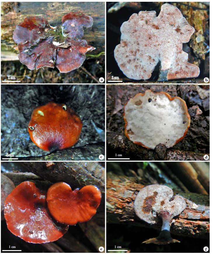
a-f. Polymorphism of Neodictyopus atlanticae. Superior and inferior views of the basidiomata. (a-b. Pagin et al. 507; c-d. Pagin et al. 543; e-f. Pagin et al. 550). Scale bars: a-f = 1 cm.
Basidiomata pileate, seasonal to perennial, laterally to eccentrically stipitate, widely flabelliform, reniform, discoid to circular, coriaceous, gregarious in small groups, sometimes cespitose, 1.5-5.5 × 1.8-3 cm, up to 2.5 mm thick. Abhymenial surface azonate, glabrous, radially striate to occasionally radially rugose when dehydrated, orange-brown, reddish-brown, dark red to purple, often darker in the central region, usually with a depression. Stipe covered by a dark cuticle, insertion at 90° to 180°, up to 2.3 cm high, up to 0.8 mm in diam. - but rarely surpassing 0.5 mm, reticulate, glabrous to slightly pubescent, base radially extended in the substrate. Margin acute to subobtuse, frequently marked by a dark line, deflexed when dehydrated. Context fibrous to occasionally pubescent, cream to grayish-cream, eventually with resinous bands, 1-3 mm thick. Tubes with up to two layers, but often one, cream, grayish-cream to brownish, up to 1 mm each layer. Hymenial surface poroid, grayish-cream, puff gray, dark gray to dark brown. Pores hardly seen to the naked eye, round to angular, 5-9(-10) per mm. Dissepiments entire, granulose to pubescent, slightly to moderately thick. Hyphal system dimitic. Trama frequently impregnated by resinous yellowish-green substance. Generative hyphae frequent, with unilateral clamps, thin-walled, tortuous, densely branched, (1.5-)2-3(-6) μm diam. Skeletal-binding hyphae dominant, aseptate, hyaline, thick-walled to solid, very tortuous, densely branched, 1-4(-5) μm diam. Cystidioles frequent, widely fusiform to narrowly utriform, with basal clamp, (8.5-)12-16 × (3-)4.7-6.5(-8) μm. Cystidia absent. Basidia clavate, 4-sterigma, hyaline, 5 μm wide. Basidiospores rare to frequent, abundant only in pores, subcylindrical, hyaline, often with refringent and ferruginous content, 5-6.9(-7.5) × (1.8-)2-3(-4) μm, Lm × Wm 6.1 × 2.3 μm, Q = (2.1-)2.2-3(-3.1), Qm = 2.6, (n = 334/6).
Material examined: Juquitiba, São Paulo Green Belt Biosphere Reserve, Zé da Paz (Parcela ESALQ), 3.X.2021, F. Pagin, FP 461, SP 528795, F. Pagin 463, SP 528796; Chama Violeta, F. Pagin, FP 507, SP 528837; Chama Violeta, 13.VII.2022, F. Pagin et al. FP 520, SP 528849; Trilha da Jaguatirica, 13.IV.2022, F. Pagin et al. FP 543, SP 528871; Chama Violeta, 13.VII.2022, F. Pagin et al. FP 550, SP 528877; Trilha da Onça Parda, 22.IX.2022, F. Pagin et al. FP 579, SP 528905; Trilha da Jaguatirica, 24.XI.2022, F. Pagin et al. FP 598, SP 528924.
Xylophagous on angiosperm wood, the species causes white rot.
The species was found in Brazil (Palacio et al. 2017). Previously recorded from the Brazilian Atlantic Forest in the state of Santa Catarina (Palacio et al. 2017). This is the first record from the Southeast of Brazil.
The type locality of Neodictyopus atlanticae is Santo Amaro de Imperatriz, Santa Catarina, Brazil (Palacio et al. 2017).
Neodictyopus atlanticae can be recognized in the field by its pileate, laterally to eccentrically stipitate, leathery basidiomata, black cuticle covering the stipe, often reddish-orange to purple, radially striate abhymenial surface, grayish-cream to brown hymenial surface, as well as by pores which are hardly seen to the naked eye.
The potential geographic distribution presented in Palacio et al. (2017) shows that Picipes dictyopus (Mont.) B.K. Cui, Xing Ji & J.L. Zhou, a morphologically similar species, is distributed in the Cerrado province, as well as in Juan Fernandez Islands, where N. atlanticae is distributed in the Atlantic province.
We observed a case of polymorphism (see paragraph 4 of the discussion) in N. atlanticae (Figs. 6-7) through morphological analysis of basidiomata. The morphological features - stipe, pileus and margin configuration as well as basidiospores size (value o Q) - used to distinguish between N. atlanticae (Atlantic province) and P. dictyopus (Cerrado province) (Palacio et al. 2017) overlapped. We observed that phenotypic traits used to distinguish these taxa had low taxonomic value when more specimens were sampled. Firstly, the unknown specimens were morphologically chimeric, exhibiting mixed features used to distinguish N. atlanticae and P. dictyopus. Thus, for the identification of N. atlanticae we used the Q value of basidiospores by means of measurements from all sampled specimens (334 basidiospores/- 6 specimens). Accounting for one subpopulation, they had higher taxonomical value, which were equal to Q = 2−3.5 matching the original description of N. atlanticae (vs. Q = 2.5-3.3 in P. dictyopus) (Palacio et al. 2017; present study). Although, we lack molecular metadata for the material examined, morphological evidence supports the hypothesis that N. atlanticae occurs in the Atlantic province,while P. dictyopus is found in the Cerrado province in Brazil (Palacio et al. 2017; present study).
6. Perenniporia brasiliensis C.R.S. de Lira, A.M.S. Soares, Ryvarden & Gibertoni, in Crous et al., Persoonia 38: 355 (2017). Figs. 8a-c; 9g

a-c. Perenniporia brasiliensis. Macromorphology of the basidiomata - a. close view of the basidioma; b. basidioma; c. hymenial surface. (a. Pagin et al. 546; b-c. Pagin et al. 552). Scale bars: a-b = 1 cm; c = 1 mm.

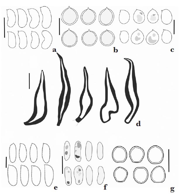
a-g. Microscopic structures of six newly recorded polypores - a. basidiospores of Fibroporia gossypium; b. basidiospores of Fomitiporia subtilissima; c. basidiospores of Fuscoporia marquesiana; d. hymenial setae of Fuscoporia marquesiana; e. basidiospores of Microporellus terrestris; f. basidiospores of Neodictyopus atlanticae; g. basidiospores of Perenniporia brasiliensis. (a. Pagin et al. 528; b. Pagin et al. 504, 548; c-d. Pagin et al. 508; e. Pagin et al. 494; f. Pagin et al. 507, 530, 543, 598; g. Pagin et al. 546, 552, 594). Scale bars: a-g = 5 μm. Drawings by Cristiano Coelho-Nascimento.
Basidiomata resupinate, seasonal to perennial, adnate to effused, coriaceous to woody, white to cream, often with orange-brown macules, difficult to separate from the substrate, often formed by fusion of smaller basidiomata, 1-6.5 × 1-5.5 cm, up to 5 mm thick. Margin sterile, thick, black to reddish-brown, glabrous, crustose, slightly zonated. Subiculum cream, less than 1 mm thick. Tubes unistratified to pluriestratified, 1-4 mm deep, eventually filled with white mycelium. Hymenial surface poroid. Pores invisible to the naked eye, round, 9-10(-11) per mm. Dissepiments slightly to moderately thick, slightly granulous, entire to slightly dentate. Hyphal system dimitic. Generative hyphae rare, with unilateral clamps, hyaline, thin- to thick-walled, 2 μm diam. Skeletal hyphae dominant, arachnoid, slightly to moderately dextrinoid, strongly dextrinoid in mass, densely branched, very tortuous, aseptate to occasionally septate, sometimes with refringent content, 1-2.5 μm diam. in lateral branches, 2-3 μm diam. in axial branches. Crystals occasional to frequent, hyaline, diamond-shaped, tiny to large. Cystidia absent. Basidia not found. Basidioles widely clavate, thin-walled, hyaline. Basidiospores frequent to abundant, subglobose to widely ellipsoid, apex truncate, thick-walled, slightly to strongly dextrinoid, hyaline, 3-4.2(-4.6) × 3-3.7(-4) μm, Lm × Wm = 3.8 × 3.3 μm; Q = 1-1.3(-1.5), Qm = 1.2 (n = 86/3).
Material examined: Juquitiba, São Paulo Green Belt Biosphere Reserve, Chama Violeta, 14.VI.2022, F. Pagin et al. FP 546, SP 528874; F. Pagin et al. FP 552, SP 528879; Trilha da Jaguatirica, 24.XI.2022, F. Pagin et al. FP 594, SP 528920.
Xylophagous on angiosperm wood, the species causes white rot.
The species was found in Brazil and Cameroon (Crous et al. 2017). Previously recorded from the Brazilian Atlantic Forest in the states of Bahia, Ceará, and Pernambuco (Crous et al. 2017); Amazon, from the state of Amapá (Crous et al. 2017). This is the first record from the Southeast of Brazil.
The type locality of Perenniporia brasiliensis is Porto Grande, Amapá, Brazil (Crous et al. 2017).
Perenniporia brasiliensis can be recognized in the field by its resupinate, white to cream, with orange-brown macules, coriaceous basidiomata, besides its pores which are hardly seen to the naked eye. In addition, other diagnostic features are its dimitic hyphal system composed by clamped generative hyphae and of dextrinoid skeletal hyphae plus the small subglobose to globose basidiospores are diagnostic. Crystals in the trama were observed for the first time in this species, although they have been reported in morphologically similar species that differ by their larger basidiospores. In Perenniporia guyanensis Decock & Ryvarden, the basidiospores measure 5.0-5.5(-6.0) μm in diameter, while in Perenniporia paraguyanensisLira & Gibertoni, they measure 4.5-5.2 μm (Ryvarden 2016; Crous et al. 2017). Perenniporia brasiliensis is unique, presenting the smallest basidiospores in the genus.
Discussion
Perspectives in basic mycological research
In this section, we discuss perspectives in basic mycological research through contextual analysis of primary and secondary metadata from inventories, monographs, and revisions of existing reference collections. We highlight the intersection between these branches in basic mycological research, which are foundational for scientific inquiries on fungal diversity, conservation and prospecting.
Our results, in consonance with metadata from inventories, monographs and revisions of existing reference collections (Drechsler-Santos et al. 2008; Wartchow et al. 2011; Abrahão et al. 2012; Baltazar et al. 2012; Pires et al. 2016; Palacio et al. 2017; Motato-Vasquez et al. 2020; Pagin-Cláudio et al. 2022; Westphalen et al. 2022; Fraga-Júnior et al. 2023, among others, some of which are compiled in Maia et al. 2015), demonstrate that the major sources from which unknown fungi emerge overlap in Brazil - (1) tropical regions and biodiversity hotspots; (2) little-explored sites and habitats; (3) morphologically and ecologically cryptic species; (4) existing reference collections (Hawksworth & Lücking 2017). For this reason, the discovery of new fungal taxa has been growing exponentially as basic mycological research advances across the country, mainly in little-explored sites, many of which still show great potential for novel discoveries compared to more studied locations (Baltazar et al. 2012; Maia et al. 2015; BFG 2021). Considering the conservation principles of vulnerability and irreplaceability at global and local scales (Grace et al. 2021), tropical regions, particularly biodiversity hotspots, are crucial for socio-environmental development. These regions are highly heterogeneous, largely unexplored, and home to endemic, cryptic, and novel taxa. They also provide all four types of ecosystem services (i.e., provisioning, regulating, cultural, and supporting), essential for the biosphere’s welfare, which is increasingly threatened by anthropogenic activities (Myers et al. 2000; Millennium Ecosystem Assessment 2005). The biodiversity crisis highlights the importance of biodiversity assessments in developing sustainable conservation plans and strategies for future biological resource use. This is notably relevant in Brazil, a developing country with biological megadiversity in Tropical America, the richest region in species on the planet (Myers et al. 2000; Mace 2004; Millennium Ecosystem Assessment 2005; Antonelli et al. 2018).
In our fungal inventory, the newly recorded Fomitiporia subtilissima, Fuscoporia marquesiana, Neodictyopus atlanticae, and Perenniporia brasiliensis, were originally described in Brazil and have an underestimated geographic distribution as they were decrypted from species complexes less than 10 years ago (Li et al. 2016; Crous et al. 2017; Palacio et al. 2017; Yuan et al. 2020). The fact that decryption detects unknown taxa, including endangered ones, needs to be acknowledge as a predictable pattern in the intersection of inventories, monographs and revisions of existing reference collections (e.g., Gómez-Montoya et al. (2017) for Trametopsis brasiliensis (Ryvarden & de Meijer) Gómez-Mont. & Robledo - vulnerable, VU, Salvador-Montoya et al. (2018) for Tropicoporus drechsleri Salvador-Montoya & Popoff - vulnerable, VU, Westphalen et al. (2018) for Steccherinum neonitidum Westphalen & Tomšovský, Alves-Silva et al. (2020) for Fomitiporia nubicola Alves-Silva, Bittencourt & Drechsler-Santos - vulnerable, VU, and Lira et al. (2021) for Megasporoporia neosetulosa C.R.S. Lira & Gibertoni). This predictable pattern also highlights that existing reference collections, particularly those from little-explored sites in biodiversity hotspots, harbor a significant number of unknown fungal species (e.g., Drechsler-Santos et al. 2008; Fraga-Júnior et al. 2023), sampled decades ago or even a century ago, in the form of vouchers requiring analytical update (BFG 2021). Goodwin et al. (2015) estimated that half of the tropical plants preserved in existing reference collections requires nomenclatural updating. From a pragmatic standpoint, plants are less diverse, less neglected, and more extensively researched than fungi. For instance, about 20% of the Earth’s flora is still unknown, whereas above 90% of the Earth’s funga remains to be known (Dawson et al. 2017; Hawksworth & Lücking 2017; Willis et al. 2018). It has also been estimated that half of the unknown plant species are already represented in existing reference collections (Bebber et al. 2010), which is at least partially true for fungal species. Consequently, revisions of existing reference collections are as important as inventories and monographs, due to their significant potential for metadata integration, as well as their availability, which positively impacts institutions, researchers and individuals worldwide (Andrew et al. 2018).
Polymorphism is a universal phenomenon in biodiversity, often neglected both epistemologically and methodologically (Wiens 1999). Here, we define it as intraspecific variation in qualitative or quantitative traits of an individual´s phenotype. The phenotypic polymorphism observed for N. atlanticae (see remarks; Fig. 6-7), may be relevant to other cryptic species; e.g., Trullella duracina complex (Pat.) Zmitr. The case for polymorphism affirms that: (1) the current set of morphological features or traits used to distinguish between cryptic species has low taxonomic value and is relative, not absolute; (2) population/subpopulation samples displays higher taxonomic value compared to individuals; (3) population/subpopulation samples from different geographic regions display higher taxonomical value than just specimens from the type locality; (4) species-level systematics are essential for distinguishing between intraspecific and interspecific variability, as closely related species share some heritable features, and may exhibit mutations (Bicudo 1988; Wiens 1999; present study). Polymorphism needs to be epistemologically and methodologically acknowledged, with the aim advancing intraspecific systematics in basic mycological research, where the current focus is on interspecific systematics. Additionally, we need to be cautious because numerous new taxa, when collections are scarce, have been described based on a single or a few specimens (e.g., Palacio et al. (2017) for N. atlanticae and Yuan et al. (2020) for Fuscoporia marquesiana), and thus may not reflect the full diversity within species.
Microporellus terrestris is known in Brazil, where it was sampled from three sites (two in the Brazilian Atlantic Forest and one in the Caatinga) (Gibertoni et al. 2004; Drechsler et al. 2007; present study). We hypothesize that M. terrestris is a rare and potentially endangered species due to its low population. We need to consider that inventories and revisions of existing reference collections can widen its population size by discovering new records. Since its original description in Gibertoni et al. (2004), M. terrestris was not sampled again until several years (Dreschsler et al. 2007; present study), which can be due to its low population, small basidiomata, and limited sampling effort. In this regard, the detection of new records positively impacts knowledge about the species population size, also increasing the availability of metadata to support education, conservation, and future planning regarding inventories, monographs and existing reference collections in basic mycological research.
Although this discussion extends beyond the primary results of the present study by incorporating secondary metadata, it highlights several key perspectives in basic mycological research. These include: (1) emphasizing the need to integrate knowledge from monographs, inventories, and existing reference collections, as these resources mutually inform each other; (2) demonstrating that major sources of unknown fungi overlap in Brazil; (3) showing that the discovery of new records can improve our understanding of species distribution and conservation status; (4) highlighting the relevance of intraspecific systematics, particularly in cases of cryptic species polymorphism; and (5) reinforcing the importance of advancing basic mycological research in a megadiverse and developing country like Brazil.
Acknowledgements
The authors kindly acknowledge the Instituto de Pesquisas Ambientais da Secretaria de Meio Ambiente, Infraestrutura e Logística (IPA/SEMIL) and the Instituto Terra Luminous (ITL), for providing structural support. They also thank the Coordenação de Aperfeiçoamento de Pessoal de Nível Superior (CAPES) and the Fundação de Amparo à Pesquisa do Estado de São Paulo (FAPESP), for providing financial support. Additionally, they express their gratitude to Cristiano Coelho-Nascimento, for the illustrations of the microstructures.
Data availability statement
In accordance with Open Science communication practices, the authors inform that all data are available within the manuscript.
References
- Abrahão MC, Gugliotta AMG & Bononi VLR (2012) Xylophilous Agaricomycetes (Basidiomycota) of the Brazilian Cerrado. Check List 8: 1102-1116.
-
Andrew C, Diez J, James TY & Kauserud H (2018) Fungarium specimens: a largely untapped source in global change biology and beyond. Philosophical Transactions of the Royal Society B 374: 1-11. DOI: 10.1098/rstb.2017.0392
» https://doi.org/10.1098/rstb.2017.0392 - Antonelli A, Ariza M, James A, Andermann T, Azevedo J, Bacon C, Faurby S, Guedes T, Hoorn C, Lohmann LG, Matos-Maraví P, Ritter CD, Sanmartín I, Silvestro D, Tejedor M, Steege H, Tuomisto H, Werneck FP, Zizka A & Edwards SV (2018) Conceptual and empirical advances in Neotropical biodiversity research. PeerJ: 1-53.
- Alves-Silva G, Reck AM, Silveira RMB, Bittencourt F, Robledo GL, Góes-Neto A & Drechsler-Santos ER (2020) The Neotropical Fomitiporia (Hymenochaetales, Basidiomycota): the redefinition of F. apiahyna s.s. allows revealing a high hidden species diversity. Mycological Progress 19: 769-790.
- Baltazar JM, Drechsler-Santos ER, Ryvarden L, Cavalcanti MAQ & Gibertoni TB (2012) Contribution to the knowledge of polypores (Agaricomycetes) from the Atlantic Forest and Caatinga, with new records from Brazil. Mycosphere 3: 267-280.
-
Bebber DP, Carine MA, Wood JR, Wortley AH, Harris DJ, Prance GT, Davidse G, Paige J, Pennington TD, Robson NKB & Scotland RW (2010) Herbaria are a major frontier for species discovery. Proceedings of the National Academy of Sciences USA 107: 22169-22171. DOI: 10.1073/pnas.1011841108
» https://doi.org/10.1073/pnas.1011841108 - BFG - The Brazil Flora Group (2021) Brazilian Flora Group: Leveraging the power of a collaborative scientific network. Taxon 71: 178-198.
- Bicudo CEDM (1988) Polymorphism in the Desmid Cosmarium abbreviatum var. minus (Zygnemaphyceae) and its taxonomic implications. Acta Botanica Brasilica 2: 1-6.
- Crous PW, Wingfield MJ, Burgess TI, Hardy GEStJ, Barber PA, Alvarado P, Barnes CW, Buchanan PK, Heykoop M, Moreno G, Thangavel R, van der Spuy S, Barili A, Barrett S, Cacciola SO, Cano-Lira JF, Crane C, Decock C, Gibertoni TB, Guarro J, Guevara-Suarez M, Hubka V, Kolařík M, Lira CRS, Ordoñez ME, Padamsee M, Ryvarden L, Soares AM, Stchigel AM, Sutton DA, Vizzini A, Weir BS, Acharya K, Aloi F, Baseia IG, Blanchette RA, Bordallo JJ, Bratek Z, Butler T, Cano-Canals J, Carlavilla JR, Chander J, Cheewangkoon R, Cruz RHSF, Silva M, Dutta AK, Ercole E, Escobio V, Esteve-Raventós F, Flores JA, Gené J, Góis JS, Haines L, Held BW, Jung MH, Hosaka K, Jung T, Jurjević Z, Kautman V, Kautmanova I, Kiyashko AA, Kozanek M, Kubátová A, Lafourcade M, la Spada F, Latha KPD, Madrid H, Malysheva EF, P Manimohan P, Manjón JL, Martín MP, Mata M, Merényi Z, Morte A, Nagy I, Normand AC, Paloi S, Pattison N, Pawłowska J, Pereira OL, Petterson ME, Picillo B, Raj KNA, Roberts A, Rodríguez A, Rodríguez-Campo FJ, Romański M, Ruszkiewicz-Michalska M, Scanu B, Schena L, Semelbauer M, Sharma R, Shouche YS, Silva V, Staniaszek-Kik M, Stielow JB, Tapia C, Taylor PWJ, Toome-Heller M, Vabeikhokhei JMC, Diepeningen ADV, Hoa NV, Van Tri M, Wiederhold NP, Wrzosek M, Zothanzama J & Groenewald JZ (2017) Fungal planet description sheets: 558-624. Persoonia - Molecular Phylogeny and Evolution of Fungi 38: 240-384.
- Dai YC & Niemelä T (2002) Changbai wood-rotting fungi 13. Antrodia sensu lato Annales Botanici Fennici 39: 257-265.
- Decock C & Ryvarden L (2002) Two undescribed Microporellus species and notes on M. clemensiae, M. setigerus, and M. subincarnatus. Czech Mycology 54: 19-30.
- Dighton J (2003) Fungi in ecosystem processes. Vol. 17. Marcel Dekker, New York. 432p.
- Drechsler-Santos ER, Vasconcellos-Neto JRT, Gibertoni TB, Góes-Neto A & Cavalcanti MAQ (2007) Notes on Navisporus: N. terrestris and N. floccosus from Brazil. Mycotaxon 101: 265-269.
- Drechsler-Santos ER, Groposo C & Loguercio-Leite C (2008) New records of lignocellulolytic fungi (Basidiomycetes) from the Atlantic Rain Forest in State of Santa Catarina, Brazil. Hoehnea 35: 57-61.
- Fidalgo O & Bononi VLR (1989) Fungos e líquens macroscópicos. In: Fidalgo O & Bononi VLR (coord.) Técnicas de coleta, preservação e herborização de material botânico. Instituto de Botânica, São Paulo. Pp. 24-26.
- Fidalgo O & Fidalgo MEPK (1967) Dicionário micológico. Instituto de Botânica, São Paulo. 232p.
-
Flora do Brasil (2020, continuously updated) Jardim Botânico do Rio de Janeiro. Available at <http://floradobrasil.jbrj.gov.br>. Access on 20 June 2023
» http://floradobrasil.jbrj.gov.br -
Fraga Júnior CAV, Gumboski EL, Feuerstein SDC, Cáceres MEDS, Aptroot A & Magnago AC (2023) Funga capixaba: Ascomycota and lichen-forming fungi. Rodriguésia 74: e00692022.
» https://doi.org/e00692022 -
Grace OM, Pérez-Escobar OA, Lucas EJ, Vorontsova MS, Lewis GP, Walker BE, Lohmann LG, Knapp S, Wilkie P, Sarkinen T, Darbyshire I, Nic Lughadha E, Monro A, Woudstra Y, Demissew S, Muasya AM, Díaz S, Baker WJ & Antonelli A (2021) Botanical monography in the Anthropocene. Trends in Plant Science 26: 433-441. DOI: 10.1016/j.tplants.2020.12.018
» https://doi.org/10.1016/j.tplants.2020.12.018 - Gibertoni TB, Ryvarden L & Cavalcanti MAQ (2004) Studies in neotropical polypores 18. New species (Basidiomycota) from Brazil. Synopsis Fungorum: 18: 44-53.
-
Goodwin ZA, Harris DJ, Filer D, Wood JR & Scotland RW (2015) Widespread mistaken identity in tropical plant collections. Current Biology 25: R1066-R1067. DOI: 10.1016/j.cub.2015.10.002
» https://doi.org/10.1016/j.cub.2015.10.002 - Gómez-Montoya N, Drechsler-Santos ER, FerreiraLopes V, Tomšovský M, Urcelay C & Robledo GL (2017) New insigths on Trametopsis Tomšovský (Polyporales Gaüm) based on phylogenetic evidences and morphological analyses of Neotropical species. Phytotaxa 311: 155-167.
- Hawksworth DL & Lücking R (2017) Fungal diversity revisited: 2.2 to 3.8 million species. Microbiology Spectrum 5: 1-17.
-
IBGE - Instituto Brasileiro de Geografia e Estatística (2012) Mapa da área de aplicação da Lei n. 11.428 de 2006 (Decreto n. 6.660, de 21 de novembro de 2008, publicado no Diário Oficial da União de 24 de novembro de 2008). Available at <https://biblioteca.ibge.gov.br/index.php/biblioteca-catalogo?view=detalhes&id=65715>. Access on 22 June 2020.
» https://biblioteca.ibge.gov.br/index.php/biblioteca-catalogo?view=detalhes&id=65715 - IF - Instituto Florestal (2020) Inventário florestal do estado de São Paulo. Mapeamento da cobertura vegetal nativa. Governo do Estado de São Paulo, São Paulo. 60p.
-
Index Fungorum (2023, continuously updated) Index Fungorum. Available at <http://www.indexfungorum.org/>. Access on 20 June 2023.
» http://www.indexfungorum.org/ -
ITL - Instituto Terra Luminous (2023, continuously updated) Terra Luminous. Available at <https://terraluminous.eco.br/>. Access on 20 June 2023.
» https://terraluminous.eco.br/ - Lewinsohn T & Prado J (2002) Biodiversidade brasileira: Síntese do estado atual do conhecimento. Vol. 1. Ed. Contexto, São Paulo. Pp. 1-239.
- Li GJ, Hyde KD, Zhao RL, Hongsanan S, Abdel-Aziz FA, Abdel-Wahab MA, Alvarado P, Alves-Silva G, Ammirati JF, Ariyawansa HA, Baghela A, Bahkali AH, Beug M, Bhat DJ, Bojantchev D, Boonpratuang T, Bulgakov TS, Camporesi E, Boro MC, Ceska O, Chakraborty D, Chen JJ, Chethana KWT, Chomnunti P, Consiglio G, Cui BK, Dai DQ, Dai YC, Dinushani A, Daranagama A, Das K, Dayarathne MC, Crop E, Oliveira RJV, Souza CAF, Souza JIS, Dentinger BTM, Dissanayake AJ, Doilom M, Drechsler-Santos ER, Ghobad-Nejhad M, Gilmore SP, Góes-Neto A, Gorczak M, Haitjema CH, Hapuarachchi KK, Hashimoto A, He MQ, Henske JK, Hirayama K, Iribarren MJ, Jayasiri SC, Jayawardena RS, Jeon SJ, Jerônimo GH, Jesus AL, Jones EBG, Kang JC, Karunarathna SC, Kirk PM, Konta S, Kuhnert E, Langer E, Lee HS, Lee HB, Li WJ, Li XH, Liimatainen K, Lima DX, Lin CG, Liu JK, Liu XZ, Liu ZY, Luangsa-ard JJ, Lücking R, Lumbsch HT, Lumyong S, Leaño EM, Marano AV, Matsumura M, McKenzie EHC, Mongkolsamri S, Mortimer PE, Nguyen TT, Niskanen T, Norphanphoun C, O’Malley MA, Parnmen S, Pawłowska J, Perera RH, Phookamsak R, Phukhamsakda C, Pires-Zottarelli CLA, Raspé O, Reck MA, Rocha SCO, Santiago ALCMA, Senanayake IC, Setti L, Shang QJ, Singh SK, Sir EB, Solomon KV, Song J, Srikitikulchai P, Stadler M, Suetrong S, Takahashi H, Takahashi T, Tanaka K, Tang LP, Thambugala KM, Thanakitpipattana D, Theodorou MK, Thongbai B, Thummarukcharoen T, Tian Q, Tibpromma S, Verbeken A, Vizzini A, Vlasák J, Voigt K, Wanasinghe DN, Wang Y, Weerakoon G, Wen HA, Wen TC, Wijayawardene NN, Wongkanoun S, Wrzosek M, Xiao YP, Xu JC, Yan JY, Yang J, Yang SD, Hu Y, Zhang JF, Zhao J, Zhou LW, Peršoh D, Phillips AJL & Maharachchikumbura SSN (2016) Fungal diversity notes 253-366: taxonomic and phylogenetic contributions to fungal taxa. Fungal Diversity 78: 253-366.
- Lira CRS, Alvarenga RLM, Soares AMS, Ryvarden L & Gibertoni TB (2021) Phylogeny of Megasporoporia s.lat. and related genera of Polyporaceae: new genera, new species and new combinations. Mycosphere 12: 1262-1289.
- Lodge DJ, Ammirath JF, O’Dell TE, Mueller GM, Huhndorf SB, Wang CJ, Stokland JN, Schmit JP, Ryvarden L, Leacock PR, Mata M, Umaña L, Wu Q & Czederpiltz DLL (2004) Terrestrial and lignicolous macrofungi. In: Mueller GM, Bills G & Foster MS (eds.) Biodiversity of fungi: inventorying and monitoring methods. Elsevier Academic Press, Boston. Pp. 128-168.
- Lombard FF (1990) A cultural study of several species of Antrodia (Polyporaceae, Aphyllophorales). Mycologia 82: 185-191.
- Mace GM (2004) The role of taxonomy in species conservation. classic taxonomy. Philosophical Transactions of the Royal Society B 359: 711-719.
- Maia LC, Carvalho AAJ, Cavalcanti LH, Gugliotta AM, Drechsler-Santos ER, Santiago ALMA, Cáceres MES, Gibertoni TB, Aptroot A, Giachini AJ, Soares AMS, Silva ACG, Magnano AC, Goto BT, Lira CRS, Montoya CAS, Pires-Zotarelli CLA, Silva DKA, Soares DJ, Rezende DHC, Luz EDMN, Gumboski EL, Wartchow F, Karstedt F, Freire FM, Coutinho FP, Melo GSN, Sotão HMP, Baseia IG, Pereira J, Oliveira JJS, Souza JF, Bezerra JL, Neta LSA, Pfenning LH, Gusmão LFP, Neves MA, Capelari M, Jaeger MCW, Pulgarín MP, Menolli NJ, Medeiros PS, Friedrich RCS, Chikowski RS, Pires RM, Melo RF, Silveira RMB, Urrea-Valencia S, Cortez VG & Silva VF (2015) Diversity of Brazilian fungi. Rodriguésia 66: 1033-1045.
- Millennium Ecosystem Assessment (2005) Millennium Ecosystem Assessment: general synthesis. Island Press, Washington D.C. Pp. 1-142.
- Motato-Vásquez V & Gugliotta AMG (2016) The genus Microporellus (Basidiomycota, Polyporales) in the Neotropics. Nova Hedwigia 103: 225-238.
- Motato-Vásquez V, Gugliotta AM, Rajchenberg M, Catania M, Urcelay C & Robledo G (2020) New insights on Bjerkandera (Phanerochaetaceae, Polyporales) in the Neotropics with description of Bjerkandera albocinerea based on morphological and molecular evidence. Plant Ecology and Evolution 153: 229-245.
- Myers N, Mittermeier RA, Mittermeier CG, Fonseca GAB & Kent J (2000) Biodiversity hotspots for conservation priorities. Nature 403: 853-858.
- Oliveira VRT & Gibertoni TB (2023) New records on Fomitiporia and Fuscoporia (Hymenochaetaceae) in areas of the Atlantic Forest in Northeastern Brazil. Lilloa 60: 41-57.
-
Pagin-Cláudio F, Gugliotta ADM & Pires RM (2022) Checklist of polypores (Agaricomycetes, Basidiomycota) reveals seventeen new records in Brazilian Atlantic Forest fragments at the Poços de Caldas Plateau, Minas Gerais. Rodriguésia. 73: e00832021. DOI: 10.1590/2175-7860202273058
» https://doi.org/10.1590/2175-7860202273058 - Palacio M, Robledo GL, Reck MA, Grassi E, Góes-Neto A & Drechsler-Santos E (2017) Decrypting the Polyporus dictyopus complex: recovery of Atroporus Ryvarden and segregation of Neodictyopus gen. nov. (Polyporales, Basidiomyocta). PLoS ONE 12: 1-26.
- Pires RM & Gugliotta AM (2016) New records of polypores (Basidiomycota) from the state of São Paulo, Brazil. Iheringia - Série Botânica 71: 124-131.
- PMGIRS - Plano municipal de gestão integrada de resíduos sólidos (2009) Aspectos gerais do município. Prefeitura Municipal de Juquitiba, Juquitiba. 148p.
- Rajchenberg M (2011) Nuclear behavior of the mycelium and the phylogeny of polypores. Mycologia 103: 677-702.
- Rajchenberg M, Gorjón SP & Pildain MB (2011) The phylogenetic disposition of Antrodia s.l. (Polyporales, Basidiomycota) from Patagonia, Argentina. Australian Systematic Botany 24: 111-120.
- Rajchenberg M (2022) An approach to the biogeography of polypore fungi (Agaricomycotina, Basidiomycota) in southern South America. Darwiniana, nueva serie, 10: 451-479.
-
RBRB - Rede Brasileira de Reservas da Biosfera (2023) Rede Brasileira de Reservas da Biosfera. Available at <https://reservasdabiosfera.org.br/>. Access on 20 july 2023.
» https://reservasdabiosfera.org.br/ - Rezende CL, Scarano FR, Assad ED, Joly CA, Metzger JP, Strassburg BBN, Tabarelli M, Fonseca GA & Mittermeier RA (2018) From hotspot to hopespot: an opportunity for the Brazilian Atlantic Forest. Perspectives in ecology and conservation 16: 208-214.
- Rick J (1960) Basidiomycetes eubasidii in Rio Grande do Sul - Brasilia. 4. Meruliaceae, Polyporaceae, Boletaceae. Iheringia Série Botânica 7: 193-295.
- Ryvarden L (1973) Some genera of resupinate polypores. Norwegian Journal of Botany 20: 7-11.
- Ryvarden L (2004) Neotropical polypores. Part 1. Introduction, Hymenochaetaceae and Ganodermataceae. Synopsis Fungorum 19: 1-227.
- Ryvarden L (2015) Neotropical polypores part 2. Polyporaceae Abortiporus-Nigroporus. Synopsis Fungorum 34: 231-443.
- Ryvarden L (2016) Neotropical polypores part 3. Polyporaceae Obba-Wrightoporia. Synopsis Fungorum 36: 445-613.
- Ryvarden L & Gilbertson RL (1993) European polypores part 1: Abortiporus-Lindtneria. Fungiflora, Oslo. 387p.
- Salvador-Montoya CA, Costa-Rezende DH, Ferreira-Lopes V, Borba-Silva MA & Popoff OF (2018) A new species in the “Inonotus linteus complex” from northern Argentina. Phytotaxa 338: 75-89.
- Spegazzini C (1899) Fungi argentini novi vel eritici. Con las laminas 4 y 5. Anales del Museo Nacional de Historia Natural de Buenos Aires 6: 169.
- Spirin W (2007) New and noteworthy Antrodia species (Polyporales, Basidiomycota) in Russia. Mycotaxon 101: 149-156.
- Vellinga EC (1988) Glossary. In: Noordelos ME, Kuyper THW & Vellinga EC (eds.) Flora Agaricina Neerlandica. Vol. 1. CRC Press, Rotterdam. Pp. 54-64.
- Yuan HS, Lu X, Dai YC, Hyde KD, Kan YH, Kušan I, He SH, Liu NG, Sarma VV, Zhao CL, Cui BK, Yousaf N, Sun G, Liu SY, Wu F, Lin CG, Dayarathne MC, Gibertoni TB, Conceição LB, Garibay-Orijel R, Villegas-Ríos M, Salas-Lizana R, Wei TZ, Qiu JZ, Yu ZF, Zeng RPM, Paloi S, Bao DF, Abeywickrama PD, Wei DP, Yang J, Manawasinghe IS, Harishchandra D, Brahmanage RS, Silva NI, Tennakoon DS, Karunarathna A, Gafforov Y, Pem D, Zhang SN, Santiago ALCMA, Bezerra JDP, Dima B, Acharya K, Alvarez-Manjarrez J, Bahkali AH, Bhatt VK, Brandrud TE, Bulgakov TS, Camporesi E, Cao T, Chen YX, Chen YY, Devadatha B, Elgorban AM, Fan LF, Du X, Gao L, Gonçalves CM, Gusmão LFP, Huanraluek N, Jadan M, Jayawardena RS, Khalid AN, Langer E, Lima DX, Lima-Júnior NC, Lira CRS, Liu JKJ, Liu S, Lumyong S, Luo ZL, Matočec N, Niranjan M, Oliveira-Filho JRC, Papp V, Pérez-Pazos E, Phillips AJL, Qiu PL, Ren Y, Ruiz RFC, Semwal KC, Soop K, Souza CAF, Souza-Motta CM, Sun LH, Xie ML, Yao YJ, Zhao Q & Zhou LW (2020) Fungal diversity notes 1277-1386: taxonomic and phylogenetic contributions to fungal taxa. Fungal Diversity 104: 1-266.
- Wartchow F, Maia L & Cavalcanti AMQ (2012) New records of Agaricales from Atlantic Forest fragments of Pernambuco, Northeast Brazil. Mycotaxon 118: 137-146.
-
Westphalen MC, Rajchenberg M, Tomsovský M & Gugliotta AM (2018) A re-evaluation of Neotropical Junghuhnia s.lat. (Polyporales, Basidiomycota) based on morphological and multigene analyses. Persoonia 41: 130-141. DOI: doi.org/10.3767/persoonia.2018.41.07
» https://doi.org/10.3767/persoonia.2018.41.07 - Westphalen MC, Motato-Vásquez V, Rajchenberg M, Gugliotta AM & Silveira RMB (2022) New insights on Flaviporus (Polyporales) in the neotropics. Mycological Progress 21: 1-16.
- Wiens JJ (1999) Polymorphism in systematics and comparative biology. Annual Review of Ecology, Evolution, and Systematics 30: 327-362.
- Willis KJ (2018) State of the world’s fungi. Royal Botanic Gardens, Kew, Richmond. 92p.
- Yuan HS & Dai YC (2008) Polypores from northern and central Yunnan Province, southwestern China. Sydowia 60: 147-159.