 Leaf anatomy of Celtis iguanaea (Cannabaceae): a contribution to the classification and taxonomy of the species
Abstract
Over the last two decades, the circumscription of Celtis iguanaea has been questioned due to the difference between the current characterization and others drawn up from specimens native to Curaçao, the type locality. This problem affects the species richness of the genus, as different species are mistakenly identified as C. iguanaea. Besides taxonomic challenges, this misinterpretation harms other anatomy, morphology, ontogeny and other study fields. To mitigate this problem, this article presents the first morpho-anatomical description of the leaves of C. iguanaea, based on specimens native to Curaçao. Herborized leaves were processed for analysis using light microscopy and scanning electron microscopy. The study identified the presence of multicellular and unicellular trichomes - the first secretory and the last non-secretory, anomocytic-type stomata, large cystoliths of silica on both leaf surfaces with apex projected beyond the epidermal surface, and dorsiventral mesophyll. Mucilaginous cells were widely distributed in the epidermis and in the cortical region of the midrib and petiole. The vascular system had collateral-type bundles. The petiole had a concave-convex shape and a single arch-shaped vascular bundle. These results can help to elucidate taxonomic problems related to C. iguanaea delimitation, and its congeners.
Key words:
Curaçao; Rosales; Taxonomy; Urticalean rosids
Resumo
Nas últimas duas décadas, a circunscrição de Celtis iguanaea tem sido questionada devido à diferença entre a caracterização atual e outras elaboradas a partir de espécimes nativos de Curaçao, localidade do tipo. Esse problema afeta a riqueza de Celtis, pois diferentes espécies são erroneamente identificadas como C. iguanaea. Além do desafio taxonômico, essa interpretação equivocada prejudica outras áreas do conhecimento, como: anatomia, morfologia, ontogenia, entre outras. Para mitigar esse problema, este artigo apresenta a primeira descrição morfo-anatômica das folhas de C. iguanaea, a qual foi realizada a partir de espécimes nativos de Curaçao. Folhas herborizadas foram processadas para análise em microscopia de luz e microscopia eletrônica de varredura. O estudo identificou a presença de tricomas multicelulares e unicelulares - o primeiro secretor e o último não secretor, estômatos do tipo anomocítico, grandes cistólitos de sílica em ambas as faces foliares - com ápice projetado além da superfície epidérmica e mesofilo dorsiventral. Células mucilaginosas estão amplamente distribuídas na epiderme e na região cortical da nervura mediana e do pecíolo. O sistema vascular possui feixes do tipo colateral. O pecíolo possui formato côncavo-convexo e um único feixe vascular em forma de arco. A partir destes resultados, espera-se contribuir para a elucidação dos problemas taxonômicos relacionados à C. iguanaea delimitação e seus congêneres.
Palavras-chave:
Curaçao; Rosales; Taxonomia; Rosídeas urticóides
Introduction
Celtis L. is the most species-rich genus of Cannabaceae, encompassing 64 species with pantropical distribution (Fu et al. 2023). Celtis can be recognized by a combination of characters: individuals are lianas, shrubs or trees, monoecious; with armed or unarmed branches; estipulate leaves, where alternate, trinervate, abaxial surface is with domatia present or not; inflorescences solitary or in pairs with diclinous (= unisexual) flowers; dialysepal calyx, five sepals; apetala; five stamens opposite to the sepals; pseudomonomerous gynoecium and bicarpellate, uniovulate, with stigma entire or bifurcate; and small drupes with a pyrene (Berg & Dahlberg 2001; Zamengo et al. 2020; Leme et al. 2021).
Celtis is classified in four subgenera: C. subg. Celtis Planch. (name invalidly published according to Art. 21.3, 22.2: ICN, Turland et al. 2018), C. subg. Mertensia Planch., C. subg. Solenostigma (Endl.) Planch., and C. subg. Sponioceltis Planch (Planchon 1848). To circumscribe the subgenera, Planchon (1848) used the distribution of the species and morphological characters. Thus, C. subg. Celtis occurs in both the northern and southern hemispheres, C. subg. Mertensia is endemic to the Neotropical region, C. subg. Solenostigma to Oceania, and C. subg. Sponioceltis to the Caribbean. The main morphological characters are the presence (C. subg. Mertensia) or absence (C. subg. Celtis, C. subg. Solenostigma and C. subg. Sponioceltis) of thorns, and flowers with stigma lobes divided (C. subg. Mertensia and C. subg. Solenostigma) or entire (C. subg. Celtis and C. subg. Sponioceltis).
Among Neotropical species (Celtis subg. Mertensia), Celtis iguanaea (Jacq.) Sarg. is the most geographically widespread, ranging from Florida, USA to Argentina (Berg & Dahlberg 2001). In their last revision of C. subg. Mertensia, Berg & Dahlberg (2001) proposed 26 synonyms for C. iguanaea, which reduced the previously suggested diversity within the genus (Planchon 1848). The synonymizations by these authors were primarily based on the morphology of thorns (straight or curved), leaf surface texture (scabrous or smooth), leaf indumentum (glabrous or velutinous), and the size and color of the drupes. The proposal by Berg & Dahlberg (2001) has been questioned due to discrepancies in the morphological characterization of C. iguanaea compared to others based on native specimens from Curaçao, the type location of the species (Jacquin 1760, 1763, 1788; Lamarck 1789; Planchon 1848; Boldingh 1913).
Recent taxonomic studies have revealed that several species previously considered synonymous with C. iguanaea (sensuBerg & Dahlberg 2001) are actually distinct species, such as C. alnifolia (Wedd.) Miq., C. spinosa Spreng., and C. spinosissima (Wedd.) Miq. (Chamorro et al. 2021; Zamengo et al. 2020, 2023). These findings emphasize the need to revise the current classification of C. iguanaea (Berg & Dahlberg 2001). Incorrectly identifying specimens as C. iguanaea not only poses challenges in taxonomy, but also adversely affects other fields of research. These misidentifications have affected studies in anatomy (Pilati & Souza 2006; Arambarri et al. 2011; Barros et al. 2023), phylogeny (Fu et al. 2023), ontogeny (Leme et al. 2020, 2021; Pedersoli et al. 2019, 2020; Teixeira et al. 2020), and pollination (Arruda & Sazima 1988).
Leaf anatomy studies contributed to the characterization of Celtis genus and species (El-Alfy et al. 2011a; Thadeo et al. 2014; Shahbaz & Sharif 2017; Arogundade & Adedeji 2019; Câmara et al. 2020; Batool et al. 2024). Therefore, detailed anatomical descriptions of leaf structure with records of collection sites can be conductive to taxonomic and systematic studies of Celtis.
The leaf of Celtis can be recognized anatomically by single-layered epidermis, dorsivental structure, mesophyll formed by one layer of palisade parenchyma and spongy parenchyma, collenchyma in the midrib and cystoliths (Metcalfe & Chalk 1957; Dottori 1976; Pilati & Souza 2006; Nughes et al. 2013). Furthermore, species of the genus can be recognized by presenting secretory or non-secretory trichomes and by the type and distribution of stomata (El-Alfya et al. 2011; Shahbaz & Sharif 2017).
Cystoliths are a synapomorphy of the Urticalean rosids clade (Sytsma et al. 2001), which is formed by four families: Cannabaceae, Moraceae, Ulmaceae and Urticaceae. Although cystoliths have been recorded in the leaf blade of Celtis species (Metcalfe & Chalk 1957; Dottori 1976; Honaine et al. 2005; Nughes et al. 2013), few studies describe their chemical composition (El-Alfya et al. 2011; Honaine et al. 2005).
The previously highlighted challenge of accurately distinguishing Celtis species (Elias 1970; Hunziker & Dottori 1976; Romanczuk & del Pero de Martinez 1978; Nee 1984; Berg & Dahlberg 2001) is largely due to their cryptic morphological characteristics (Chamorro et al. 2021). For instance, after extensive morphological analysis, Celtis ehrenbegiana (Klotzsch) Liebm. was found to involve three distinct entities (Asmus et al. 2018). Recent studies have emphasized the importance of pyrenes in delineating these species (Hunziker & Dottori 1976; Sattarian & Van Der Maesen 2006; Zarafshar et al. 2010; Heo 2020; Zamengo et al. 2020; Chamorro et al. 2021). While pyrenes are significant for taxonomy, species such as C. iguanaea and C. spinosissima can be challenging to differentiate because of their similar pyrene profiles (Zamengo et al. 2020; Chamorro et al. 2021). In such scenarios, leaf anatomy serves as a valuable tool to distinguish similar taxa (Alves et al. 2002; Carvalho et al. 2017; Shahbaz & Sharif 2017; Santos et al. 2020).
Anatomical studies of Neotropical Celtis species are limited, especially with regard to specific variability (Dottori 1990, 1991, 1994; Marchiori & Freitas 1993; Pilati & Souza 2006; Nughes et al. 2013; García et al. 2016; Pedersoli et al. 2019; Nascimento et al. 2022; Barros et al. 2023). Generally, descriptive studies are related to medicinal species, such as C. ehrenbergiana (Arambarri et al. 2011) and C. iguanaea (Paula et al. 2010; Arambarri et al. 2011), whereas taxonomical studies are very rare (Maiti et al. 2016). Therefore, our work fills a gap by describing anatomical and micromorphological analyses of C. iguanaea leaves from Curaçao, identifying characters that aid in the characterization of the species. Furthermore, we compare our results with other anatomical studies of specimens identified as C. iguanaea, as well as other Celtis species. This contribution holds significant importance for Celtis taxonomy by calling for a reassessment of the current classification of C. iguanaea (Berg & Dahlberg 2001).
Materials and Methods
Plant material
Dried leaves of Celtis iguanaea were obtained from the type locality of the species, from specimens E.A.T. Houtepen 1 and E.A.T. Houtepen 2 (RB herbarium numbers 815396, 815397 and barcode numbers 01443256, 01443257), are native to Curaçao in the Caribbean. The leaves were photographed under a stereomicroscope for morphological characterization. Afterwards, five herborized leaves of each voucher were processed for analysis under light microscopy (LM) and scanning electronic microscopy (SEM). The leaves were rehydrated in 1:1 solution of 70% ethanol and pure glycerin (Sosnovsky et al. 2017, modified), dehydrated in ethanolic series, and preserved in 70% ethanol to be processed for anatomical analysis.
Anatomical procedures
To conduct the anatomical analysis, the rehydrated leaves were cut in the middle third of the leaf blade, including the intercostal portion, main vein, leaf margin and petiole. The samples were dehydrated in an up to 95% series of ethanol and embedded in glycol-methacrylate (Historesin Leica®) according to the manufacturer’s instructions. The transverse sections were sectioned at 6-10 µm thickness in a rotary microtome model RM-2145 (Leica Microsystem, Germany), stained with 0.1% toluidine blue pH 4.4 (O’Brien et al. 1964), and mounted with water and coverslip. The epidermis surface was analyzed by dissociation with epidermal peels prepared using Franklin solution (hydrogen peroxide and glacial acetic acid 1:1 v/v; Franklin 1945), stained in 0.25% basic fuchsin dye dissolved in 50% ethanol and mounted on slides in 50% glycerin. Photomicrographs were taken using a MoticamPro 252B digital camera coupled to a Nikon Eclipse Ci light microscope. Leaf cross sections were treated with ruthenium red (Gregory & Baas 1989) and toluidine blue (modified from O’Brien et al. 1964) to identify mucilage. In order to identify the composition of the crystals (druses and cystholits), we submitted the sections to 5% sulfuric acid and hydrochloric acid to dissolve calcium oxalate crystals following the methods of Strasburger (1924). However, as this method did not successfully dissolve the cystholits, we used energy dispersive X-ray spectroscopy (EDS) (Oxford Instruments, Model: Xplore) to detect their chemical composition.
For SEM analysis, samples were dried in stove apparatus, mounted on a metal support with carbon adhesive tape, and coated with gold for 360 s using a Denton Vacuum Desk III instrument (Moorestown, Nova Jersey, EUA). Electron micrographs were obtained with a JEOL JSM - 6380 Scanning Electron Microscope (SEM) operated at an accelerated voltage of 10 kV.
Results
Leaf blade
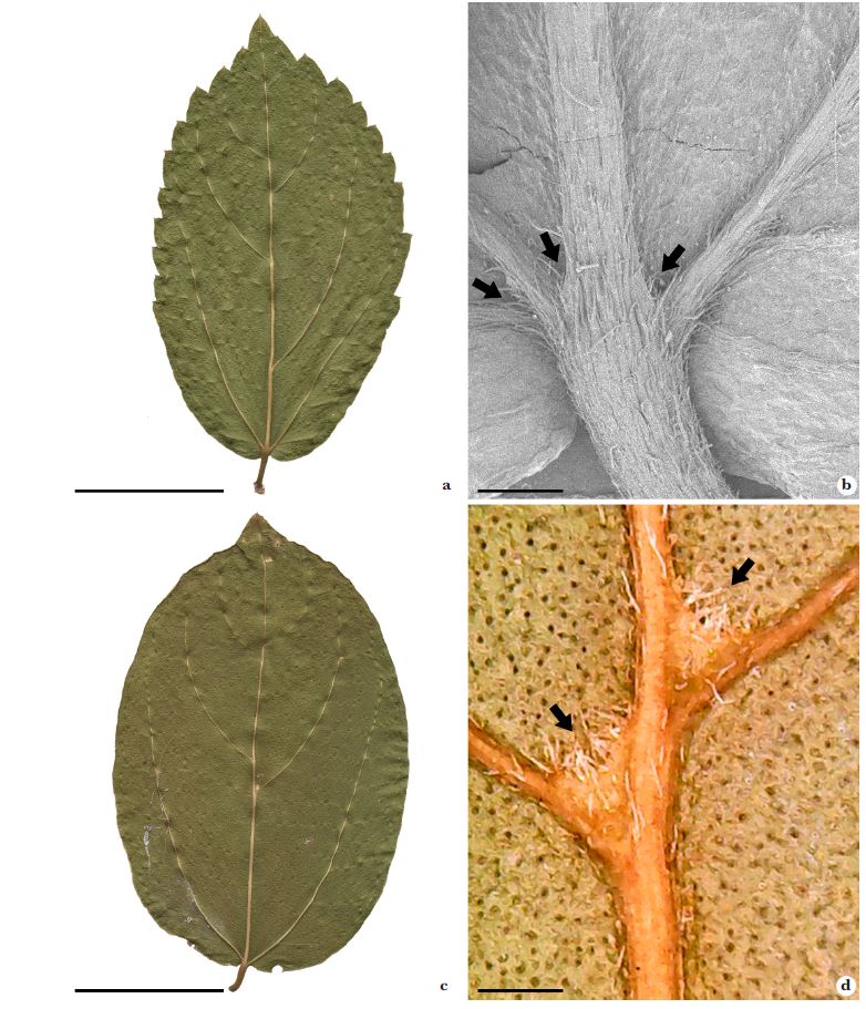
Young leaves of Celtis iguanaea possessed toothed margin and a midrib with inconspicuous domatia (Fig. 1a,b), while mature leaves had entire margin and visible domatia (Fig. 1c,d).
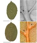
a-d. Leaves and domatias in the abaxial face of Celtis iguanaea (Cannabaceae) - a. young leaf, abaxial surface; b. base of the young leaf, inconspicuous domatias (black arrow) (SEM); c. mature leaf, abaxial surface; d. domatias of mature leaf (black arrow) (photograph). Scale bars: a = 1 cm; b, d = 1 mm; c = 3 cm.
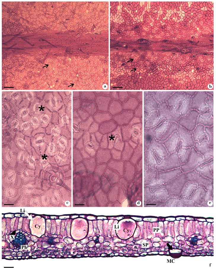
Anatomically, Celtis iguanaea leaves were covered by a single-layered epidermis composed of common epidermal cells, stomata, secretory and non-secretory trichomes, and crystal idioblasts (Figs. 2; 3). In the frontal view, the common epidermal cells had straight or curved anticlinal cell wall on both surfaces (Fig. 2a-d). In the midrib region, the common epidermal cells had straight anticlinal walls, with an elongated shape (Fig. 2a-b).

a-f. Epidermis of Celtis iguanaea (Cannabaceae) under light microscopy - a. epidermis on the abaxial face, stomata in all surfaces (black arrow); b. epidermis on the adaxial surface, stomata near of the midrib (black arrow); c. note the stomata (*) scattered throughout the epidermis on the abaxial surface; d. few stomata (*) in the epidermis of the adaxial surface; e. anomocytic stomata; f. cross section of the leaf. Note epidermis with lithocysts (white arrow) and mucilaginous cell (black arrow); collateral vascular bundles with phloem and xylem. Symbols: Cy = cystolith; Li = lithocyst; MC = mucilaginous cell; Ph = phloem; PP = palisade parenchyma; SP = spongy parenchyma; X = xylem. Scale bars: a, b = 100 μm; c, d, e, f = 10 μm.
Anomocytic stomata type occurs on both leaf surfaces in different distributions (Fig. 2c-e). On the adaxial surface the stomata occurred near the midrib region and on secondary veins, whereas on the abaxial side, they were distributed throughout the surface (Fig. 2b,d). In cross-section, the epidermis was covered by a thin cuticle layer and showed mucilaginous cells (Fig. 2f).
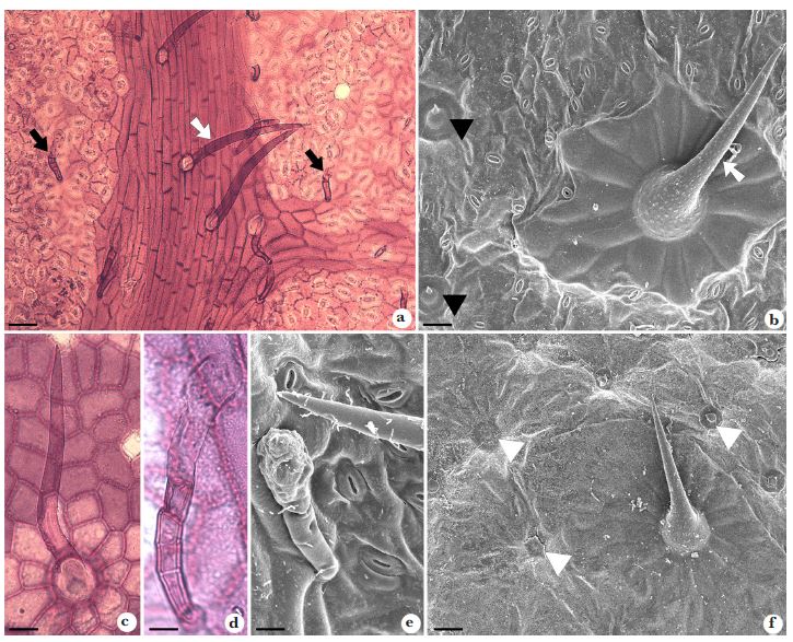
Two types of trichomes were observed: unicellular non-secretory trichomes, and pluricellular secretory trichomes (Fig. 3a-e). The length of the non-secretory trichomes varied from short and spine-like to long and filiform, both having strongly thickened cell walls (Fig. 3a-c). The multicellular secretory trichomes were of the capitate type, composed of a uniseriate peduncle consisting of 2-5 tabular cells, with an elongated multicellular secretory head (Fig. 3d,e). The secretory trichomes generally occurred on the abaxial surface in the region covering the midrib (Fig. 3a).

a-f. Trichomes and cystolith protrusion of Celtis iguanaea (Cannabaceae) in light microscopy (a, c, d) and scanning electronic microscopy (b, e, f) - a. midrib with non-secretory (white arrow) and secretory trichomes (black arrow) on the abaxial surface of the leaf; b. non-secretory trichomes short (black arrowhead) and long (white arrow); c. non-secretory trichomes long; d-e. secretory trichomes; f. protrusion of the cystolith of the lithocyst (white arrowhead). Scale bars: a = 100 μm; b, d, e, f = 10 μm; c = 50 μm.
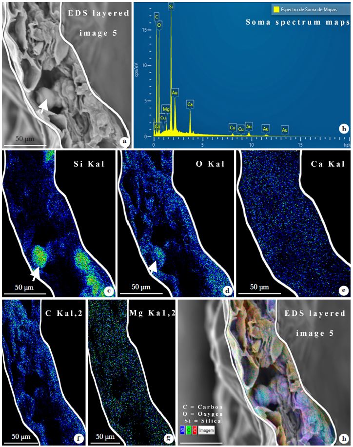
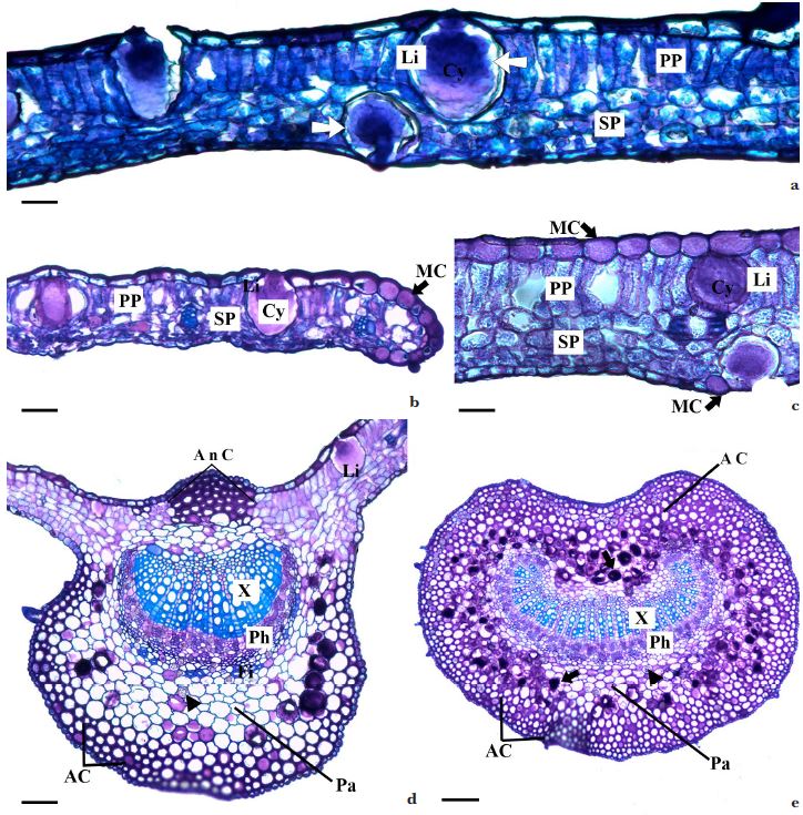
Lithocysts were detected in the epidermis on both leaf surfaces containing a large cystolith of approximately circular-to-elongate form, with a rough surface (Figs. 2f; 4; 5a-d). The cystoliths formed a tiny apex projecting towards the leaf surface and forming a small protrusion visible in frontal view (Fig. 3f). Traditionally used histochemical tests performed with acids did not result in the dissolution of these structures, indicating that they were not made of calcium carbonate or calcium oxalate. Next, EDS analyses were performed, identifying high silica concentration in the cystoliths (Fig. 4a-h), visible in the graph (Fig. 4b), as well as by image (Fig. 4c). In addition to silica, carbon, oxygen and calcium were also identified in significant quantities (Fig. 4b,d-g). The high concentration of silica and oxygen indicates that the main component of the cystolith is silicon dioxide, marked in blue-green on the EDS image (Fig. 4h). The presence of gold originated from the coating of the Denton Vacuum Desk III instrument.
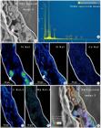
a-h. EDS analysis - a. cross section of Celtis iguanaea leaf in SEM, the cystolith indicated by the arrow; b. graph of the main chemical components identified in the leaf; c. detection of silica in green in the leaf; d. detection of oxygen in green; e-g. detection of Ca, C and Mg in green, note the uniform distribution of these components throughout the mesophyll; h. EDS image in layered showing the presence of O, Si and C. Symbols: Ca = calcium; C = carbon; O = oxygen; Si = silica; Mg = magnesium; Cu = copper; Au = gold.
a-e. Anatomy of the leaf blade of Celtis iguanaea (Cannabaceae) under light microscope - a. dorsiventral mesophyll. Note the lithocyst on both face (white arrow); b. leaf edge with mucilaginous cells in the epidermis (black arrow); c. mucilaginous cells (black arrow); d. midrib of the leaf. Note angular collenchyma in the cortical region and annular collenchyma restricted to the central region below adaxial epidermis; e. petiole. Symbols: AC = angular collenchyma; AnC = annular collenchyma; Cy = cystolith; Dr = druses; Fi = fiber; Li = lithocyst; MC = mucilaginous cells; Ph = phloem; PP = palisade parenchyma; SP = spongy parenchyma; X = xylem. Scale bars: a = 50 μm; b = 100 μm; c, e = 10 μm; d = 100 μm.
The mesophyll of C. iguanaea was dorsiventral (Figs. 2f; 5a-c). The palisade parenchyma was composed of one layer of elongated cells, interrupted by lithocysts (Figs. 2f; 5a-c). The spongy parenchyma presented up to four cell layers with rare idioblasts containing druse crystals (Figs. 2f; 5a-c). The vascular system consisted of collateral vascular bundles (Fig. 2f). The leaf margin flexed downwards, and the epidermal cells were round in the cross section, and more voluminous compared to the others, while also storing mucilage (Fig. 5b).
In the midrib the cortical region of the was formed by angular collenchyma underlying the epidermis, consisting of 3-5 cell layers restricted to the abaxial face (Fig. 5d). Under the adaxial epidermis, the annular collenchyma was restricted to a small central region (Fig. 5d). The space between the collenchyma and the main vascular bundle was filled by six layers of regular parenchyma formed by voluminous polygonal cells. Mucilaginous cells and idioblasts with druse crystals were detected in this region, mainly in the abaxial side (Fig. 5d). The vascular system consisted of a single collateral vascular bundle with a flat concave shape surrounded by small groups of fibers in the abaxial side (Fig. 5d).
Petiole
The adaxial surface of the petiole was concave (Fig. 5e). The epidermis was single-layered, with few stomata on the adaxial surface. The cortical region was occupied by four to six layers of angular collenchyma, and internally by six layers of regular parenchyma. Mucilaginous cells and idioblasts with druse crystals were observed in the cortical region, dispersed around of the vascular system (Fig. 5e). The vascular system was composed of a single collateral vascular bundle, with a semi-circular shape and ends facing the center (Fig. 5e).
Discussion
Our study revealed distinctive features of the leaves of Celtis species, including single-layered epidermis with stomata predominantly on the abaxial surface, lithocysts, unicellular and multicellular trichomes, and dorsiventral mesophyll. Some characters of C. iguanaea leaves diverged from previously published descriptions, suggesting that leaf anatomy could contribute to a more accurate identification of specimens of this species.
The leaves of Celtis species have a single-layered epidermis, composed of common epidermal cells with a variable, straight-to-sinuous contour (Metcalfe & Chalk 1950; El-Alfy et al. 2011b; García et al. 2016). For some taxa, the contour of the epidermal cell wall may vary according to the growth conditions of the plant, as observed in C. ehrenbergiana (sensu Berg & Dahlberg 2001), which presents cells with sinuous walls when exposed to sunlight, compared to those that have epidermal cells with straight walls (Nughes et al. 2013). The influence of light conditions on cell contour needs to be further studied to confirm the potential use of this attribute in taxonomic studies. In the leaves of C. iguanaea evaluated here, we observed a predominance of straight-to-curved anticlinal walls, corroborating the variations cited for the genus.
The epidermis leaves of C. iguanaea from Curaçao is amphistomatic and have a single-layered epidermis as cited by studies describing specimens from other localities (Pilati & Souza 2006; Paula et al. 2010). Although stomata occur on both surfaces, on the adaxial surface the stomates are restricted to the midrib. Amphistomatic leaves, with more stomata on the abaxial side, have been described for several species of Celtis (García et al. 2016; Arambarri et al. 2011; Maiti et al. 2016). The presence of stomata only on the abaxial surface of the leaf blade is reported for C. australis (El Alfi et al. 2011). The most frequently described stomata types are paracytic, anisocytic and anomocytic (Metcalfe & Chalk 1950; El Alfy et al. 2011; García et al. 2016; Maiti et al. 2016). In our samples of C. iguanaea the anomocytic type was observed.
The leaf anatomy of Celtis species can be very similar. Nevertheless, certain characters, particularly trichomes, are useful to differentiate these species. Non-secretory trichomes are the most frequently described for Celtis species and they can be micropapilate or smooth (Tobe & Takaso 1996). Non-secretory trichomes in C. iguanaea are unicellular and smooth, and may be short and prickle-like or long (as in the present study), whereas the trichomes of “C. iguanaea” from Goiás (Brazil) have unicellular and pluricellular non-secretory trichomes (Paula et al. 2010). Furthermore, Paula et al. (2010) did not observe secretory trichomes. In fact, the specimen analyzed by Paula et al. (2010) was different from the C. iguanaea from Curaçao.
Although cystolytic trichomes (i.e., trichomes presenting cystoliths at the base) often occur in Cannabaceae and are frequently used in forensic examinations of Cannabis sativa L. (Dayanandan & Kaufman 1976) or Celtis species (Gangadhara & Inamdar 1977; Maiti et al. 2011; Shahbaz & Sharif 2017), we did not observe this trichome type in C. iguanaea in the present study.
In a revision of secretory trichomes in Urticalean species, Nascimento et al. (2022) cite two types of secretory trichomes for Celtis species: 1) filiform long (uniseriate and more than four cells long) for Celtis sp., C. pubescens Spreng. (sensuTorres & Luca 2005), C. boninensis Koidz., C. sinensis Pers., and C. spinosa Spreng. (sensu Torres & Luca 2005), and 2) capitate (uniseriate stalk and pluricellular head) (C. pubescens sensu Torres & Luca 2005). However, in our samples of C. iguanaea, we found only one type of secretory trichomes, i.e., C. iguanaea did not show the trichome secretor filiform described by Nascimento et al. (2022). Our samples of Celtis iguanaea presented capitate secretory trichomes with uniseriate stalk and multicellular head, similar to those described in C. pubescens (Nascimento et al. 2022). Other studies describing the leaf anatomy of C. iguanaea did not cite secretory trichomes neither in seedlings (Pilati & Souza 2006), nor in leaves (Paula et al. 2010).
Lithocysts are epidermal idioblasts that usually contain calcium carbonate or silica and store calcium or carbon dioxide (Honaine et al. 2023). The presence of cystoliths in leaves appears to be a condition shared by the entire Urticalean clade (Sytsma et al. 2002). Cystoliths may vary in location, frequency, and dimensions in the leaf epidermis, on one or both sides of the leaf, and these variations can be used to distinguish some species. In a comparative study, Shahbaz & Sharif (2017) identified smaller and less frequent lithocysts in C. tournefortii Lam. compared to C. australis. For C. occidentalis L., silica cystoliths have been reported, occurring predominantly on the adaxial surface (El-Alfy et al. 2011a, 2011b). In the samples of C. iguanaea evaluated here, cystoliths were observed on both sides of the leaf, forming tiny projections towards the adaxial surface as seen in the frontal view (Fig. 3f), possibly constituting an additional anatomical taxonomic character for C. iguanaea.
Celtis iguanaea has lithocysts on both leaf surfaces, another characteristic that differs from the specimen studied by Paula et al. (2010), which possessed lithocysts only on the adaxial leaf side. Cystoliths vary in location, frequency, and dimensions in the leaf epidermis and can be observed on one or both sides of the leaf. These variations could represent additional taxonomic information. In a comparative study, Shahbaz & Sharif (2017) found that lithocysts in C. tournefortii were smaller and less frequent than in those in C. australis. Pilati & Souza (2006) described cystoliths on both sides of C. iguanaea seedling leaves (eophyll).
Cystolith may be composed primarily of calcium carbonate (Metcalfe 1985; Watt et al. 1987), soluble with weak acidic solution, but the presence of silica as silicon dioxide or calcium silicate has also been found forming the phytolith, or at least the initial stalk-like protrusion that connects the cystolith to the outer cell wall (Metcalfe 1985; Watt et al. 1987, Gal et al. 2012). Silica was also identified in the cystoliths of C. tala Gillies ex Planch. (Honaine et al. 2005), C. australis and C. occidentalis (El-Alfya et al. 2011). The chemical composition was evaluated by energy-dispersive X-ray spectroscopy (EDS or EDX), confirming silica as the main component of the photoliths observed in the large epidermal cells in C. iguanaea (this study).
While C. iguanaea and other specimens native to South America differed in some characters, they had similar dorsiventral leaf blades with a layer of palisade parenchyma, idioblasts with druse, lithocysts and three layers of spongy parenchyma - as described from specimens from the Brazilian states of Goiás, Mato Grosso do Sul and Paraná (Pilati & Souza 2006; Paula et al. 2010). The vascular bundle parenchyma sheath of C. iguanaea has also been described for specimens from Argentina (Arambarri et al. 2011). Dorsiventral mesophyll and a vascular system consisting of collateral vascular bundles are features cited for the genus (Metcalfe & Chalk 1950; García et al. 2016; Maiti et al. 2016).
The midrib of Celtis iguanaea leaves is biconcave, with a vascular bundle surrounded by cortical parenchyma with druse crystals, collateral vascular bundle in the convex plane shape, phloem with druses and a strip of fibers facing the abaxial part. Similar characteristics have been observed in the specimens studied by Paula et al. (2010).
Regarding the leaf margin, Paula et al. (2010) described the specimens from Goiás state (Brazil) as having similar morphology, with small vascular bundles surrounded by regions of collenchyma. However, in the samples evaluated in the present study, the leaves present margins flexed towards the abaxial region, and vascular bundles surrounded by a parenchyma sheath.
Similar to the specimens studied by Paula et al. (2010), Celtis iguanaea has petioles with a concave-convex outline, uniseriate epidermis, a cortical region with angular collenchyma and crystalliferous idioblasts containing forming druses. Despite these similarities, the samples of C. iguanaea evaluated in the present study have stomata and mucilage idioblasts dispersed both in the cortical region and the vascular system, characteristics not mentioned previously (Paula et al. 2010).
Even though the Celtis iguanaea specimen from Curaçao has anatomical similarities with other species native to South America (Pilati & Souza 2006; Paula et al. 2010; Arambarri et al. 2011; Bento 2019; Nascimento et al. 2022), significant differences were observed, as presence of only a type of secretory trichome, absence of filiform uniseriate secretory trichome and cystoliths with protrusions. Our results support recently raised questions (Chamorro et al. 2021; Zamengo et al. 2020, 2023b) about the current circumscription of C. iguanaea (Berg & Dahlberg 2001), and we emphasize the need for a new revision of this species. Our work could also support correct identification of other Celtis species. From the comparative analysis of leaf anatomy with other species, we were able to highlight some characteristics that we consider relevant for the recognition of C. iguanaea: common epidermal cells with straight-to-curved walls, anomocytic stomata; amphistomatic leaves; stomata on the adaxial surface of the leaf blade distributed exclusively in the regions over veins; lithocysts containing silica cystoliths with circular-to-elongate shape, rough surface, occupying up to half the thickness of the leaf; secretory trichomes of the capitate type, with pluricellular uniseriate peduncle, and multiseriate head; spine-shaped to filiform non-secretory trichomes ; mucilaginous idioblasts in the epidermis, mesophyll, and cortical region of the midrib and petiole.
Although many specimens of Celtis have been identified as C. iguanaea in several regions of Brazil, these specimens probably refer to other species of Celtis, such as C. alnifolia (Wedd.) Miq., C. clausseniana (Wedd.) Miq., C. serratissima Zamengo, Torres, Gaglioti & Romaniuc and C. spinosissima (Wedd.) Miq. (Zamengo et al. 2020). We emphasize the importance of further studies of the leaf anatomy of other, mainly Neotropical Celtis species, focusing on trichome types, cystolith shape and composition, stomata types and distribution, and presence of mucilage cells to support the correct taxonomic classification of several species synonymized by Berg & Dahlberg (2001).
Acknowledgements
Author HBZ (scholarships CAPES/BRASIL, 88887.721972/2022-00) thanks Paul Stokkermans (The Caribbean Research and Management of Biodiversity CARMABI), for sending materials ofCeltis iguanaea; and Patrick Scannell, for enabling the partnership with collector E.A.T. Houtepen. DAFS thanks the Conselho Nacional de Desenvolvimento Científico e Tecnológico; for the research grant (IC-CNPq/UFMS). The authors thank Luiz Felipe Placa Vargas and Laboratório Multiusuário de Análises de Materiais (MULTILAM) by SEM and EDS analyses, and Judit Szabo by English review. This work was supported by Fundação Universidade Federal de Mato Grosso do Sul-UFMS/MEC-Brazil and Coordenação de Aperfeiçoamento de Pessoal de Nível Superior-Brasil (CAPES)-Finance Code 001.
Data availability statement
In accordance with Open Science communication practices, the authors inform that all data are available within the manuscript.
References
-
Alves MV, Estelita MEM, Wanderley MGL & Thomas WW (2002) Aplicações taxonômicas da anatomia foliar das espécies brasileiras de Hypolytrum Rich. (Cyperaceae). Brazilian Journal of Botany 25: 1-9. DOI: 10.1590/S0100-84042002000100002
» https://doi.org/10.1590/S0100-84042002000100002 - Arambarri AM, Novoa MC, Bayón ND, Hernández MP, Colares MN & Monti C (2011) Anatomía foliar de arbustos y árboles medicinales de la región chaqueña semiárida de la Argentina. Dominguezia 27: 5-24.
- Arruda VLV & Sazima M (1988) Polinização e reprodução de Celtis iguanaea (Jacq.) Sarg. (Ulmaceae), uma espécie anemófila. Brazilian Journal of Botany 11: 113-122.
-
Asmus J, Chamorro D, Mogni VY, Oakley LJ & Prado DE (2018) Identidad taxonómica de los “talas”: Análisis morfológico de Celtis tala y Celtis pallida (Celtidaceae). Boletín de la Sociedad Argentina de Botánica 53: 653-664. DOI: 10.31055/1851.2372.v53.n4.22117
» https://doi.org/10.31055/1851.2372.v53.n4.22117 -
Barros TC, Leite VG, Pedersoli GD, Leme FM, Marinho CR & Teixeira SP (2023) Mucilage cells in the flower of Rosales species: reflections on morphological diversity, classification, and functions. Protoplasma 260: 1135-1147. DOI: 10.1007/s00709-023-01836-8
» https://doi.org/10.1007/s00709-023-01836-8 - Bento KBD (2019) Estratégias anatômicas adaptativas do complexo epidérmico de espécies arbóreas no sul da Amazônia. Dissertação de Mestrado. Universidade do Estado de Mato Grosso, Alta Floresta. 2019. 96 p.
-
Berg CC & Dahlberg SV (2001) A revision of Celtis subg. Mertensia (Ulmaceae). Brittonia 53: 66-81. DOI: 10.1007/BF02805398
» https://doi.org/10.1007/BF02805398 - Boldingh I (1913) Flora voor de Nederlandsch West-Indische eilanden. J.H. de Bussy, Amsterdam. 450p.
-
Carvalho R, Pellissari LCO, Pace MR, Scremin-Dias E, Arruda RCO & Farinaccio MA (2017) Leaf morphoanatomy of Araujia and Morrenia (Asclepiadoideae, Apocynaceae): phylogenetic implications and species key. Botanical Journal of the Linnean Society 183: 1-14. DOI: 10.1093/botlinnean/bow004
» https://doi.org/10.1093/botlinnean/bow004 -
Chamorro DC, Zamengo HB, Mogni VY, Torres RB, Gaglioti AL, Da-Silva PR, Leme FM, Prado DE & Oakley LJ (2021) Morfología comparada de diez taxones del género Celtis (Cannabaceae) del Cono Sur Sudamericano. Darwiniana 9: 217-244. DOI: 10.14522/darwiniana.2021.91.936
» https://doi.org/10.14522/darwiniana.2021.91.936 -
Dayanandan P & Kaufman PB (1976) Trichomes of Cannabis sativa L. (Cannabaceae). American Journal of Botany 63: 578. DOI: 10.2307/2441821
» https://doi.org/10.2307/2441821 - Dottori NM (1976) Morfología foliar en Celtis tala y C. pallida con especial referencia a los domacios. Kurtziana 9: 63-80.
- Dottori N (1990) Anatomia reproductiva en Ulmaceae “sensu lato” II. Estrectura y desarrollo del fruto de Celtis tala y Trema micrantha Boletín Sociedad Argentina de Botánica 26: 247-257.
- Dottori N (1991) Anatomia reproductiva en Ulmaceae sensu lato. III. Esporangios, esporogenesis y gametogénesis de Phyllostylon rhamnoides y Celtis tala Kurtziana 21: 81-110.
- Dottori N (1994) Anatomia reproductiva en Ulmaceae sensu lato IV. Fertilización, ontogenia de la semilla y plántula en Phyllostylon rhamnoides y Celtis tala Kurtziana 23: 27-54.
- El-Alfy TSMA, El-Gohary HMA, Sokkar NM, Hosny M & Al-Mahdy DA (2011a) A new flavonoid C-Glycoside from Celtis australis L. and Celtis occidentalis L. leaves and potential antioxidant and cytotoxic activities. Scientia Pharmaceutica 79: 963-975.
-
El-Alfy TSMA, El-Gohary HMA, Sokkar NM, El-Tawab SA & Al-Mahdy DAM (2011b) Botanical and genetic characteristics of Celtis australis L. and Celtis occidentalis L. grown in Egypt. Bulletin of Faculty of Pharmacy, Cairo University 49: 37-57. DOI: 10.1016/j.bfopcu.2011.07.007
» https://doi.org/10.1016/j.bfopcu.2011.07.007 - Elias TS (1970) The genera of Ulmaceae in the Southern United States. Journal of the Arnold Arboretum 51: 18-40.
- Franklin GL (1945) Preparation of thin sections of synthetic resins and wood-resin composites, and a new macerating method for wood. Nature 155: 51.
-
Fu XG, Liu SY, Velzen R, Stull GW, Tian Q, Li YX, Folk RA, Guralnick RP, Kates HR, Jin JJ, Li ZH, Soltis DE, Soltis PS & Yi TS (2023) Phylogenomic analysis of the hemp family (Cannabaceae) reveals deep cyto-nuclear discordance and provides new insights into generic relationships. Journal of Systematics and Evolution 61: 806-826. DOI: 10.1111/jse.12920
» https://doi.org/10.1111/jse.12920 - Gal A, Hirsch A, Siegel S, Li C, Aichmayer B, Politi Y & Addadi L (2012) Plant cystoliths: a complex functional biocomposite of four distinct silica and amorphous calcium carbonate phases. Chemistry - A European Journal 18: 10262-10270.
- Gangadhara M & Inamdar JA (1977) Trichomes and stomata, and their taxonomic significance in the Urticales. Plant Systematic and Evolution 127: 121-137.
- García MG, Arrúa RD & Oakley LJ (2016) Morfoanatomía de la hoja de Celtis chichape (Wedd.) Miq. (Cannabaceae). Rojasiana 15: 41-46.
- Gregory M & Baas P (1989) A survey of mucilage cells in vegetative organs of the dicotyledons. Israel Journal of Botany 38: 125-174.
- Heo T (2020) A systematic study on the genus Celtis L. (Cannabaceae) in Korea. PhD thesis. Kangwon National University. Chuncheon, South Korea. 2020. 164 p.
- Honaine FM, Zucol AF & Osterrieth ML (2005) Biomineralizaciones de sílice en Celtis tala (Celtidaceae). Boletín de la Sociedad Argentina de Botánica 40: 229-239.
- Hunziker AT & Dottori NM (1976) Contribución al conocimiento sobre los talas (Celtis - Ulmaceae) de Argentina, com especial referencia a la región mediterrânea. I. Sobre uma espécie Austro-sudamericana y sobra otra de área disyunta. Kurtziana 9: 103-140.
- Jacquin NJ (1760) Enumeratio systematica plantarum. Theodorus Haak, Leiden. 41p.
- Jacquin NJ (1763) Selectarum Stirpeum Americanarum Historia. Vienna. Krausiana, Vienna. 74 p.
- Jacquin NJ (1788) Selectarum Stirpeum Americanarum Historia. Ed. 3. Vienna. Manhemii, Vienna. 94p.
- Lamarck JBABM (1789) Encyclopedie methodique. botanique. Vol. 3. Panckoucke, Paris.
-
Leme FM, Schönenberger J, Staedler YM & Teixeira SP (2020) Comparative floral development reveals novel aspects of structure and diversity of flowers in Cannabaceae. Botanical Journal of the Linnean Society 193: 64-83. DOI: 10.1093/botlinnean/boaa004
» https://doi.org/10.1093/botlinnean/boaa004 -
Leme FM, Staedler YM, Schönenberger J & Teixeira SP (2021) Floral morphogenesis of Celtis species: implications for breeding system and reduced floral structure. American Journal of Botany 108: 1-17. DOI: 10.1002/ajb2.1724
» https://doi.org/10.1002/ajb2.1724 - Lillie RD (1965) Histopathologic technic and practical histochemistry. McGraw-Hill, New York. 501p.
-
Maiti R, Gonzáles-Rodriguez H, Díaz JCG, Tijerina HAD, Flores AAC, Kumari A & Sarkar NC (2016) Comparative wood anatomy of 20 woody plant species in Northeastern Mexico and its significance. International Journal of Bio-resource and Stress Management 7: 229-246. DOI: 10.23910/IJBSM/2016.7.2.1549
» https://doi.org/10.23910/IJBSM/2016.7.2.1549 -
Marchiori JNC & Freitas AM (1993) Anatomia da madeira de Celtis pallida Torrey (Ulmaceae). Ciência e Natura, Santa Maria 15: 137-147. DOI: 10.5902/2179460X26378.
» https://doi.org/10.5902/2179460X26378 - Metcalfe CR & Chalk L (1950) Anatomy of the dicotyledons. Vol. 1. Clarendon Press, Oxford. Pp. 243-245.
- Metcalfe CR (1985) Secreted mineral substances, Crystals. In: Metcalfe CR & Chalk L (eds.) Anatomy of the dicotyledons. Vol 2. Claredon Press, Oxford. Pp. 82-97.
-
Nascimento IC, Leme FM & Teixeira SP (2022) Morphological diversity of glandular trichomes in urticalean rosids. Acta Botanica Brasilica 36: 1-15. DOI: 10.1590/0102-33062022abb0061
» https://doi.org/10.1590/0102-33062022abb0061 - Nee M (1984) Celtis L. Flora de Vera Cruz, fascículo 40 (Ulmaceae): 11-20.
-
Nughes L, Colares M, Hernandéz M & Aranbarri A (2013) Morfo-anatomía de las hojas de Celtis ehrenbergiana (Celtidaceae) desarrolladas bajo condiciones naturales de sol y sombra. Bonplandia 22: 159-170. DOI: 10.30972/bon.2221245
» https://doi.org/10.30972/bon.2221245 -
O’Brien TP, Feder N & McCully ME (1964) Polychromatic staining of plant cell walls by toluidine blue O. Protoplasma 59: 368-373. DOI: 10.1007/BF01248568
» https://doi.org/10.1007/BF01248568 - Paula MA, Couto RO, Bara MTF, Rezende MH, Paula JR & Costa EA (2010) Caracterização farmacognóstica de Celtis iguanaea (Jacq.) Sargent. Latin American Journal of Pharmacy 29: 526-533.
-
Pedersoli GD, Leme FM, Leite VG & Teixeira SP (2019) Anatomy solves the puzzle of explosive pollen release in wind-pollinated urticalean rosids. American Journal of Botany 106: 489-506. DOI: 10.1002/ajb2.1254
» https://doi.org/10.1002/ajb2.1254 - Pedersoli GD & Teixeira SP (2020) Flower morphogenesis and gametophyte fertility attest to the rare condition of gynomonoecy in Parietaria debilis (Urticaceae). Botany 98: 703-715.
-
Pilati R & Souza LA (2006) Morfoanatomia da plântula de Celtis iguanaea (Jacq.) Sarg. (Ulmaceae). Acta Scientiarum. Biological Sciences 28: 1-6. DOI: 10.4025/actascibiolsci.v28i1.1051
» https://doi.org/10.4025/actascibiolsci.v28i1.1051 - Planchon JE (1848) Sur les Ulmacées (Ulmacées et Celtidées de quelques auteurs) considerées comme tribu de la famille des Urticées. Annales des Sciences Naturelles, Botanique, sér. 3, 10: 244-341.
- Romanczuk MC & del Pero de Martínez MA (1978) Las especies del género Celtis (Ulmaceae) en la flora argentina. Darwiniana 21: 541-577.
-
Santos JVC, Oliveira MFV, Filho FSS, Silva LNNS & Araújo JS (2020) The taxonomic value of leaf anatomy for species Byrsonima: a difficult genus of Malpighiaceae Juss. Acta Botanica Brasilica 34: 570-579. DOI: 10.1590/0102-33062020abb0144
» https://doi.org/10.1590/0102-33062020abb0144 - Sattarian A & Van der Maesen LJG (2006) Endocarp morphology of African Celtis (Celtidaceae/Ulmaceae). Blumea 51: 389-397.
-
Shahbaz SE & Sharif NM (2017) Anatomical and palynological characters of Celtis L. in Kurdistan-Iraq. Science Journal of University of Zakho 5: 57-63. DOI: 10.25271/2017.5.1.302
» https://doi.org/10.25271/2017.5.1.302 -
Sosnovsky Y, Nachychko V, Prokopiv A & Honcharenko V (2017) Leaf architecture in Rhododendron subsection Rhododendron (Ericaceae) from the Alps and Carpathian Mountains: taxonomic and evolutionary implications. Flora 230: 26-38. DOI: 10.1016/j.flora.2017.03.003
» https://doi.org/10.1016/j.flora.2017.03.003 - Strasburger E (1924) Handbook of practical botany. 5th ed. George Allen & Nonwin, London. 533p.
-
Sytsma KJ, Morawetz J, Pires JC, Nepokroeff M, Conti E, Zjhra M, Hall JC & Chase MW (2002) Urticalean rosids: circumscription, rosid ancestry, and phylogenetics based on rbcL, trnL-trnF, and ndhF sequences. American Journal of Botany 89: 1531-1546. DOI: 10.3732/ajb.89.9.1531
» https://doi.org/10.3732/ajb.89.9.1531 - Teixeira SP, Pedersoli GD, Leme FM, Leite VG & Basso-Alves JP (2020) Development resolves the uncommon floral construction of urticalean rosids. Plant ontogeny: studies, analyses and evolutionary implications. Chapter 7. Nova Science Publishers, New York. Pp.181-205.
- Tobe H & Takaso T (1996) Trichome micromorphology in Celtidaceae and Ulmaceae (Urticales). Acta Phytotaxonomica et Geobotanica 47: 153-168.
- Torres RB & Luca AQ (2005) Ulmaceae. In: Wanderley MGL, Shepherd G, Giulietti AM & Melhem TS (eds.) Flora fanerogâmica do estado de São Paulo. Instituto de Botânica, São Paulo. Vol. 4, pp. 361-370.
- Turland NJ, Wiersema JH, Barrie FR, Greuter W, Hawksworth DL, Herendeen PS, Knapp S, Kusber WH, Li DZ, Marhold K, May TW, McNeill J, Monro AM, Prado J, Price MJ & Smith GF (2018) International code of nomenclature for algae, fungi, and plants (Shenzhen Code) adopted by the Nineteenth International Botanical Congress Shenzhen, China, July 2017. Regnum Vegetabile 159. Koeltz Botanical Books, Glashütten. 254p.
-
Zamengo HB, Gaglioti AL, Chamorro D, Mogni V, Oakley L, Prado D, Torres RB, Mattos L, Da-Silva PR & Romaniuc-Neto S (2020) Nomenclatural novelties in Celtis (Cannabaceae) and a preliminary phylogeny of the genus with emphasis on the South American species. Brazilian Journal of Botany 43: 947-960. DOI: 10.1007/s40415-020-00656-x
» https://doi.org/10.1007/s40415-020-00656-x -
Zamengo HB, Gaglioti AL & Pederneiras LC (2023) Celtis flumeniana (Cannabaceae): a new species from Brazil and reestablishment of Celtis alnifolia Phytotaxa 620: 251-263. DOI: 10.11646/phytotaxa.620.4.1
» https://doi.org/10.11646/phytotaxa.620.4.1 -
Zarafshar M, Akbariia M & Sattarian A (2010) Endocarp morphology of Iranian Celtis (Celtidaceae-Cannabaceae). International Journal of Plant Production 4: 73-78. DOI: 10.22069/IJPP.2012.684
» https://doi.org/10.22069/IJPP.2012.684 - Watt WM, Morrell CK, Smith DL & Steer MW (1987) Cystolith development and structure in Pilea cadieri (Urticaceae) Annals of Botany 60: 71-84.