Effects of ionizing radiation on healing after experimental periodontitis
Resumo
Introdução Adoença periodontal é uma condição inflamatória crônica causada pela colonização de biofilmes subgengivais altamente complexos nas superfícies dentárias, que afeta o periodonto e o osso alveolar.
Objetivo Investigar os efeitos da radiação ionizante na reparação dos tecidos periodontais após tratamento da periodontite experimental em ratos.
Material e método A doença periodontal foi induzida por ligaduras de seda ao redor dos segundos molares inferiores por sete dias, utilizando o lado contralateral como controle. Vinte e oito ratos Wistar machos foram distribuídos em quatro grupos (n=7): não ligado e não irradiado, não ligado e irradiado, ligado e não irradiado, e ligado e irradiado. Após a remoção das ligaduras, os grupos irradiados receberam dose única de 30 Gy aplicada na região mandibular aos 7 e 20 dias. A eutanásia foi realizada após o período experimental. A microtomografia computadorizada avaliou perda óssea alveolar e fração de volume ósseo. Análises histológicas coradas em hematoxilina e eosina consideraram infiltrado inflamatório, vascularização, hiperemia, lacunas de Howship, fibrose e lacunas osteocíticas vazias. Os dados foram analisados com testes estatísticos (p<0,05).
Resultado Animais com doença periodontal apresentaram maior perda óssea alveolar em comparação aos controles. Irradiados mostraram perda adicional em relação aos não irradiados, independentemente do tempo. Aos 20 dias, o grupo ligado e irradiado apresentou redução significativa da fração de volume ósseo. A análise histológica evidenciou infiltrado inflamatório, alterações vasculares, fibrose e maior número de lacunas osteocíticas vazias, sugerindo atraso ou comprometimento da reparação tecidual.
Conclusão A radiação ionizante afetou negativamente a reparação periodontal, intensificando a perda óssea alveolar e a inflamação. Esses efeitos podem comprometer a cicatrização a longo prazo e interferir na recuperação óssea e de tecidos moles.
Descritores:
Doenças periodontais; microtomografia por Raio-X; perda do osso alveolar
Abstract
Introduction Periodontal disease is a chronic inflammatory condition caused by the colonization of highly complex, subgingival biofilms on tooth surfaces, that affects the periodontium and alveolar bone.
Objective This study aimed to investigate the effects of ionizing radiation on the repair of periodontal tissues after treatment of experimental periodontitis in rats.
Material and method Periodontal disease was induced by silk ligatures placed around the second mandibular molars for seven days, with the contralateral side serving as non-ligated control. Twenty-eight male Wistar rats were randomly allocated into four groups (n=7): non-ligated and non-irradiated (control), non-ligated and irradiated, ligated and non-irradiated, and ligated and irradiated. After ligature removal, animals in the irradiated groups received a single dose of 30 Gy of ionizing radiation applied to the mandibular region at 7 and 20 days. Euthanasia was performed after the experimental period. Micro-computed tomography was used to evaluate alveolar bone loss and bone volume fraction. Histological analyses with hematoxylin and eosin included inflammatory infiltrate, vascularization, hyperemia, Howship’s lacunae, fibrosis, and empty osteocytic lacunae. Data were analyzed with statistical tests, significance set at p<0.05.
Result Animals with periodontal disease showed greater alveolar bone loss compared to controls. Irradiated animals exhibited additional loss relative to non-irradiated ones, regardless of time point. At 20 days, the ligated and irradiated group displayed a significant reduction in bone volume fraction. Histological analysis revealed inflammatory infiltrate, vascular changes, fibrosis, and higher numbers of empty osteocytic lacunae in irradiated groups, suggesting delayed or impaired repair mechanisms.
Conclusion Ionizing radiation negatively affected periodontal repair by intensifying alveolar bone loss and inflammation. These effects may compromise long-term periodontal healing and interfere with bone and soft tissue recovery.
Descriptors:
Periodontal diseases; X-Ray microtomography; alveolar bone loss
INTRODUCTION
Periodontal disease (PD) is a chronic inflammatory condition caused by the colonization of highly complex, subgingival biofilms on tooth surfaces, that affects the periodontium and alveolar bone1,2. Standard treatment for periodontitis is based on the mechanical removal of bacterial plaques by scaling and root planing3. Several modifiable and non-modifiable factors increase the risk for PD, including cigarette smoking, medications, such as steroids and anti-epilepsy drugs, cancer therapy drugs, and systemic diseases, including diabetes mellitus, cardiovascular disease, and osteoporosis4.
According to the International Agency for Research on Cancer, the worldwide incidence of cancer is projected to reach 27.5 million by 20405. Although adjuvant radiotherapy (RT) is often a critical component of treatment for head and neck cancer6, it is associated with collateral effects in the surrounding healthy tissues. The RT in the head and neck region has a direct impact on the periodontium, leading to clinical and structural changes that compromise its integrity7. There is significant clinical attachment loss, gingival recession, and increased periodontal inflammation, especially when the radiation field directly includes the maxilla or mandible8. In addition to clinical manifestations, histological alterations such as hypovascularization, fibrosis, and disorganization of periodontal fibers are evident, reducing the regenerative capacity of periodontal tissues. Biomarkers such as matrix metalloproteinase (MMP)-8 and MMP-9 remain elevated even after the end of radiotherapy, suggesting ongoing collagenolytic activity and an increased risk of periodontal destruction8.
Despite advances in understanding the mechanisms underlying periodontitis and its tissue destruction, an attention has been given to the effects of ionizing radiation (IR) on periodontal healing after treatment9. It remains unclear how the periodontium responds to high-dose fractions of IR. Alterations in cellularity, vascularity, and repair potential of the periodontium have been reported; nevertheless, the literature is scarce10. As such, the aim of the present study was to investigate the effects of IR on periodontal repair after the treatment of experimentally induced PD in rat model animals. This study aimed to evaluate whether a single dose of IR (30 Gy) influenced periodontal repair after ligature removal in an animal model of periodontitis. The null hypothesis was that RT would not affect periodontal repair in rats after treatment of experimentally induced PD.
MATERIAL AND METHOD
Animals
The experimental protocol was approved by the Institutional Ethics Committee on the Use of Animals (CEUA 103/19) according to the recommendations of the Brazilian National Council for the Control of Animal Experimentation (CONCEA). The animals were acclimated in plastic boxes in groups of 4 in a controlled environment (mean [± SD] temperature, 23 ± 3°C; with ad libitum access to rat food and water. The study included 28 male rats (Rattus, Norvergius, Albinus, and Wistar), approximately 9 weeks of age and weighing 275–325 g.
Ligation-Induced Experimental Periodontitis
Experimental PD was induced by the accumulation of bacterial biofilm through ligature of the lower second molar11. The animals were subjected to intramuscular anesthesia combining 0.1 mL of ketamine hydrochloride (Ketamina Agener, Agener União Ltda, São Paulo, SP, Brazil) along with 0.05 mL of xylazine hydrochloride (Rompum, Bayer SA, São Paulo, SP, Brazil) per 100 g body weight. Ligatures (3–0 silk suture; Ethicon, Johnson & Johnson, Somerville, NJ, USA) were placed submarginally12 around the second mandibular molar on the right side.
To facilitate ligature placement, a slight separation of the interproximal area was performed using a periodontal probe (Hu-Friedy, Chicago, IL, USA)11. Ligatures were maintained in position to permit biofilm accumulation during the 7-day experimental induction of PD11,13. The second molars on the left side were left non-ligated to serve as controls14.
Periodontal Treatment
Periodontal treatment was performed with the animals under general anesthesia, with ligature removal after 7 days13. Subsequent to the development of experimental periodontitis, ligatures were removed and the rats were randomly assigned into four groups: Control group (nPDnIr), rats were non-ligated and nonirradiated; Irradiated group (nPDIr), rats were non-ligated and irradiated; Periodontal disease group (PDnIr), rats were ligated and nonirradiated; Periodontal disease + irradiated group (PDIr), rats were ligated and irradiated. In addition, the groups were evaluated at two time points: 7 and 20 days after periodontitis treatment (i.e., ligature removal) (n=7 each group). Removal of the ligatures enables investigation of the resolution of inflammation and the healing response14.
Ionizing Radiation
After 7 days (nPDIr7d; PDIr7d) and 20 days (nPDIr20d; PDIr20d) of PD treatment by removing the ligature, the animals in the irradiated group received a single dose of IR (30 Gy)15,16 to the mandible bilaterally through a medical linear accelerator 6MeV (600-C, Varian Medical System Inc Palo Alto, CA, USA) with a source-to-skin distance of 60 cm and field size of 15 × 15 mm16. The animals were subjected to general anesthesia, positioned with only their mandible in the radiation field, and immobilized using adhesive tape. A wax bolus measuring 1.5 cm was placed over the mandible to increase the thickness of soft tissues and concentrate the maximal dose on the bone. The radiation dose was selected based on previous studies conducted by the authors’ research group. Animals in the non-irradiated group were anesthetized only (sham method).
Sample Collection
After 7 days of RT, all animals were euthanized using an intraperitoneal overdose of thiopental (150 mg/kg). The mandibles of each animal were dissected and made hemispherical. The hemimandibles were removed, immediately fixed in 4% phosphate-buffered paraformaldehyde solution for 48 h then stored in ethanol (70%). The samples were first scanned using X-ray microcomputed tomography (micro-CT) for morphometric analysis of alveolar bone loss and subsequently decalcified in 4% EDTA for 8 weeks under agitation at room temperature, dehydrated using an ethanol gradient and, finally, embedded in paraffin wax. Longitudinal histological sections (5 μm thick) were obtained from the bucco-lingual direction (frontal plane), always in the long axis of the tooth, and stained with hematoxylin and eosin (H&E) for histological analysis.
Microcomputed Tomography (micro-CT) Analysis
The samples were scanned using a micro-CT scanner (SkyScan 1272, Bruker, Kontich, Belgium). Mandibles were scanned at 12 μm resolution (X-ray source 80 kV, 125 μA) using an aluminum filter of 1 mm thickness and exposure of 400 ms with averaging of 2 frames. The reconstructions were performed using nRecon version 1.6.10.1 (SkyScan, Bruker, Belgium), following the parameters of smoothing of 0, ring artifact reduction of 4, and beam hardening of 0% for all samples. Three-dimensional volume viewing and analysis software (DataViewer, CT-volume and CT-analyzer, SkyScan, Bruker, USA) was used to visualize and quantify the two-dimensional data.
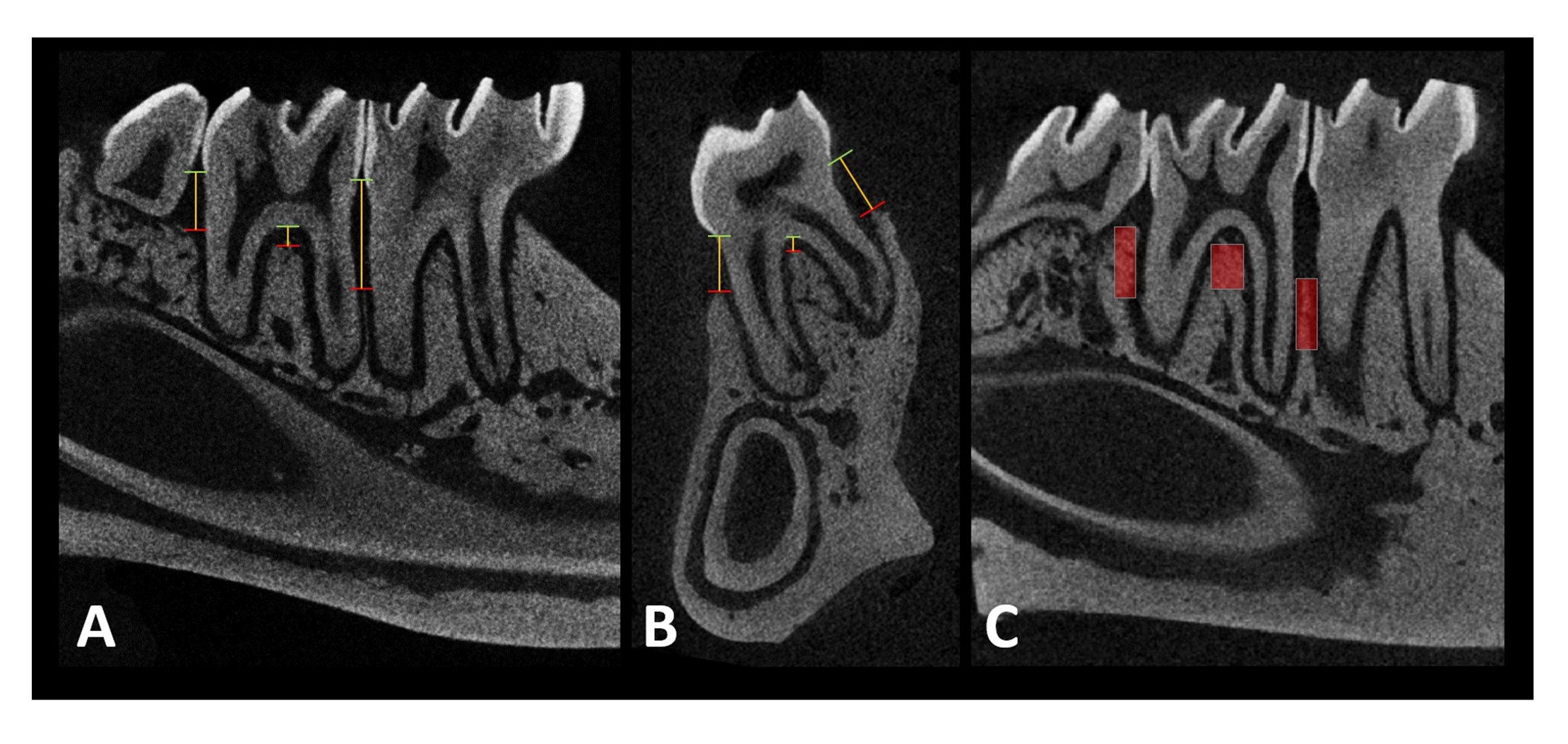
Alveolar bone loss (ABL) was evaluated by a single experienced operator, and the measurements were determined as the distance between the CEJ and the bone crest (BC)17 in the mesial and distal root of the mandibular second molar teeth, and the furcation ceiling (FC) to BC in the furcation region (Figure 1A,B) using DataViewer software. The mean values of the measurements (in millimeters [mm]) were calculated for statistical analysis. Measurements were performed after intra-examiner calibration by evaluating 10 images.
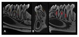
Reconstructed 3D micro-CT images. A. Linear measurements of alveolar bone loss (ABL) determined of distance between the cemento-enamel junction (CEJ) to bone crest (BC) in the mesial and distal root of the mandibular second molar teeth and the furcation ceiling (FC) to bone crest (BC) in the furcation region (green line: JCE, red line: CO, yellow line: distance (mm)); coronal plane measurements. B. Linear measurements in the sagittal plane. C. Coronal view, a rectangular region was selected as the ROI to evaluate the ratio of the bone volume fraction (BV/TV).
For volumetric analysis, a standardized region of interest (ROI) of was defined as 1400 × 500 × 540 μm (length × height × thickness), including the mesial, distal region and furcation of the second molar (Figure 1C) and a standardized gray-scale (63–255) value was used. The bone volume fraction (BV/TV [in %]), defined as the percentage of the ROI filled with bone volume, was calculated using CT Analyzer software version 1.12.4.0 (Bruker microCT, Skyscan, Belgium) by a trained examiner who was blinded to the experimental conditions of each sample.
Histomorphometric Analysis
The H&E-stained sections were analyzed using a scanner (ScanScope AT Turbo, Leica Biosystems, Nussloch, Germany) with the 20× objective and semiquantitative histopathological scoring was performed. Six sections per specimen (42 sections for each experimental condition from 7 animals) were evaluated.
Inflammatory cell infiltration (ICI), vascularization, hyperemia, Howship lacunae and fibrosis, in the infrapapillary region and periodontal ligament were analyzed in the interproximal (M-D) and furcation region of the second molar by semiquantitative histopathological scoring as according to the number of blood vessels, number of Howship lacunae (counted in the cementum region) and number of vessels with hyperemia6: Score 0 = absent; Score 1 = 1–5; Score 2 = 6–9; Score 3 = ≥10. The intensity of the inflammatory reaction was assessed as follows18: Score 0 = none; Score 1 = mild; Score 2 = moderate; Score 3 = extensive. The fibrotic component was scored as follows19: Score 0 = no fibrosis; Score 1= slightly visible fibrosis; Score 3= dense fibrosis.
The total number of empty osteocytic lacunae in the interproximal (M-D) and furcation regions of the alveolar bone was measured. Moreover, ORN was considered to be present when the bone presented with a loss of > 5 contiguous osteocytes with confluent areas of empty gaps20.
Statistical Analysis
Jamovi version 1.6 21 was used to perform statistical analysis. The effect of the independent variables in this study (induction of experimental periodontitis, irradiation, and experimental period) on the study outcomes (ABL, BV/TV%, and empty osteocytic lacuna count) was evaluated using three-way ANOVA, complemented by the Tukey test to compare the groups (p < 0.05). The results are expressed as mean ± standard deviation or percentage. The Kruskal-Wallis test was used for non-parametric data for the histological scores, followed by Dunn’s test (p < 0.05). Values are summarized as the highest values expressed.
RESULT

The means and standard deviations of linear parameters are listed in Table 1. Figure 2. illustrates the images of microCT analysis. Micro-CT analysis revealed that PD, RT, and the experimental period significantly influenced CEJ-BC values. Regarding data from linear analyses (alveolar bone loss), groups with PD exhibited higher CEJ-BC values, regardless of the evaluated region and evaluation period (p < 0.05). The animals subjected to RT also exhibited higher values of these parameters than the groups that did not undergo RT (nIr), regardless of the experimental period (p < 0.05), except for the furcation region. In the proximal and furcation regions, there was a significant difference (p < 0.05) in the experimental period for the group with irradiated PD (i.e., PDIr), with higher ABL values over 20 days.
Mean data and standard deviation of linear measurements by micro-CT performed in all groups and experimental periods (n=7 per group)
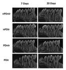
Alveolar bone loss in the mandible in the region of the mandibular second molar observed by micro-CT. The groups induced to experimental periodontitis and ionizing radiation showed higher values of CEJ-BC compared to the other experimental groups (p<0.05). Groups: nPDnIr: control group (non-ligated and nonirradiated at 7 and 20 days); nPDIr: irradiated group (non-ligated and irradiated at 7 and 20 days); PDnIr - periodontal disease group (ligated and nonirradiated at 7 and 20 days); PDIr: periodontal disease + irradiated group (ligated and irradiated at 7 and 20 days).
The means and standard deviations of BV/TV parameter are listed in Table 2. BV/TV% data were influenced by RT and PD induction. Regarding the BV/TV% data, it was verified that in the proximal regions, the PDIr group presented lower values at both evaluation periods than the control-nPDnIr group at both evaluation periods (p < 0.05) and at 20 days (p < 0.05). Additionally, the nDPIr group had lower BV/TV% values in the proximal region than the control (i.e., nDPnIr) group on day 20 (p<0.05). Regarding BV/TV% values in the furcation region, the PDIr20d group presented the lowest values (p < 0.05).
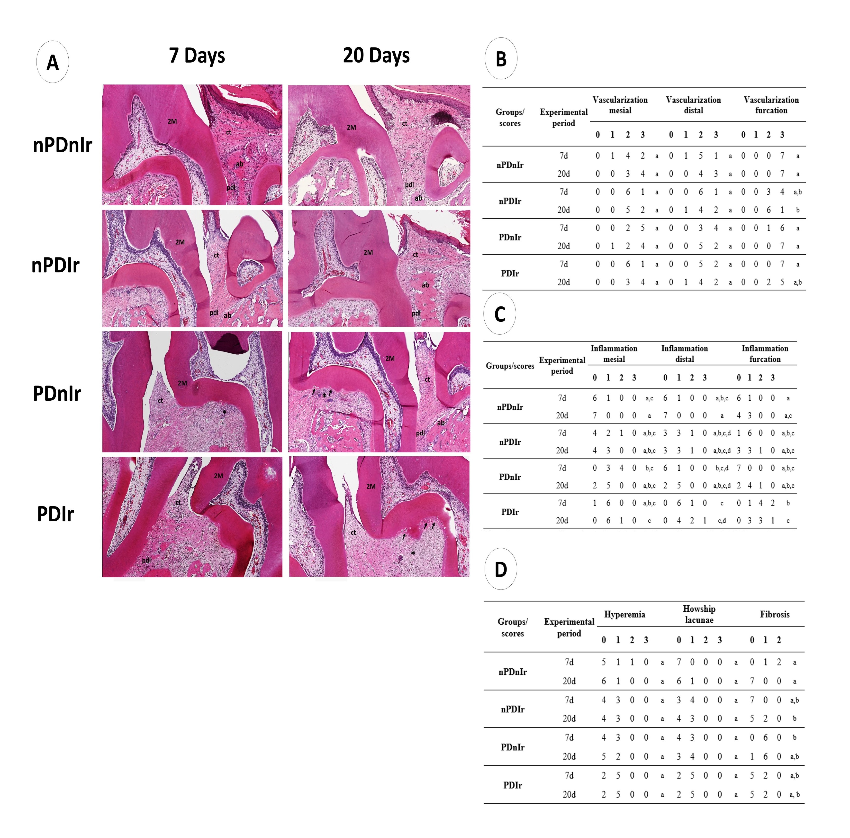
H&E staining results are shown in Figure 3A. In the nPDnIr and nPDIr groups, the subepithelial connective tissue appeared normal, regardless of the experimental period. Fibrous connective tissue of the periodontal ligament appeared normal. Inflammatory infiltrates were absent or very few inflammatory cells were observed in the completely organized connective tissue. Howship lacunae were not observed on the surface of the cementum. In the PDnIr group, regardless of the experimental period, greater degeneration of the gingival epithelium and ICI into the subepithelial connective tissue were observed. Small Howship lacunae were observed on the surface of the cementum. In the PDIr group, increased bone remodeling was observed regardless of the experimental period. Moderate ICI was observed, and small Howship lacunae were observed. No visual differences were observed between the 7- and 20-day groups in all groups.
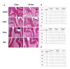
Histological analysis of the periodontal tissue of the left mandibular molar (H&E stained). A. The histological images of nPDnIr and nPDIr groups at 7 and 20 days showing an organized connective tissue, the inflammatory infiltrate was absent or very few inflammatory cells and howship lacunae were not observed. In the PDnIr group at 7 and 20 days, a greater degeneration of gingival epithelium and inflammatory cell infiltration were observed with the presence of Howship lacunae. In the PDIr group at 7 and 20 days, an increased bone remodeling, a moderate inflammatory cell infiltration with the presence of Howship lacunae. Abbreviation: ab: alveolar bone, pdl: periodontal ligament; CT: connective tissue; M2: mandibular second molar; *: infiltrate inflammatory; black arrows: howship lacunae. Magnification: 4 ×. B. Results of semi-quantitative analysis for vascularization. Abbreviations score 0 = absent, score 1 = 1–5, score 2 = 6–9, score 3 = 10≤. C. Results of semi-quantitative analysis for Hyperemia, Howship lacunae and Fibrosis. Abbreviations score: score 0 = absent, score 1 = 1–5, score 2 = 6–9, score 3 = 10≤. Fibrosis: score 0 = no fibrosis, score 1 = slightly visible fibrosis, score 2 = dense fibrosis. D. Results of semi-quantitative analysis for Inflammation. Abbreviations: score 0 = none, score 1 = mild, score 2 = moderate, score 3 = extensive. Different letters represent different levels of statistically significant differences. Kruskal-Wallis complemented by Dunn´s.
For the semi-quantitative measurements the results are show in Figure 3 (B, C and D). No statistically significant differences were observed among the groups in terms of mesial region vascularization, distal region vascularization, hyperemia, and Howship lacunae values (p > 0.05). A significant increase in inflammation was observed in the PDIr group on days 7 and 20 (p < 0.05). Regarding inflammation values in the mesial region, the nPDnIr group at 7 and 20 days was significantly different (p = 0.000092) from the PDnIr group at 7 days, with higher values for PDnIr at 7 days. In the distal region, the nPDnIr group at days 7 and 20 differed from PDIr at 20 days, with higher inflammation values for PDIr (p = 0.00015). In the furcation region, the nPDnIr group on day 7 was significantly different from the PDIr group on days 7 and 20 (p = 0.000069). Significant differences were also observed between the nPDnIr group on day 20 and the PDIr group on day 7.
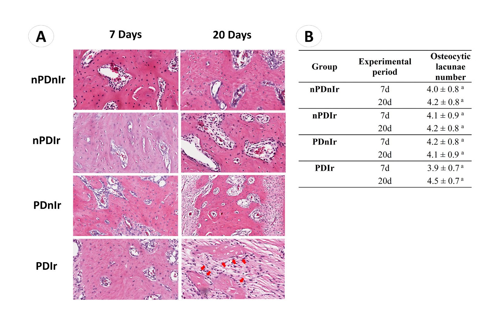
The presence of osteocytes within the osteocytic lacunae of the alveolar bone with the absence of osteonecrotic areas was observed between the groups, except for the PDIr group at 20 days, which presented a loss of >5 contiguous osteocytes with confluent areas of empty gaps in the images of 3 animals in the group (Figure 4A). There was no significant difference in the empty osteocytic lacuna counts between the groups (p > 0.05) (Figure 4B).
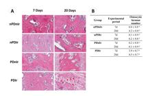
Presence of osteocytes result. A. Histological aspects of presence of osteocytes within the osteocytic lacunae of the alveolar bone in region furcation (H&E stained). Magnification: 15×. B. Mean and standard deviation data of empty osteocytic lacunae count. The red arrow indicates the presence of empty lacunae, demonstrating osteonecrotic areas. Different letters represent different levels of statistically significant differences. Three-way Anova complemented by Tukey, p < 0.05.
DISCUSSION
This study showed that a single dose of irradiation (30 Gy) impaired bone healing, with alterations in the woven bone microstructure. Micro-CT analysis revealed greater bone loss in the periodontitis groups, especially those exposed to radiation, as well as lower bone volume (BV/TV%) in the PDIr group. Histological analysis confirmed increased inflammation and signs of exacerbated bone remodeling in these animals. Thus, the null hypothesis―that RT does not affect periodontal repair after experimental periodontitis in rats―was rejected.
Experimental animal models of PD have provided important insights into the pathogenesis of this disease11. Among the various methods used to mimic PD in animals, ligature induction is the most common11. The use of a ligature throughout the cervical region of teeth leads to the accumulation of biofilms and bacterial colonization, resulting in apical epithelial migration and bone loss, and symptoms similar to those observed in the clinical setting11. Bone loss occurs over a period of 7 to 15 days in an animal model of rats11,22. In the present study, inflammatory cells infiltrating the connective tissue were observed histologically in rats with experimental periodontitis. In addition, micro-CT results revealed an increased CEJ-BC distance in the PD group compared with that in the control group (nPD). Other studies have reported gingival inflammation and bone loss 7 days after induction of experimental periodontitis11,22. Results of the present study indicate that an experimental periodontitis model was successfully established.
The periodontium is affected by the secondary effects of irradiation, and morphological and histological changes are observed after RT12. The effects of high-dose RT on the periodontium result in decreased vascularity and cellularity of the periodontal membrane and an increased risk for periodontal attachment loss10. Hypovascularity in the alveolar bone is related to endarteritis caused by RT and results in an greater risk to ORN, especially in patients with periodontitis10,12. These effects can lead to the progression of PD, causing greater ABL and an increased risk for ORN10.
IR exerts multifactorial deleterious effects on mineralized tissues, negatively impacting the bone repair process23. IR directly compromises the osteocyte lacunar network by increasing the prevalence of empty lacunae and reducing the number of viable osteocytes, which indicates cellular apoptosis and disruption of the signaling pathways involved in bone remodeling23. This cellular alteration is associated with an exacerbated inflammatory microenvironment, promoting a chronic inflammation pattern23. Additionally, IR induces narrowing of blood vessels and a reduction in local vascularization, impairing the delivery of nutrients and reparative cells and contributing to ischemia and hyperemia in the adjacent tissues24. These vascular and inflammatory alterations further contribute to the development of fibrosis, given the impairment in collagen deposition and maturation, with an increase in immature and disorganized collagen fibers in irradiated areas23. The literature also highlights that IR hampers the recruitment and function of immune cells involved in tissue homeostasis, thereby negatively affecting periodontal and peri-implant regeneration25. Thus, the effects of IR on inflammation, vascularization, and fibrosis represent a major obstacle to proper bone repair in clinical contexts involving radiotherapy.
In the present study, we evaluated the impact of irradiation in terms of ABL. Animals subjected to RT exhibited greater ABL than those that did not undergo RT, regardless of the experimental period. Moreover, the lowest mean bone mineralization (i.e., BV/TV%) value was observed in the interproximal and furcation regions in the PDIr group at day 20 compared to the other experimental groups and control. Results of this study corroborate those of other investigations finding that RT increased periodontal attachment levels and ABL in rats with experimental periodontitis12. This reflects the unwanted long-term effects of irradiation on the periodontium after treatment for PD. Moreover, it was possible to observe the effects of irradiation on bone remodeling. This may represent a potentially negative effect on turnover26 in experimentally induced PD in rats, even after ligature removal.
Moreover, no significant difference was observed in the data obtained from the BV/TV in the furcation region between the evaluated groups and their respective controls at 7 and 20 days, except for the PDIr 20-day group. These results provide a comprehensive understanding of the entire secondary molar region. The induction and treatment of PD during the experimental period may have been sufficient to improve the damage caused to the bone tissue in the furcation region27.
Regarding inflammation values, a similar pattern was observed in all groups and periods evaluated for the mesial, distal, and furcation regions, with a significant increase in inflammation for groups with experimental PD (i.e., PDIr) at 7 and 20 days and PDnIr at 7 days compared with the control group (i.e., nPDnIr) at 7 and 20 days. This is associated with the stimulation of osteoclastogenesis, increasing the resorptive capacity of mature osteoclasts in bone tissue28, which was also confirmed by the higher CEJ-BC values for these groups observed in micro-CT analysis in the present study.
The effects of irradiation on osteogenic cells has revealed a significant decrease in osteocytes and an increase in empty lacunae in murine mandibles29. In our study, there was no significant difference in the number of empty osteocytic lacunae between the groups, reflecting the absence of ORN. Furthermore, the number of osteocytes in the bone tissue appeared to be normal among all groups. High doses of RT are associated with the development of ORN in sockets after tooth extraction30. In fact, this event appears to be more related to bone repair after tooth extraction than to periodontitis treatment in an irradiated animal model.
IR leads to a decrease in cellularity and vascularity, and resorption lacunae are observed in the bone tissue16. In our study, no differences were found in the vessel counts in the interproximal region, vessels with hyperemia, or Howship lacunae. These controversial results may be related to variations in the experimental models used in different studies12.
However, animal models of human disease have limitations. Although molars in rats are similar to humans, they are are smaller, which makes periodontal treatment difficult19, such as scaling and root planning. This procedure is performed on the animal can lead to destruction of the gingival tissue and, consequently, affect histological processing31. A further limitation of the experimental model used is that in small animals, there may be mechanical trauma caused by the ligatures, which contributes to bone loss11. Regardless of the condition, biofilm accumulation and bacterial colonization are major factors in the induction of bone loss14, reflected by the increase in ICI in the PD group in this study.
It was noted that there is an apparent increased risk for ABL in patients treated for PD who subsequently undergo RT, which demonstrates the importance of oral health care in these patient. Despite these findings, this study was carried out in an animal model, and its results cannot necessarily be directly applied to humans. New studies are needed to better elucidate the effects of radiation on the periodontium. A limitation of the present study is the absence of specific staining for inflammatory and fibrotic markers, which may have limited a more in-depth analysis of the periodontal tissue response. Future investigations should include these analyses to clarify the underlying mechanisms. The results of this study can serve as a basis for new research, new therapies, as well as periodontal treatment and reduce damage to the periodontium in patients undergoing radiation to the head and neck.
CONCLUSION
The results of the present study revealed that ionizing radiation modulated the extent of alveolar bone loss in rats with induced periodontitis. In the long term (20 days), ionizing radiation increased alveolar bone loss in an experimental periodontitis rat model, even after treatment for periodontal disease. Furthermore, ionizing irradiation appears to upregulate the inflammatory response, and this effect may be associated with the impairment of periodontal repair.
ACKNOWLEDGMENTS
The authors are very grateful to Rede de Biotérios de Roedores of Federal University of Uberlandia, to Francisco Américo Silveira Marcelino and Adriano Luiz Balthazar Bianchini from the Department of Radiology of the Federal University of Triângulo Mineiro (UFTM) for the animals’s radiotherapy, to Jéssica Afonso Ferreira of Federal University of Uberlandia for his help in handling the animals, to Centro de Pesquisa Odontológico - Biomecânica, Biomateriais e Biologia celular (CPBIO) and Débora Oliveira Santos from the Pathology Laboratory of the Federal University of Uberlandia for her support in the histological processing.
-
How to cite:
Borges JS, Paula IS, Alves RO, Oliveira GJPL, Cardoso SV, Spin-Neto R, et al. Effects of ionizing radiation on healing after experimental periodontitis. Rev Odontol UNESP. 2025;54:e20250034. https://doi.org/10.1590/1807-2577.20250034
-
FUNDING
This study was supported by grants from the Fundação de Amparo à Pesquisa de Minas Gerais (FAPEMIG) (APQ-03238-24; RED-00204-23), the Coordenação de Aperfeiçoamento de Pessoal de Nível Superior- Brazil (CAPES) Finance Code 001 and Conselho Nacional de Desenvolvimento Científico e Tecnológico (CNPq); INCT Saúde Oral e Odontologia - Grants n. 406840/2022-9.
-
DATA AVAILABILITY
The contents will be made available at the time of publication of the article.
REFERENCES
-
1 Kinane DF, Stathopoulou PG, Papapanou PN. Periodontal diseases. Nat Rev Dis Primers. 2017 Jun;3(1):17038. https://doi.org/10.1038/nrdp.2017.38 PMid:28805207.
» https://doi.org/10.1038/nrdp.2017.38 -
2 Pereira SRA, de Oliveira ICV, Vieira RC, Silva MML, Branco-de-Almeida LS, Rodrigues VP. Effect of photobiomodulation therapy as an adjunct to scaling and root planing in a rat model of ligature-induced periodontitis: a histological and radiographic study. Lasers Med Sci. 2020 Jun;35(4):991-8. https://doi.org/10.1007/s10103-020-02952-0 PMid:31955304.
» https://doi.org/10.1007/s10103-020-02952-0 -
3 Sinha S, Sonoo PR, Siddhartha R, Singh SK, Singh A. Effect of conventional periodontal treatment (Scaling and Root Planing) on type-2 diabetic patient with moderate generalized chronic periodontitis: a clinical study. J Pharm Bioallied Sci. 2021 Jun;13(Suppl 1):S706-10. https://doi.org/10.4103/jpbs.JPBS_692_20 PMid:34447186.
» https://doi.org/10.4103/jpbs.JPBS_692_20 -
4 Bhuyan R, Bhuyan SK, Mohanty JN, Das S, Juliana N, Juliana IF. Periodontitis and its inflammatory changes linked to various systemic diseases: a review of its underlying mechanisms. Biomedicines. 2022 Oct;10(10):2659. https://doi.org/10.3390/biomedicines10102659 PMid:36289921.
» https://doi.org/10.3390/biomedicines10102659 -
5 Daniel M, Luby AO, Buchman L, Buchman SR. Overcoming nuclear winter: the cutting-edge science of bone healing and regeneration in irradiated fields. Plast Reconstr Surg Glob Open. 2021 Jun;9(6):e3605. https://doi.org/10.1097/GOX.0000000000003605 PMid:34235033.
» https://doi.org/10.1097/GOX.0000000000003605 -
6 Chen HHW, Kuo MT. Improving radiotherapy in cancer treatment: Promises and challenges. Oncotarget. 2017 Jun;8(37):62742-58. https://doi.org/10.18632/oncotarget.18409 PMid:28977985.
» https://doi.org/10.18632/oncotarget.18409 -
7 Sroussi HY, Epstein JB, Bensadoun RJ, Saunders DP, Lalla RV, Migliorati CA, et al. Common oral complications of head and neck cancer radiation therapy: mucositis, infections, saliva change, fibrosis, sensory dysfunctions, dental caries, periodontal disease, and osteoradionecrosis. Cancer Med. 2017 Dec;6(12):2918-31. https://doi.org/10.1002/cam4.1221 PMid:29071801.
» https://doi.org/10.1002/cam4.1221 -
8 Brandt E, Keskin M, Räisänen IT, Tervahartiala T, Mäkitie A, Harmankaya İ, et al. Induction of collagenolytic MMP-8 and -9 tissue destruction cascade in mouth by head and neck cancer radiotherapy: a cohort study. Biomedicines. 2023 Dec;12(1):27. https://doi.org/10.3390/biomedicines12010027 PMid:38275388.
» https://doi.org/10.3390/biomedicines12010027 -
9 Zambrano LMG, Brandao DA, Rocha FRG, Marsiglio RP, Longo IB, Primo FL, et al. Local administration of curcumin-loaded nanoparticles effectively inhibits inflammation and bone resorption associated with experimental periodontal disease. Sci Rep. 2018 Apr;8(1):6652. https://doi.org/10.1038/s41598-018-24866-2 PMid:29703905.
» https://doi.org/10.1038/s41598-018-24866-2 -
10 Irie MS, Mendes EM, Borges JS, Osuna LG, Rabelo GD, Soares PB. Periodontal therapy for patients before and after radiotherapy: A review of the literature and topics of interest for clinicians. Med Oral Patol Oral Cir Bucal. 2018 Sep;23(5):e524-30. https://doi.org/10.4317/medoral.22474 PMid:30148466.
» https://doi.org/10.4317/medoral.22474 -
11 de Molon RS, Park CH, Jin Q, Sugai J, Cirelli JA. Characterization of ligature-induced experimental periodontitis. Microsc Res Tech. 2018 Dec;81(12):1412-21. https://doi.org/10.1002/jemt.23101 PMid:30351474.
» https://doi.org/10.1002/jemt.23101 -
12 Köse O, Arabaci T, Kizildag A, Erdemci B, Özkal Eminoğlu D, Gedikli S, et al. Melatonin prevents radiation-induced oxidative stress and periodontal tissue breakdown in irradiated rats with experimental periodontitis. J Periodontal Res. 2017 Jun;52(3):438-46. https://doi.org/10.1111/jre.12409 PMid:27510437.
» https://doi.org/10.1111/jre.12409 -
13 Garcia VG, Longo M, Gualberto Júnior EC, Bosco AF, Nagata MJ, Ervolino E, et al. Effect of the concentration of phenothiazine photosensitizers in antimicrobial photodynamic therapy on bone loss and the immune inflammatory response of induced periodontitis in rats. J Periodontal Res. 2014 Oct;49(5):584-94. https://doi.org/10.1111/jre.12138 PMid:24206053.
» https://doi.org/10.1111/jre.12138 -
14 Abe T, Hajishengallis G. Optimization of the ligature-induced periodontitis model in mice. J Immunol Methods. 2013 Aug;394(1-2):49-54. https://doi.org/10.1016/j.jim.2013.05.002 PMid:23672778.
» https://doi.org/10.1016/j.jim.2013.05.002 -
15 Borges JS, Paula IS, de Araújo Reis NT, de Lima Oliveira AP, Soares CJ, Soares PBF. Effects of different doses of ionizing radiation on alveolar bone repair in post-extraction tooth socket: an experimental study in rats. Clin Oral Investig. 2023;27(12):7583-93. https://doi.org/10.1007/s00784-023-05348-w PMid:37906304.
» https://doi.org/10.1007/s00784-023-05348-w -
16 Sønstevold T, Johannessen AC, Stuhr L. A rat model of radiation injury in the mandibular area. Radiat Oncol. 2015;10(1):129. https://doi.org/10.1186/s13014-015-0432-6 PMid:26050968.
» https://doi.org/10.1186/s13014-015-0432-6 -
17 Cheng WC, Huang RY, Chiang CY, Chen JK, Liu CH, Chu CL, et al. Ameliorative effect of quercetin on the destruction caused by experimental periodontitis in rats. J Periodontal Res. 2010 Dec;45(6):788-95. https://doi.org/10.1111/j.1600-0765.2010.01301.x PMid:20663021.
» https://doi.org/10.1111/j.1600-0765.2010.01301.x -
18 Aydinyurt HS, Sancak T, Taskin C, Basbugan Y, Akinci L. Effects of ınjectable platelet-rich fibrin in experimental periodontitis in rats. Odontology. 2021 Apr;109(2):422-32. https://doi.org/10.1007/s10266-020-00557-1 PMid:33068206.
» https://doi.org/10.1007/s10266-020-00557-1 -
19 Toker H, Ozan F, Ozer H, Ozdemir H, Eren K, Yeler H. A morphometric and histopathologic evaluation of the effects of propolis on alveolar bone loss in experimental periodontitis in rats. J Periodontol. 2008 Jun;79(6):1089-94. https://doi.org/10.1902/jop.2008.070462 PMid:18533788.
» https://doi.org/10.1902/jop.2008.070462 -
20 Leite de Marcelos PGC, Perez DEDCP, Soares DM, de Araújo SS, Evêncio LB, Pontual MLDA, et al. The effects of zoledronic acid on the progression of experimental periodontitis in rats: histological and microtomographic analyses. J Periodontal Implant Sci. 2021 Aug;51(4):264-75. https://doi.org/10.5051/jpis.2001100055 PMid:34387046.
» https://doi.org/10.5051/jpis.2001100055 -
21 The Jamovi Project. Jamovi (version 1.6) [Internet]. Sydney: The jamovi project; 2025 [cited 2025 Oct 22]. Available from: https://www.jamovi.org
» https://www.jamovi.org -
22 Molon RS, Mascarenhas VI, de Avila ED, Finoti LS, Toffoli GB, Spolidorio DM, et al. Long-term evaluation of oral gavage with periodontopathogens or ligature induction of experimental periodontal disease in mice. Clin Oral Investig. 2016 Jul;20(6):1203-16. https://doi.org/10.1007/s00784-015-1607-0 PMid:26411857.
» https://doi.org/10.1007/s00784-015-1607-0 -
23 Mendes EM, Irie MS, Rabelo GD, Borges JS, Dechichi P, Diniz RS, et al. Effects of ionizing radiation on woven bone: influence on the osteocyte lacunar network, collagen maturation, and microarchitecture. Clin Oral Investig. 2020;24(8):2763-71. https://doi.org/10.1007/s00784-019-03138-x PMid:31732880.
» https://doi.org/10.1007/s00784-019-03138-x -
24 Alves RO, Oliveira GJPL, Oliveira RC, Limirio PHJO, Claudino M, Zanetta-Barbosa D, et al. Ionizing radiation effects on osseointegration: a pre-clinical study. Braz Oral Res. 2024 Dec;38:e112. https://doi.org/10.1590/1807-3107bor-2024.vol38.0112 PMid:39661793.
» https://doi.org/10.1590/1807-3107bor-2024.vol38.0112 -
25 Marchesan J, Girnary MS, Jing L, Miao MZ, Zhang S, Sun L, et al. An experimental murine model to study periodontitis. Nat Protoc. 2018 Oct;13(10):2247-67. https://doi.org/10.1038/s41596-018-0035-4 PMid:30218100.
» https://doi.org/10.1038/s41596-018-0035-4 -
26 Mendes EM, Irie MS, Rabelo GD, Borges JS, Dechichi P, Diniz RS, et al. Effects of ionizing radiation on woven bone: influence on the osteocyte lacunar network, collagen maturation, and microarchitecture. Clin Oral Investig. 2020 Aug;24(8):2763-71. https://doi.org/10.1007/s00784-019-03138-x PMid:31732880.
» https://doi.org/10.1007/s00784-019-03138-x -
27 Macedo PD, Corbi ST, de Oliveira GJPL, Perussi JR, Ribeiro AO, Marcantonio RAC. Hypericin-glucamine antimicrobial photodynamic therapy in the progression of experimentally induced periodontal disease in rats. Photodiagnosis Photodyn Ther. 2019 Mar;25:43-9. https://doi.org/10.1016/j.pdpdt.2018.11.003 PMid:30399457.
» https://doi.org/10.1016/j.pdpdt.2018.11.003 -
28 Miessi DMJ, Garcia VG, Ervolino E, Scalet V, Nuernberg MAA, Dos Santos Neto OM, et al. Lactobacillus reuteri associated with scaling and root planing in the treatment of periodontitis in rats submitted to chemotherapy. Arch Oral Biol. 2020 Sep;117:104825. https://doi.org/10.1016/j.archoralbio.2020.104825 PMid:32622257.
» https://doi.org/10.1016/j.archoralbio.2020.104825 -
29 Tchanque-Fossuo CN, Monson LA, Farberg AS, Donneys A, Zehtabzadeh AJ, Razdolsky ER, et al. Dose-response effect of human equivalent radiation in the murine mandible: part I. A histomorphometric assessment. Plast Reconstr Surg. 2011 Jul;128(1):114-21. https://doi.org/10.1097/PRS.0b013e31821741d4 PMid:21701328.
» https://doi.org/10.1097/PRS.0b013e31821741d4 -
30 Bléry P, Espitalier F, Hays A, Crauste E, Demarquay C, Pilet P, et al. Development of mandibular osteoradionecrosis in rats: importance of dental extraction. J Craniomaxillofac Surg. 2015 Nov;43(9):1829-36. https://doi.org/10.1016/j.jcms.2015.08.016 PMid:26433771.
» https://doi.org/10.1016/j.jcms.2015.08.016 -
31 Oliveira GJ, Paula LG, Souza JA, Spin-Neto R, Stavropoulos A, Marcantonio RA. Effects of avocado/soybean unsaponifiables (ASU) on the treatment of ligature-induced periodontitis in rats. Braz Oral Res. 2017 Apr;31:e28. https://doi.org/10.1590/1807-3107bor-2017.vol31.0028 PMid:28403331.
» https://doi.org/10.1590/1807-3107bor-2017.vol31.0028
Edited by
-
Edited by
Editor: Rosemary Adriana Chierici Marcantonio
The contents will be made available at the time of publication of the article.