Digestive system and gill morphology of the female doublespotted queenfish, Scomberoides lysan (Carangidae: Actinopterygii)
ABSTRACT
The doublespotted queenfish, Scomberoides lysan (Forsskål, 1775), is an economically important coastal teleost with aquaculture potential. Although aspects of growth and reproduction have been investigated, the digestive physiology of this species remains poorly characterized, representing a critical gap for the development of evidence-based aquaculture protocols. This study provides a morphological and histological characterization of the digestive system of female S. lysan (mean total length = 57.0 ± 0.5 cm) obtained from aquaculture ponds. We examined the entire alimentary canal, from the oral cavity to the hindgut, with special attention to gill structures relevant to feeding mechanics. Distinctive morphological features included heterogeneous gill rakers, with elongated, spine-like structures on the first gill arch contrasting with shorter, blunt rakers on subsequent arches, potentially facilitating opportunistic feeding across different prey types. The intestinal coefficient (0.20-0.36; mean = 0.27 ± 0.05) confirmed carnivorous feeding habits. Histologically, the digestive system exhibited the typical four-layer organization (mucosa, submucosa, muscularis, and serosa) throughout, with region-specific specializations. Notably, gastric glands were significantly more abundant in the cardiac region than in the pyloric stomach (p < 0.01), suggesting functional compartmentalization of digestive processes. Specialized mucus-secreting cells were distributed throughout the digestive system, with variable densities among regions. These findings provide foundational insights into the digestive biology of female S. lysan, establishing an anatomical framework for future studies on digestive morphophysiology in emerging aquaculture species.
KEYWORDS:
Digestive habit; histology; intestinal coefficient
INTRODUCTION
The digestive system of fish, while sharing fundamental similarities with that of other vertebrates, exhibits remarkable adaptations shaped by species-specific feeding ecologies and evolutionary histories (Wilson and Castro 2010, Huang et al. 2020). These adaptations are reflected in the structural and functional variation of the alimentary canal, which in teleosts typically comprises four histological layers: mucosa, submucosa, muscularis, and serosa (Jeamah et al. 2023, Sukkhee et al. 2024). Understanding such adaptations has important implications for aquaculture, as they directly inform feed formulation and strategies aimed at optimizing growth performance and production efficiency (Jiao et al. 2023).
Although primarily associated with respiration, gills also provide valuable insights into feeding ecology through the morphology of gill rakers, which vary according to diet and prey size. Planktivorous fishes generally possess elongated gill rakers adapted for filtering small particles, whereas carnivorous species tend to exhibit shorter more robust rakers suitable for capturing larger prey (Almeida et al. 2013, Alsafy et al. 2023). These morphological specializations are key to understanding trophic strategies; however, the relationship between gill structure and feeding behavior remains poorly explored for many commercially important taxa.
The doublespotted queenfish, Scomberoides lysan (Forsskål, 1775), is an economically important carangid distributed throughout the Indo-Pacific region (Sommer et al. 1996) and has attracted increasing interest for aquaculture diversification. Adults are predominantly carnivorous, whereas juveniles exhibit specialized dentition adapted for feeding on the scales and skin of other fish (Yoshida et al. 2013). Despite its economic relevance, research on S. lysan has focused mainly on growth dynamics and reproductive biology (Thulasitha and Sivashanthini 2012), while detailed information on the morphology and histology of its digestive system remains scarce. This lack of anatomical and functional knowledge constrains the development of species-specific feeding protocols.
The present study investigates the morphological and histological characteristics of the digestive system of female S. lysan, encompassing structural adaptations of the alimentary canal from the oral cavity to the intestine, with particular emphasis on gill rakers and gastric glands. Female specimens were selected due to their central role in aquaculture production systems; as broodstock, they represent the highest-value individuals in commercial operations. Their nutritional status directly influences egg quality, fecundity, and overall reproductive success, with significant consequences for farm productivity (Bromage and Roberts 1995, Izquierdo et al. 2001). By establishing detailed anatomical baselines for the digestive system of female S. lysan, this study aims to support the development of evidence-based feeding strategies and provide a comparative framework applicable to other emerging aquaculture species.
MATERIAL AND METHODS
Six healthy adult female S. lysan specimens (mean total length = 38.35 ± 2.72 cm), were randomly collected in September 2020 from a rearing pond at the Faculty of Science, Rajamangala University of Technology Srivijaya, Trang Campus, Thailand. From these, six individuals were selected for detailed histological analyses. The outdoor rearing pond measured 30 m in width, 60 m in length, and 2.5 m in depth, covering a total area of 1,800 m2.
In the aquaculture system, wild juvenile S. lysan (approximately 7.5 cm total length) were initially obtained from local fishermen along the Andaman Seacoast, Thailand. These juveniles were transferred to the pond and reared for 18 months prior to sampling. The stocking density comprised approximately 150 juveniles and 100 adults, corresponding to an average density of 4.2 m2 per fish.
The sampled fish were fed exclusively with fresh chopped yellowtail fish, Selaroides leptolepis (Cuvier, 1833), without the use of commercial pellets. Feeding was performed manually twice daily (08:00 am and 04:00 pm), at a feeding rate equivalent to 8% of body weight per day. All S. lysan were maintained under controlled conditions, with continuous monitoring of water quality parameters. Salinity ranged between 25 and 30 ppt, water temperature was maintained between 28 and 30 °C, dissolved oxygen levels remained above 3 mg/L, and ammonia concentrations did not exceed 0.02 mg/L.
Fish were euthanized by rapid cold-shock immersion in an ice-water bath (2-4 °C), following the protocol of Wilson et al. (2009). Total length was measured to the nearest 0.01 mm using digital vernier calipers. After ventral dissection, the complete digestive tract was excised, and intestinal length was measured to calculate the intestinal coefficient (IC = intestinal length/total body length). All specimens were processed within 30 minutes after euthanasia to minimize post-mortem tissue degradation.
Samples of digestive organs and gills were fixed in Davidson’s solution (Dietrich and Krieger 2009) for 48 hours at room temperature. Tissues were then processed using standard histological procedures (Presnell et al. 1997, Suvarna et al. 2019), including dehydration through a graded ethanol series (70-100%), clearing in xylene, and paraffin embedding at 58 °C. Paraffin blocks were sectioned both transversely and longitudinally at a thickness of 4 μm. Three representative sections per sample were selected and stained with Harris’s hematoxylin and eosin (H&E).
Histological slides were examined and photographed using a 3DHISTECH Panoramic Viewer (3DHISTECH, Hungary).
Gastric gland height was measured from digitized images using ImageJ software (version 1.53; U.S. National Institutes of Health). For each specimen, measurements were obtained from three different sections of both the cardiac and pyloric stomach regions, with ten measurements per section, totaling 30 measurements per region per individual.
Quantitative data are presented as mean ± standard deviation (SD). Statistical analyses were performed using GraphPad Prism version 5.0 (GraphPad Software Inc., San Diego, CA, USA). Differences in gastric gland height between cardiac and pyloric stomach regions were assessed using an unpaired Student’s t-test. When multiple comparisons were required, one-way analysis of variance (ANOVA) followed by Tukey-Kramer post hoc tests were applied. Statistical significance was set at p < 0.05.
All experimental procedures complied with institutional guidelines for animal research and were approved by the Animal Care and Use Committee of Rajamangala University of Technology Srivijaya, Thailand (approval ID: IAC 13-12-64).
RESULTS
Morphology of the digestive system
Scomberoides lysan exhibited an elongated, laterally compressed body and a large, slightly oblique terminal mouth. The digestive tract followed the typical teleost configuration, comprising the esophagus, stomach, intestine, and accessory organs (Fig. 1A-D). The esophagus was a short, thin-walled tubular structure with a longitudinally folded mucosa, connecting the pharynx to the stomach. The stomach was distinctly J-shaped, with tubular pyloric caeca located at the junction between the stomach and the intestine (Fig. 1A-D).
The intestine was the longest component of the digestive tract, measuring 7.8-10.5 cm (mean = 10.26 ± 2.05 cm). The intestinal coefficient ranged from 0.20 to 0.36 (0.27 ± 0.05). The liver was the largest accessory digestive organ, displaying a dark brown to reddish coloration (Fig. 1A-B). Cross-sectional and sagittal sections revealed a continuous organization of the digestive tract from the oral cavity to the anus (Fig. 1A-J).

The digestive system of Scomberoides lysan is shown photographed and illustrated in a longitudinal view. (A-B) Digestive tract including the esophagus (Es), stomach (St), pyloric caeca (Pc) and intestine (In) throughout the anus (An) is shown. The liver (Li) is clearly visible. (E-J) Features of the digestive system. Scale bars: 10 mm.
Gill structure and specialization
Four pairs of gills extended from the floor to the roof of the buccal cavity (Fig. 2A-B). Each gill consisted of three main components: gill arches, gill filaments, and gill rakers (Fig. 2C-H). The first gill arch exhibited markedly elongated, spine-like gill rakers (Fig. 2E), whereas the rakers on the second through fourth gill arches were significantly shorter and more robust (Fig. 2H).
Despite these morphological differences, gill rakers across all arches shared similar epithelial characteristics, consisting of stratified epithelium interrupted by tooth-like structures (Fig. 2I-K). Gill filaments were composed of primary and secondary lamellae, forming the respiratory surface (Fig. 2L).
On the fourth gill arch, paired pharyngeal tooth plates were observed, showing clear morphological differentiation between dorsal and ventral elements. The dorsal pharyngeal tooth plate displayed three distinct zones with pronounced curvature, whereas the ventral plate was triangular in shape and composed of two zones (Fig. 2M-P).

Morphology and histology of the gills and pharyngeal plates of Scomberoides lysan. (A-B) The gills in the oral cavity are composed of four paired structures (1st to 4th gills). (C-H) The three main gill parts include gill filaments (Gf), gill rakers (Gr), and gill arches (Ga). The raker of the first gill are spine-like (C-E) while the rakers of the second to fourth gill rakers (Ga) are shorter and blunt (F-H). (I-K) All gill rakers are lined with stratified epithelium (Ep) and teeth (Te). (L) The gill filament is composed of primary lamella (PL) and secondary lamella (SL). (M-N) Triangular ventral pharyngeal plate with two zones. (O-P) Large dorsal pharyngeal plate with three zones. The sectioned positions for figures 4C-D are showed. (Dt) Dentin layer, (LPT) Lower Pharyngeal teeth, (UPT) Upper Pharyngeal teeth. Scale bars: 10 mm.
Oral cavity and buccopharyngeal cavity
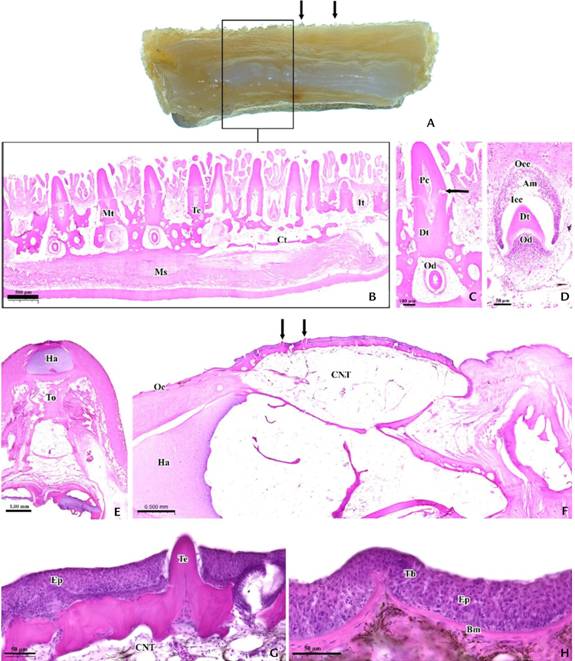
The oral cavity comprised teeth and tongue structures (Fig. 3). The teeth of S. lysan were classified into two main types: jaw teeth and tooth patches. Jaw teeth exhibited a canonical shape on both upper and lower jaws (Fig. 3A-D) and were classified into mature and immature stages based on histological characteristics. Mature teeth consisted of elongated structures protruding beyond the epithelium, with distinct crown and pedicel regions separated by an annular dividing zone (Fig. 3C). The crown projected into the oral cavity, whereas the pedicel was implanted in the dentigerous bone of the oral floor. These teeth exhibited a cellular dentin layer and a pulp cavity lined with odontoblast-like cells (Fig. 3C).
Immature teeth, embedded within the oral epithelium, consisted of an enamel organ and a dental papilla (Fig. 3D). During tooth development, the enamel organ assumed a cup-shaped structure enclosed by a single epithelial layer. The enamel epithelium was differentiated into outer and inner layers: the outer epithelium comprised cuboidal cells, whereas the inner epithelium consisted of columnar cells capable of differentiating into ameloblasts. The dental papilla was located beneath the enamel organ and consisted of a condensation of odontoblasts (Fig. 3D).

Stained slides and light microscope images show the oral cavity of Scomberoides lysan with teeth and tongue. A-B: Representative images showing mature (Mt) and immature teeth (It) teeth (Te, arrows) in the lower jaw. C: The elongated mature tooth has a crown (Cr) and pedicel (Pe), with an annular division (arrows). A cellular dentin layer (Dt), odontoblast-like cells (Od) and a pulp cavity (Pc) are also present. D: The immature tooth is an enamel organ composed of odontoblast-like cells (Od), a dentin layer (Dt) and an ameloblast (Am) having two sub-layers, an inner enamel epithelium (Iee) and an outer enamel epithelium (Oee). E-F: The tongue (To) with teeth (arrows). G-H: High magnification shows the existence of teeth (Te) and taste buds (Tb) among the epithelium of the tongue (Ep). (Bm) basement membrane, (CNT) connective tissue, (Ct) jaw bone, (Ha) hyaline cartilage, (Oe) oral epithelium, (Ms) muscularis, arrow: an annular dividing zone.
The tongue exhibited three distinct layers: mucosa, submucosa, and hyaline cartilage (Fig. 3E-F). The mucosal layer consisted of stratified squamous epithelium containing scattered taste buds and teeth, whereas the submucosa was composed of connective tissue (Fig. 3G-H).
Tooth patches, located at the posterior limit of the oral region (Fig. 4A-B), exhibited similar histological features in both upper and lower patches, including the presence of mature and immature teeth (Fig. 4C-F). These structures were lined by epithelium with several longitudinal folds and contained developing and mucus-secreting cells. At higher magnification, the epithelium appeared as a stratified layer of polygonal squamous cells interspersed with mucus-secreting cells (Fig. 4H-K), which reacted positively to PAS staining (Fig. 4F-L).
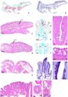
Images of stained slides show the end of oral cavity (A-B), longitudinal views of upper (C) and lower (D) pharyngeal teeth of Scomberoides lysan. E-F: Cross-sectional images of the posterior zone of lower pharyngeal teeth (Te), showing a mature tooth (Mt) among epithelium (Ep). G-J: Mature tooth. M-N: Immature tooth. G: Mucus-secreting cells (Msc) and basal cells (arrow) among epithelium (Ep). H-K: High magnification image shows an oval mucus-secreting cell (Msc) with elongated, eccentric nucleus (arrow). The apical structure in the cell is identified (triple asterisks). L: The positive areas of Msc (asterisk) are also found. (CNT) connective tissue, (Od) odontoblast-like cells, (Dt) dentin layer, the annular division (long arrows).
Esophagus
The esophagus comprised four distinct histological layers: mucosa, submucosa, muscularis, and serosa, and was continuous with the pharynx (Fig. 5A-B). The mucosa was markedly folded and lined with stratified squamous epithelium (Fig. 5C-F), containing interspersed oval mucus-secreting cells (MSCs) and a lamina propria with a greenish appearance due to the abundance of blood vessels (Fig. 5A-E). Aggregations of highly pigmented phagocytes, as well as melanomacrophage centers, were also observed (Fig. 5E).
Mucus-secreting cells were not reactive to hematoxylin and eosin staining (Fig. 5F), were weakly stained with Masson’s trichrome (Fig. 5G), but reacted intensely to PAS staining (Fig. 5H). The submucosa consisted of loose and dense connective tissue, whereas the muscularis was composed of prominent smooth muscle arranged into an inner circular layer and an outer longitudinal layer (Fig. 5A).

Light microscope images of stained slides show the buccopharyngeal region in association with the esophagus (Es) of Scomberoides lysan. A: The buccopharyngeal region (Ph) and esophagus (Es). B: A longitudinal fold (Lf) of the esophagus with epithelium (Ep) and lamina propria (Lp). C-D: The mucosal layer with prominent epithelium (Ep). E-G: A high magnification image shows simple squamous epithelium (double arrows, Ep) and mucus-secreting cells (Msc) in the esophagus. The Msc (asterisk) with its histochemical reaction are also shown. (Bc) basal cell, (Lp) lamina propria, (Mu) mucosa, (Sm) submucosa, (MMC) melanomacrophage center, (Ms) muscularis, arrows: blood vessel, N = Nucleus.
Stomach
The esophagogastric junction was characterized by an abrupt transition from stratified squamous epithelium with mucous cells to simple columnar epithelium containing gastric glands (Fig. 6A-E). The stomach wall exhibited numerous longitudinal folds (gastric rugae), which were absent in the esophagus (Fig. 6B-C). Proximal regions of the stomach were lined by tall columnar epithelial cells, whereas distal regions exhibited shorter columnar cells (Fig. 6F-G).
The stomach was divided into cardiac (anterior) and pyloric (posterior) regions. Both regions consisted of four layers: mucosa, submucosa, muscularis, and serosa (Fig. 6H-L). The mucosa was lined with simple columnar epithelium that reacted positively to PAS staining (Fig. 6L), and together with the submucosa supported the formation of gastric rugae (Fig. 6K). The muscularis comprised two smooth muscle sublayers: an inner circular layer and an outer longitudinal layer (Fig. 6H).
Gastric glands were tubular and present within the lamina propria of both stomach regions (Fig. 6M). Quantitative analysis revealed that gastric glands were significantly longer in the cardiac stomach compared to the pyloric region (p < 0.01) (Fig. 6N).

Light microscope images of stained slides and a schematic diagram show the transition between the esophagus (Es) and stomach (St) of Scomberoides lysan. (A-B) The transition between the esophagus (Es) and stomach (St) features a change in the type of cells present. (C) The esophageal stomach region presents mucus-secreting cells (arrows) in the epithelium (Ep). (D-F) The stratified squamous epithelium with mucous cells in the esophagus gives way to a simple columnar epithelium with mucus-secreting cells in the stomach. (G) A schematic diagram shows the alteration in cell type between the esophagus (Es) and stomach (St). (H) The stomach wall is composed of four layers: mucosa (Mu), submucosa (Sm), muscularis (Ms), and serosa (Se). (I) Prominent ruga (Ru) juxtaposed to the mucosa (Mu) and submucosa (Sm). (K) The abundance of gastric glands (Gg) in the lamina propria (Lp) in the cardiac stomach. (M) A longitudinal view shows a gastric gland (Gg) in cross-section, and a gastric cell (Gc) containing a vacuole (asterisk). (N) The bar chart shows the average height of gastric glands in the cardiac (anterior) and pyloric stomachs. Values represent means ± SE. **Significant difference (p < 0.01). (ESJ) esophagus-stomach junction, (Lu) lumen, (Msc) mucus-secreting cells.
Intestine and pyloric caeca
The intestine and pyloric caeca shared a similar histological organization, with all regions comprising mucosa, submucosa, muscularis, and serosa (Fig. 7A-G). The mucosa exhibited narrow longitudinal folds lined with simple columnar epithelium bearing microvilli (Fig. 7B). These folds were extensive (Fig. 7B-C), and numerous PAS-positive goblet cells were distributed throughout the epithelium (Fig. 7E).
The submucosa consisted of loose connective tissue (Fig. 7C-D, G). The muscularis was composed of two thin smooth muscle layers arranged circumferentially as an inner circular layer and an outer longitudinal layer (Fig. 7A).
An incidental finding of tumor-like cellular proliferation in the intestine was documented in a single specimen (Fig. 7I-J).
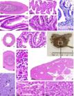
Light microscope images of stained slides show the intestine and morpho-histology of accessory organs of Scomberoides lysan. Cross-sectional views A-G: The intestinal structure throughout the region. H-I: An intestinal tumor-like (asterisks) cellular proliferation. J: Liver morphology (Li.). KI: The liver lobule (Li) contains the large hepatic vein (Hv), closely located to the pancreas showing an exocrine gland of the exocrine pancreas (Eg) and islets of Langerhans (IL). L: The large hepatic vein enclosed by the hepatic stroma (arrows). M: The hepatic parenchyma showing hepatocytes (Hp) and hepatic sinusoids (Hs). N: The pancreatic islets of Langerhans (Pi) and capillaries (arrows) are identified. (Cv) central vein, arrowheads = glycogen, arrows = cell proliferation, (Ep) epithelium, (Lf) longitudinal fold, (Msc) mucus-secreting cell, (Mu) mucosa, (Sm) submucosa, (Ms) muscularis, (Se) serosa.
Liver and pancreas
The liver exhibited a lobular organization surrounded by a capsule of loose connective tissue (Fig. 7L). The hepatic parenchyma consisted of hepatocytes arranged in hepatic cords separated by sinusoids (Fig. 7M). The central vein and components of the portal system were embedded within the hepatic stroma (Fig. 7M). Hepatocytes were irregular in shape and displayed centrally located nuclei (Fig. 7N).
The pancreas presented both exocrine and endocrine components (Fig. 7L). The exocrine pancreas consisted of acini composed of pyramidal acinar cells with basally positioned nuclei, closely associated with blood vessels. The endocrine pancreas was represented by pancreatic islets of Langerhans, composed of endocrine cells interspersed with blood vessels (Fig. 7O).
DISCUSSION
The architecture of the fish digestive system exhibits remarkable plasticity, shaped by phylogeny, feeding ecology, and environmental adaptations (Senarat et al. 2013, Purushothaman et al. 2016). Consequently, histological investigations of the digestive tract provide valuable biological insights into the physiological and pathological conditions of a species (Okuthe and Bhomela 2020). Such knowledge contributes to the development of species-specific feeding protocols and management strategies, with direct implications for conservation and aquaculture practices (Xiong et al. 2011, Germano et al. 2014, Purushothaman et al. 2016).
Oral cavity and gill specializations
This study demonstrated that S. lysan, a carnivorous carangid with aquaculture potential, exhibits several notable morphological adaptations. The large, oblique mouth and expansive upper jaw facilitate whole-prey ingestion, consistent with predatory feeding strategies described for other carnivorous teleosts (Pusey et al. 2004, Yoshida et al. 2013). The presence of multiple patches of villiform teeth throughout the oropharyngeal cavity likely serves a dual role in prey capture and in guiding food toward the esophagus (Santos et al. 2015). This dental configuration is consistent with patterns reported for other carnivorous fishes, although confirmation of its functional significance would benefit from complementary stomach content analyses.
A particularly noteworthy finding was the heterogeneity in gill raker morphology, with elongated, spine-like rakers on the first gill arch contrasting with shorter rakers on the subsequent arches. This pattern differs from the more uniform gill raker morphology typically observed in strictly carnivorous species (Almeida et al. 2013, Chen et al. 2023), but resembles the configuration described for Boops boops (Alsafy et al. 2023). The observed dimorphism in gill raker structure may represent an evolutionary adaptation that enables S. lysan to handle prey of varying sizes, with longer rakers facilitating the retention of smaller food items and shorter rakers accommodating larger prey (Magnuson and Heitz 1971, O’Brien 1987, Salman et al. 2005). Unlike species with highly specialized feeding mechanisms, this distinctive gill architecture may support opportunistic feeding behavior across multiple trophic levels (Amundsen et al. 2004, Almeida et al. 2013, Alsafy et al. 2023). This interpretation represents a testable hypothesis that could be further evaluated through integrated analyses of diet composition and functional morphology.
Digestive tract specializations
The esophagus of S. lysan exhibited a robust anatomical organization characterized by prominent longitudinal folds, stratified squamous epithelium rich in mucus-secreting cells, and well-developed musculature-features commonly observed in predatory teleosts that must accommodate large prey items (Domeneghini et al. 1999, Purushothaman et al. 2016). This pattern is consistent with previous observations in several teleost species, including the white sturgeon (Domeneghini et al. 1999), black scorpionfish (Nazlić et al. 2014), and Japanese flathead (Jeamah et al. 2023).
The densely packed epithelial layer likely provides protection against both physical and chemical damage during food intake (Machado et al. 2013, Santos et al. 2015). The abundance of mucus-secreting cells suggests multiple physiological roles beyond lubrication, including defense against pathogen invasion (Diaz et al. 2003) as well as protection from chemical and mechanical abrasion during food passage (Machado et al. 2013, Okuthe and Bhomela 2020). In addition, the presence of mucosal folds combined with well-developed musculature allows for substantial esophageal distension during the ingestion of large prey (Okuthe and Bhomela 2020).
Although the stomach of S. lysan did not exhibit marked external compartmentalization, histological analyses revealed distinct cardiac and pyloric regions. This organization is commonly observed in carnivorous teleosts (Westneat 2001), although it is less pronounced than the clear regional differentiation reported in some centropomid fishes, such as species of Centropomus and Lates. (Machado et al. 2013, Jeamah et al. 2023). Histochemical analyses revealed PAS-positive epithelial surfaces, indicating glycoprotein production essential for mucosal protection (Cao and Wang 2009). Gastric glands and surface mucus-secreting cells were more abundant in the cardiac region than in the pyloric stomach, suggesting functional specialization, with the cardiac stomach optimized for chemical digestion and enzyme secretion, whereas the pyloric region likely functions primarily as a temporary storage chamber (Purushothaman et al. 2016). This pattern of functional compartmentalization has been reported in several predatory teleosts, including walking catfish, red-bellied piranha (Raji and Norouzi 2010), black scorpionfish (Nazlić et al. 2014), Asian seabass (Purushothaman et al. 2016), and Japanese flathead (Jeamah et al. 2023).
Intestinal specialization and trophic ecology
Intestinal length relative to body size is a phenotypically plastic trait strongly correlated with feeding strategy. The intestinal coefficient calculated for S. lysan (0.20-0.36) falls well within the range reported for carnivorous species (0.2-2.5; Ward-Campbell et al. 2005) and is considerably lower than values typically observed in herbivorous fishes (0.8-15.0) (Xiong et al. 2011, Santos et al. 2015). This morphometric evidence strongly supports a carnivorous feeding habit in S. lysan, although trophic classification should ideally be based on multiple lines of evidence rather than morphology alone.
The histological organization of the intestine of S. lysan, characterized by the typical layered structure from mucosa to serosa, is consistent with patterns described for other carnivorous teleosts, such as Larimichthys crocea (Kalhoro et al. 2018) and Tilapia sparrmanii (Okuthe and Bhomela 2020). The intestine plays a central role in nutrient digestion, absorption, and transport (Oliveira Ribeiro and Fanta 2000). In the present study, mucus-secreting goblet cells were distributed along the entire intestinal length, similar to observations in black scorpionfish (Nazlić et al. 2014) and centropomid species (Machado et al. 2013). These cells likely contribute to both nutrient absorption and protection of the intestinal epithelium from mechanical and chemical stress (Machado et al. 2013).
Implications for aquaculture
The present findings on the digestive histology of female S. lysan have direct relevance for the development of commercial aquaculture protocols. Female broodstock represent the most valuable individuals in production systems, and their nutritional management directly influences reproductive performance, egg quality, and overall farm profitability (Bromage and Roberts 1995, Izquierdo et al. 2001). The carnivorous adaptations observed throughout the digestive tract-from heterogeneous gill rakers to a relatively short intestine-indicate a requirement for high-protein diets (40-45% crude protein) with particle sizes compatible with oral and pharyngeal morphology (Xiong et al. 2011, Purushothaman et al. 2016).
The region-specific distribution of mucus-secreting cells and gastric glands suggests heightened sensitivity to feed formulation, particularly during vitellogenesis, when nutritional demands increase substantially (Rønnestad et al. 2013). Current aquaculture practices often rely on empirically derived feeding strategies, including the use of trash fish or generic commercial pellets, which may not fully meet the physiological requirements of female broodstock. The histological evidence presented here supports the development of specialized diets with appropriate acidity levels and fiber content tailored to female digestive physiology (Cao and Wang 2009, Diaz et al. 2003).
The functional differentiation of gastric regions may also influence optimal feeding frequency and meal size. The apparent storage capacity of the pyloric stomach suggests that female S. lysan may benefit from less frequent but larger meals during reproductive periods, potentially reducing labor costs while improving nutritional efficiency (Machado et al. 2013, Nazlić et al. 2014). Understanding these female-specific digestive traits provides a scientific foundation for refining broodstock feeding protocols aimed at maximizing egg production and quality while minimizing feed costs (Izquierdo et al. 2001).
Furthermore, the widespread occurrence of specialized mucosal cells throughout the digestive tract indicates potential sensitivity to immunological challenges under intensive culture conditions. This insight may guide the formulation of functional feeds containing immunostimulants targeted to these regions, thereby enhancing disease resistance in high-value female broodstock (Rønnestad et al. 2013).
Methodological contributions and future applications
This study establishes a comprehensive methodological framework for investigating digestive tract morphohistology in marine fishes with emerging aquaculture potential (Wilson and Castro 2010). The integration of gross morphological observations with detailed histological and histochemical analyses provides a robust and replicable model applicable across diverse teleost taxa (Adamek-Urbańska et al. 2023).
Future applications of this framework may extend beyond descriptive morphology to include functional analyses of enzyme activity, nutrient transport mechanisms, and host-microbiome interactions. Integrating these approaches with the morphological baseline established here would enable a more comprehensive understanding of digestive physiology in cultured teleosts and support the development of species-specific nutrition protocols essential for sustainable aquaculture expansion (Wilson and Castro 2010).
ACKNOWLEDGEMENTS
We would like to thank the Department of Marine Science and Environment, Faculty of Science and Fisheries Technology, Rajamangala University of Technology Srivijaya, Trang Campus, Thailand for their technical support in the laboratory. Histochemical procedures and staining were supported by the Microtechnique Laboratory (MIC-LAB), Division of Biological Science, Faculty of Science, Prince of Songkla University. This research was also partially supported by Chiang Mai University.
LITERATURE CITED
-
Adamek-Urbańska D, Kamaszewski M, Wiechetek W, Wild R, Boczek J, et al. (2023) Comparative Morphology of the Digestive Tract of African Bush Fish (Ctenopoma acutirostre) and Paradise Fish (Macropodus opercularis) Inhabiting Asian and African Freshwaters. Animals 13(16): 2613. https://doi:org/10.3390/ani13162613
» https://doi:org/10.3390/ani13162613 -
Alsafy MA, El-Gendy SA, El-Bakary NE, Kamal BM, Derbalah A, Roshdy K (2023) Morphological comparison of the detailed structure of gill rakers from three different feeding habits of marine fish species. Zoomorphology 142(1): 87-97. https://doi.org/10.1007/s00435-022-00574-4
» https://doi.org/10.1007/s00435-022-00574-4 -
Almeida APG, Behr ER, Baldisserotto B (2013) Gill rakers in six teleost species: influence of feeding habit and body size. Ciência Rural 43(12): 2208-2214. https://doi.org/10.1590/S0103-84782013005000142
» https://doi.org/10.1590/S0103-84782013005000142 -
Amundsen PA, Bøhn T, Våga GH (2004) Gill raker morphology and feeding ecology of two sympatric morphs of European whitefish (Coregonus lavaretus). Annales Zoologici Fennici 41: 291-300. https://hdl.handle.net/10037/19535
» https://hdl.handle.net/10037/19535 - Bromage NR, Roberts RJ (1995) Broodstock Management and Egg and Larval Quality. Blackwell Science, Oxford.
-
Cao XJ, Wang WM (2009) Histology and mucin histochemistry of the digestive tract of Yellow catfish, Pelteobagrus fulvidraco Anatomia, Histologia, Embryologia 38(4): 254-261. https://doi:org/10.1111/j.1439-0264.2009.00932.x
» https://doi:org/10.1111/j.1439-0264.2009.00932.x -
Chen X, Liu S, Ding Q, Teame T, Yang Y, et al. (2023) Research advances in the structure, function, and regulation of the gill barrier in teleost fish. Water Biology and Security 2(2): 100139. https://doi.org/10.1016/j.watbs.2023.100139
» https://doi.org/10.1016/j.watbs.2023.100139 -
Diaz AO, Garcia AM, Devincenti CV, Goldemberg AL (2003) Morphological and histochemical characterization of the mucosa of the digestive tract in Engraulis anchoita (Hubbs and Marini, 1935). Anatomia, Histologia, Embryologia 32(6): 341-346. https://doi:org/10.1111/j.1439-0264.2003.00490.x
» https://doi:org/10.1111/j.1439-0264.2003.00490.x - Dietrich D, Krieger HO (2009) Histological analysis of endocrine disruptive effects in small laboratory fish. John Wiley & Sons, New Jersey, 396 pp.
- Domeneghini C, Arrighi S, Radaelli G, Bosi G, Mascarello F (1999) Morphological and histochemical peculiarities of the gut in the white sturgeon, Acipenser transmontanus European Journal of Histochemistry 43(2): 135-145.
-
Germano RM, Stabille SR, De Britto Mari R, Pereira JNB, et al. (2014) Morphological characteristics of the Pterodoras granulosus digestive tube (Valenciennes, 1821) (Osteichthyes, Doradidae). Acta Zoologica 95(2): 166-175. https://doi:org/10.1111/azo.12016
» https://doi:org/10.1111/azo.12016 -
Huang L, Yang L, Liu J, Cao X (2020) Comparative histological analysis of intestines of loach, grass carp and catfish provide insights into adaptive characteristics in air-breathing fish. Croatian Journal of Fisheries 78(2): 91-98. https://doi:org/10.2478/cjf-2020-0009
» https://doi:org/10.2478/cjf-2020-0009 -
Izquierdo MS, Fernandez-Palacios H, Tacon AGJ (2001) Effect of broodstock nutrition on reproductive performance of fish. Aquaculture 197(1-4): 25-42. https://doi.org/10.1016/S0044-8486(01)00581-6
» https://doi.org/10.1016/S0044-8486(01)00581-6 -
Jeamah A, Sudtongkong C, Kenthao A, Imsonpang S, Angsujinda K, et al. (2023) Microanatomy of the digestive tract and accessory organs of the Japanese flathead (Inegocia japonica Cuvier, 1829) (Scorpaeniformes, Platycephalidae). Acta Zoologica 104(4): 575-585. https://doi:org/10.1111/azo.12441
» https://doi:org/10.1111/azo.12441 -
Jiao F, Zhang L, Limbu SM, Yin H, Xie Y, et al. (2023) A comparison of digestive strategies for fishes with different feeding habits: Digestive enzyme activities, intestinal morphology, and gut microbiota. Nature Ecology & Evolution 13(9): e10499. https://doi:org/10.1002/ece3.10499
» https://doi:org/10.1002/ece3.10499 -
Kalhoro H, Tong S, Wang L, Hua Y, Volatiana JA, Shao Q (2018) Morphological study of the gastrointestinal tract of Larimichthys crocea (Acanthopterygii: Perciformes). Zoologia 35: e25171. https://doi.org/10.3897/zoologia.35.e25171
» https://doi.org/10.3897/zoologia.35.e25171 -
Machado MRF, Souza HDO, Souza VLD, Azevedo AD, Goitein R, Nobre AD (2013) Morphological and anatomical characterization of the digestive tract of Centropomus parallelus and Centropomus undecimalis Acta Scientiarum. Biological Sciences 35(4): 467-474. https://doi:org/10.4025/actascibiolsci.v35i4.14352
» https://doi:org/10.4025/actascibiolsci.v35i4.14352 - Magnuson JJ, Heitz JG (1971) Gill raker apparatus and food selectivity among mackerels, tunas, and dolphins. Fishery Bulletin 69(2): 361-370.
- Nazlić M, Paladin A, Bočina I (2014) Histology of the digestive system of the black scorpionfish Scorpaena porcus L. Acta Adriatica 55: 65-74.
- O’Brien WJ (1987) Planktivory by freshwater fish: Thrust and parry in the pelagia. In: Kerfoot WC, Sih A (Eds) Predation: direct and indirect effects on aquatic communities. University Press of New England, New Hampshire, 3-16.
-
Okuthe GE, Bhomela B (2020) Morphology, histology and histochemistry of the digestive tract of the Banded tilapia, Tilapia sparrmanii (Perciformes: Cichlidae). Zoologia 37: 1-14. https://doi.org/10.3897/zoologia.37.e51043
» https://doi.org/10.3897/zoologia.37.e51043 -
Oliveira Ribeiro CA, Fanta E (2000) Microscopic morphology and histochemistry of the digestive system of a tropical freshwater fish Trichomycterus brasiliensis (Lütken) (Siluroidei, Trichomycteridae). Revista Brasileira de Zoologia 17(4): 953-971. https://doi.org/10.1590/S0101-81752000000400007
» https://doi.org/10.1590/S0101-81752000000400007 - Presnell JK, Schreibman MP (1997) Humason’s animal tissue techniques. Johns Hopkins University Press, Baltimore, 5th ed., 600 pp.
-
Purushothaman K, Lau D, Saju JM, Musthaq SkS, Lunny DP, et al. (2016) Morpho-histological characterisation of the alimentary canal of an important food fish, Asian seabass (Lates calcarifer). PeerJ 4: e2377. https://doi:org/10.7717/peerj.2377
» https://doi:org/10.7717/peerj.2377 - Pusey B, Kennard M, Arthington AH (2004) Freshwater fishes of north-eastern Australia. CSIRO, Melbourne, 400 pp.
- Raji A, Norouzi E (2010) Histological and histochemical study on the alimentary canal in Walking catfish (Claris batrachus) and piranha (Serrasalmus nattereri). Iranian Journal of Veterinary Research 11: 255-261.
- Salman NA, AL-Mahdawi GJ, Heba HM (2005) Gill rakers morphology and filtering mechanism in some marine teleosts from Red Sea coasts of Yamen. Egyptian Journal of Aquatic Research 31: 286-296
-
Santos ML, Arantes FP, Pessali TC, Santos JE (2015) Morphological, histological and histochemical analysis of the digestive tract of Trachelyopterus striatulus (Siluriformes: Auchenipteridae). Zoologia 32(4): 296-305. https://doi.org/10.1590/S1984-46702015000400005
» https://doi.org/10.1590/S1984-46702015000400005 - Senarat S, Yenchum W, Poolprasert P (2013) Histological study of the intestine of Stoliczkae’s barb Puntius stoliczkanus (Day, 1871) (Cypriniformes: Cyprinidae). Agriculture and Natural Resources 47(2): 247-251.
- Sommer C, Schneider W, Poutiers JM (1996) The living marine resources of Somalia. Food and Agriculture Organization of the United Nations, Rome, 378 pp.
-
Sukkhee N, Senarat S, Charoenphon N, Kaneko G, Kettratad J, et al. (2024) Histological structure of the digestive tract, liver, and pancreas of Ambassis nulua (Hamilton, 1822) with ultrastructural details of the gastric gland. Journal of Fish Biology 104(1): 113-124. https://doi:org/10.1111/jfb.15566
» https://doi:org/10.1111/jfb.15566 -
Suvarna KS, Layton C, Bancroft JD (2019) Bancroft’s theory and practice of histological techniques. Elsevier Health Sciences, Amsterdam, 8th ed. https://doi.org/10.1016/C2015-0-00143-5
» https://doi.org/10.1016/C2015-0-00143-5 -
Thulasitha WS, Sivashanthini K (2012) Growth pattern and length-weight relationship of Scomberoides lysan (Pisces: Carangidae) from the northern waters of Sri Lanka. Journal of Fisheries and Aquatic Science 7(1): 57-64. https://doi:org/10.3923/jfas.2012.57.64
» https://doi:org/10.3923/jfas.2012.57.64 -
Westneat MW (2001) Ingestion in Fish. Encyclopedia of Life Sciences, John Wiley & Sons. https://doi.org/10.1038/npg.els.0001834
» https://doi.org/10.1038/npg.els.0001834 -
Wilson JM, Castro LFC (2010) Morphological diversity of the gastrointestinal tract in fishes. Fish Physiology and Biochemistry 30: 1-55. https://doi:org/10.1016/S1546-5098(10)03001-3
» https://doi:org/10.1016/S1546-5098(10)03001-3 - Wilson JM, Bunte RM, Carty AJ (2009) Evaluation of rapid cooling and tricaine methanesulfonate (MS222) as methods of euthanasia in zebrafish (Danio rerio). Journal of the American Association for Laboratory Animal Science 48: 785-789.
-
Xiong D, Zhang L, Yu H, Xie C, Kong Y, et al. (2011) A study of morphology and histology of the alimentary tract of Glyptosternum maculatum (Sisoridae, Siluriformes): The alimentary tract of Glyptosternum maculatum Acta Zoologica 92(2): 161-169. https://doi:org/10.1111/j.1463-6395.2010.00458.x
» https://doi:org/10.1111/j.1463-6395.2010.00458.x - Yoshida T, Motomura H, Musikasinthorn P, Matsuura K (2013) Fishes of northern gulf of Thailand. National Museum of Science and Nature, Tsukuba.
-
Rønnestad I, Yúfera M, Ueberschär B, Ribeiro L, Sæle Ø, Boglione C (2013) Feeding behaviour and digestive physiology in larval fish: current knowledge, and gaps and bottlenecks in research. Reviews in Aquaculture 5(Suppl. s1): S59-S98. https://doi.org/10.1111/raq.12010
» https://doi.org/10.1111/raq.12010
ADDITIONAL NOTES
- ZooBank register
-
Data Availability
Datasets generated or analyzed in this study are available from the corresponding author on reasonable request.
-
Funding
This research received no external funding.
-
How to cite this article
Tongtako W, Phinrub W, Kenthao A, Thaochan N, Iida A, Nganvongpanit K, Sornying P, Imsonpang S, Prakobkarn A, Senarat S (2026) Digestive system and gill morphology of the female doublespotted queenfish, Scomberoides lysan (Carangidae: Actinopterygii). Zoologia 43: e25035. https://doi.org/10.1590/S1984-4689.v43.e25035
-
Published by
Sociedade Brasileira de Zoologia at Scientific Electronic Library Online - https://www.scielo.br/zool
Datasets generated or analyzed in this study are available from the corresponding author on reasonable request.