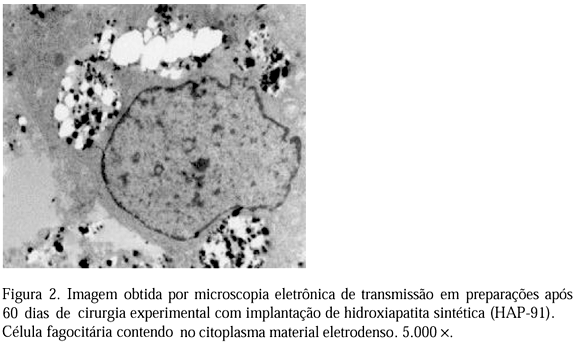
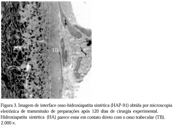
With the objective of studying the synthetic hydroxyapatite (HAP-91) as a bone substitute, eight healthy mongrel adult dogs were used. Following the habitual anesthetic and surgical protocol, a bone defect was provoked in the proximal diafisis of the left and right tibias, being implanted the graft of HAP-91 just in the right tibia. The animals, two at each time, were sacrificed at the 8th, 30th, 60th and 120th days after the surgery, when lesion samples were obtained for histopathology, submitted to the double coloration in 1% uranil acetate solution and in lead citrate solution. These sections were examined and photographed in an electronic transmission microscope. The bone tissue components were identified both in the control and treated tibia. The absorption of HAP-91 was characterized by the presence of multinuclear cells in the interface between the hydroxyapatite and the bone, morphologically considered as osteoclasts. In addition, the concomitant presence of HAP-91, with the adjacent formation of new bone was found, which suggests that the osteointegration of HAP-91 is similar to the bone reabsorption apposition normal process.
Dog; synthetic hydroxyapatite; bone substitute


