ABSTRACT
Purpose: to verify the occurrence of posterior or submucosal lingual frenulum and evaluate the efficiency of a special maneuver for its visual inspection.
Methods: an experimental study including 1,715 healthy infants, in which prematurity, perinatal complications, craniofacial anomalies neurological disorders, and visible genetic syndromes were the exclusion criteria. A clinical examination was performed by means of a maneuver that consisted in rising the lateral margins of the tongue to visualize the anatomical characteristics of the lingual frenulum. In some of the infants, a special maneuver was performed to assist visualization of posterior lingual frenulum, since its visualization was not possible. The maneuver consisted in two simultaneous actions: elevating and pushing the tongue back.
Results: 558 infants (32.54%), out of the 1,715 had posterior frenulum, which required the special maneuver that consisted in both elevating and pushing the tongue back, simultaneously.
Conclusion: the occurrence of posterior lingual frenulum was high and the special maneuver consisted in elevating and pushing the tongue back proved to be efficient to visualize the posterior lingual frenulum.
Keywords: Lingual Frenulum; Speech, Language and Hearing Sciences; Anatomy, Ankyloglossia; Tongue
RESUMO
Objetivos: verificar a ocorrência do frênulo lingual posterior ou submucoso, bem como, avaliar a eficiência de uma manobra que possibilita sua visualização.
Métodos: estudo experimental realizado com 1715 lactentes saudáveis, sendo considerados como critérios de exclusão a prema turidade, as complicações perinatais, a presença de anomalias craniofaciais, as doenças neurológicas e as síndromes genéticas. Exame clínico realizado por meio da manobra de elevação da língua para observar as características anatômicas do frênulo lingual. Nos lactentes cuja visualização do frênulo não foi possível apenas com a referida manobra, pois o mesmo se encontrava recoberto por uma cortina de mucosa, foi utilizada uma manobra simultânea de elevação e posteriorização da língua.
Resultados: dos 1715 lactentes avaliados, em 558 não foi possível visualizar o frênulo lingual somente por meio da manobra de elevação, pois os lactentes apresentaram frênulo posterior ou submucoso, havendo necessidade de realizar uma manobra simultânea de posteriorização da língua para visualização das características anatômicas.
Conclusão: a ocorrência do frênulo posterior ou submucoso foi alta, sendo que a manobra de elevação e posteriorização da língua se mostrou eficiente para evidenciar o frênulo recoberto por cortina de mucosa.
Descritores: Freio Lingual; Fonoaudiologia; Anatomia; Anquiloglossia; Língua
Introduction
Although lingual frenulum is a widely discussed subject1-7, its anatomical characteristics have not been extensively studied. Differentiating anatomical variations of lingual frenulum requires deep knowledge of the anatomy of both the tongue and the floor of the mouth.
The posterior ankyloglossia or submucosal tongue-tie, which consists of the presence of abnormal collagen fibers in a sub mucosal location surrounded by tight mucous membrane under the front of the tongue is a variation poorly described in the literature8.
There are a few studies in the literature on posterior lingual frenulum and all of them classify it as posterior ankyloglossia9-13. Nevertheless, Douglas14 states that the published studies regarding posterior tongue-tie do not provide clear evidence that the diagnosis of posterior tongue-tie has validity, or that frenotomy is an effective treatment. The author also reports that often, photographs of the frenula purported to show posterior tongue-tie are indistinguishable from normal frenulum variants. According to the author, data are either unreliable or interpreted through the lens of posterior tongue-tie when multiple other potential factors could explain the results. Douglas claims that health professionals should be extremely cautious given the absence of reliable evidence or historical precedence to support the efficacy of frenotomy other than for anterior tongue-tie.
In a study including 1,084 healthy newborns, Martinelli et al.8 concluded that 35% of newborns had posterior lingual frenulum. However, this type of lingual frenulum did not interfere with breastfeeding; therefore, surgery was not recommended. There are few studies concerning the absence of lingual frenulum15-19.
A study published recently described the phenotypic spectrum of congenital Zika syndrome, and the absence of lingual frenulum was one of the characteristics observed in this syndrome19. However, Fonteles et al. reported that the absence of lingual frenulum was not observed in Brazilian infants with congenital Zika syndrome. Many of those newborns had submucosal frenulum, what could be misinterpreted as absent frenulum20.
This study aimed to verify the occurrence of posterior or submucosal lingual frenulum and evaluate the efficiency of a maneuver for its visual inspection.
Methods
This experimental study, which included 1,715 healthy infants, was approved by the Ethic Committee of CEFAC under the number CAAE 47613115.9.0000.5538. All the participants were informed about the procedures and signed the Free Informed Term of Consent (FITC).
Prematurity, perinatal complications, craniofacial anomalies neurological disorders, and visible genetic syndromes were the exclusion criteria. The clinical examination was performed by a Speech Language Pathologist (SLP) specialist in Orofacial Myofunctional Disorders, who was trained and calibrated to administer the validated Neonatal Tongue Screening Test21 in the first 48 hours after birth, before hospital discharge. All assessments were registered in patient records and filmed.
The mothers of the infants were requested to cradle hold their babies holding the infant´s hands during the assessment. Visual inspection was conducted by performing a maneuver that consisted of rising the lateral margins of the tongue using the right and left gloved index fingers to visualize the anatomical characteristics of lingual frenulum.
In some of the infants, a special maneuver was then performed to assist visualization of posterior lingual frenulum, since its visualization was not possible by simply elevating the lateral margins of the tongue. The maneuver consisted in two simultaneous actions: elevating and pushing the tongue back. Both thickness and place of attachment of lingual frenulum could be visualized by means of the maneuver22.
Posteriorly the images of the assessments were analyzed independently by two SLPs experienced in lingual frenulum assessment. There was agreement between both SLPs regarding the findings. The data were submitted to descriptive statistical analysis.
Results
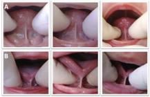
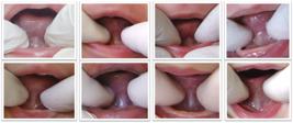
Of the 1,715 infants, 1,157 (67.46%) had the lingual frenulum visualized by the simple maneuver consisting in elevating the lateral margins of the tongue (Figure 1). 558 infants (32.54%) had posterior frenulum, which required the special maneuver that consisted in both elevating and pushing the tongue back simultaneously (Figure 2).
The special maneuver allowed the visualization of the thickness and place of attachment of lingual frenulum in 549 infants (98.39%) who had posterior lingual frenulum (Figure 3). In nine infants (1.61%), lingual frenulum could not be visualized by means of the maneuver before hospital discharge. In these cases, the visualization of lingual frenulum was possible within the first 3 months after birth.
Regarding the gender, out of the 558 infants who had posterior lingual frenulum, 302 (54.12%) were females and 256 (45.88%), males.
(A) normal lingual frenulum; (B) altered lingual frenulum. Both were visualized by elevating the lateral margins of the tongue (simple maneuver)
Posterior frenulum not visualized by elevating the lateral margins of the tongue (simple maneuver)
(A) posterior frenulum not visualized by elevating the lateral margins of the tongue (simple maneuver); (B) same lingual frenulum visualized by means of the special maneuver consisting in elevating and pushing the tongue back, simultaneously
Discussion
There is a great variation of anatomical characteristic of lingual frenulum reported in the literature including both healthy2-7 subjects and individuals with syndromes23-28. However, the literature about posterior lingual frenulum is scarce8-13.
By performing the special maneuver, this study observed the occurrence of 32.54% of posterior frenulum in the sample. Those results indicate that this anatomical variation is not rare. These findings are consistent with another study also conducted with healthy infants, which reported a 35% occurrence of posterior lingual frenulum8. Regarding subjects with syndromes, there are only a few studies reporting on the absence of lingual frenulum, based exclusively on visual inspection without clear criteria for the diagnosis15-19.
Published in 2017, a study conducted by medical geneticists and pediatric neurologists observed the absence of lingual frenulum in 4 (36.36%) out of 11 infants with the congenital Zika syndrome19. Conversely, a study conducted by SLPs reported that in a total of 54 infants with the congenital Zika syndrome, lingual frenulum visibility required a specific maneuver to retract the tongue in 20 (37%), since it was covered by mucous tissue20. The authors suggested the terminology “absent lingual frenulum” used by Del Campo et al.19 be replaced for “submucous lingual frenulum”20.
In 98.39% of the 558 infants diagnosed with posterior lingual frenulum, the special maneuver consisting in elevating and pushing the tongue back allowed the visualization of the thickness and place of attachment of lingual frenulum. Therefore, the maneuver may be performed to visualize these aspects of lingual frenulum when it is covered by mucous tissue. However, in 1.61% lingual frenulum could not be visualized by means of the maneuver before hospital discharge. The visualization could be performed within the first 3 months after birth. This may be explained by the fact that due to the small size of some infants´ oral cavity, pushing the tongue back was not possible immediately after birth. However, those findings cannot be compared to those in the, literature since no studies about it were found.
This study observed that females showed a higher occurrence (54.12%) of this anatomical variation of lingual frenulum. This finding is in agreement with another study found in the literature that reported higher occurrence of posterior lingual frenulum among females, observed that this anatomical variation did not interfere with the movements of the tongue - sucking and swallowing - during breastfeeding, and concluded that this variation cannot be classified as ankyloglossia8. It is important to highlight that ankyloglossia - an anomaly that restricts the movements of the tongue - is predominantly observed in males2,29.
Thus, posterior lingual frenulum is an anatomical variation that may be present in both healthy infants8 and infants with syndromes20. The special maneuver consisting of elevating and pushing the tongue back simultaneously is efficient, easy, and does not require any device for lingual frenulum assessment.
An important contribution for further understanding of this anatomical characteristic and its impact on the functions of the tongue may be given by future studies performing this special maneuver to assess subjects diagnosed with different syndromes characterized by absent lingual frenulum.
Conclusion
In this sample, the occurrence of posterior lingual frenulum was high. The special maneuver consisting in elevating and pushing the tongue back was efficient in visualizing the posterior lingual frenulum.
References
- 1 Ghaheri BA, Cole M, Fausel SC, Chuop M, Mace JC. Breastfeeding improvement following tongue-tie and lip-tie release: a prospective cohort study. Laryngoscope. 2017;127(5):1217-23.
- 2 Martinelli RLC, Marchesan IQ, Berretin-Felix G. Protocol for infants: relationship between anatomic and functional aspects. Rev. CEFAC. 2013;15(3):599-609.
- 3 Haham A, Marom R, Mangel L, Botzer E, Dollberg S. Prevalence of breastfeeding difficulties in newborns with a lingual frenulum: a prospective cohort series. Breastfeeding Med. 2014;9(9):438-41.
- 4 Berry J, Griffiths M, Westcott C. A double-blind, randomized, controlled trial of tongue-tie division and its immediate effect on breastfeeding. Breastfeed Med. 2012;7(3):189-93.
- 5 Buryk M, Bloom D, Shope T. Efficacy of neonatal release of ankyloglossia: a randomized trial. Pediatrics. 2011;128(2):280-8.
- 6 Emond A, Ingram J, Johnson D, Blair P, Whitelaw A, Copeland M et al. Randomised controlled trial of early frenotomy in breastfed infants with mild-moderate tongue-tie. Arch Dis Child Fetal Neonatal. 2014;99(3):F189-95.
- 7 Hogan M, Westcott C, Griffiths M. Randomized, controlled trial of division of tongue-tie in infants with feeding problems. J Paediatr Child Health. 2005;41(5-6):246-50.
- 8 Martinelli RLC, Marchesan IQ, Berretin-Felix G. Posterior lingual frenulum and breastfeeding. Int J Orofacial Myology. 2016;42:49-54.
- 9 Chu MW, Bloom DC. Posterior ankyloglossia: a case report. Int J Pediatr Otorhinolaryngol. 2009;73(6):881-3.
- 10 Hong P, Lago D, Seargeant J, Pellman L, Magit AE, Pransky SM. Defining ankyloglossia: a case series of anterior and posterior tongue ties. Int J Pediatr Otorhinolaryngol. 2010;74(9):1003-6.
- 11 Knox I. Tongue tie and frenotomy in the breastfeeding newborn. NeoReviews. 2010;11(9):513-9.
- 12 Pradhan S, Yasmin E, Mehta A. Management of posterior ankyloglossia using the Er,Cr: YSGC Laser. Int J Laser Dentistry. 2012;2(2):41-6.
- 13 Pransky SM, Lago D, Hong P. Breastfeeding difficulties and oral cavity anomalies: the influence of posterior ankyloglossia and upper-lip ties. Int J Pediatr Otorhinolaryngol. 2015;79(10):1714-7.
- 14 Douglas PS. Rethinking "posterior" tongue-tie. Breastfeed Med. 2013;8(6):503-6.
- 15 De Felice C, Toti P, Di Maggio G, Parrini S, Bagnoli F. Absence of the inferior labial and lingual frenula in Ehlers-Danlos syndrome. Lancet. 2001;357(9267):1500-2.
- 16 Machet L, Hüttenberger B, Georgesco G, Doré C, Jamet F, Bonnin-Goga B et al. Absence of inferior labial and lingual frenula in Ehlers-Danlos syndrome: a minor diagnostic criterion in French patients. Am J Clin Dermatol. 2010;11(4):269-73.
- 17 Tulika W, Kiran A. Ehlers-Danlos syndrome. J Dent Res Ver. 2015;2(1):42-6.
- 18 Mitakides J, Tinkle BT. Oral and mandibular manifestations in the Ehlers-Danlos syndromes. Am J Med Genet C Semin Med Genet. 2017;175(1):220-5.
- 19 Del Campo M, Feitosa IM, Ribeiro EM, Horovitz DD, Pessoa AL, França GV et al. Zika Embryopathy Task Force-Brazilian Society of Medical Genetics ZETF-SBGM. The phenotypic spectrum of congenital Zika syndrome. Am J Med Genet A. 2017;173(4):841-57.
-
20 Fonteles CSR, Marques RE, Sales ASM, Ferreira PLR, Sales AG, Monteiro AJ et al. Lingual frenulum phenotypes in brazilian infants with congenital Zika syndrome. Cleft Palate Craniofac J. 2018 Jan 1:10.1177/1055665618766999.
» https://doi.org/10.1177/1055665618766999 - 21 Martinelli RLC, Marchesan IQ, Lauris JR, Honório HM, Gusmão RJ, Berretin-Felix G. Validity and reliability of the neonatal tongue screening test. Rev. CEFAC. 2016;18(6):1323-31.
-
22 Martinelli RLC, Marchesan IQ, Berretin-Felix G. Manobra para visualização do frênulo lingual em bebês. Anais da XIX Jornada Fonoaudiológica de Bauru. 2012. Disponível em: http://www.cofab.fob.usp.br/wp-content/uploads/Anais-2012.pdf
» http://www.cofab.fob.usp.br/wp-content/uploads/Anais-2012.pdf - 23 Llames S, Recuero I, Romance A, Garcia E, Peña I, Valle AF et al. Tissue-engineered oral mucosa for mucosal reconstruction in a pediatric patient with hemifacial microsomia and ankyloglossia. Cleft Palate-Craniofacial Journal. 2014;51(2):246-51.
- 24 Soman C, Lingappa A. Robinow syndrome: a rare case report and review of literature. Int J Clin Pediatr Dent. 2015;8(2):149-52.
- 25 Al-Qattan MM, Javed K. Variability of expression of oral-facial-digital syndrome type I in 15 Saudi girls: Why is there a high rate of median cleft lip in the phenotype? Plast Surg (Oakv). 2014;22(4):229-32.
- 26 Kaul B, Mahajan N, Gupta R, Kotwal B. The syndrome of pit of the lower lip and its association with cleft palate. Contemp Clin Dent. 2014;5(3):383-5.
- 27 Shetty P, Shetty D, Priyadarshana PS, Bhat S. A rare case report of Ellis Van Creveld syndrome in indian patient and literature review. J Oral Biol Craniofac Res. 2015;5(2):98-101.
- 28 Singh A, Bhatia HP, Sood S, Sharma N, Mohan A. A novel finding of oligodontia and ankyloglossia in a 14-year-old with Floating-Harbor syndrome. Spec Care Dentist. 2017;37(6):318-21.
- 29 Hogan M, Westcott C, Griffiths M. Randomized, controlled trial of division of tongue-tie in infants with feeding problems. J Paediatr Child Health. 2005;41(5-6):246-50.
Publication Dates
-
Publication in this collection
Jul-Aug 2018
History
-
Received
22 Apr 2018 -
Accepted
01 July 2018

 Posterior lingual frenulum in infants: occurrence and maneuver for visual inspection


