Atrial Myxoma as a Rare Cause of Stroke: Case Report
Keywords
Myxoma; Heart Neoplasms; Stroke
Palavras-chave
Mixoma; Neoplasias Cardíacas; Acidente Vascular Cerebral
Keywords
Myxoma; Heart Neoplasms; Stroke
Palavras-chave
Mixoma; Neoplasias Cardíacas; Acidente Vascular Cerebral
Introduction
Atrial myxoma (AM) is the most common primary benign tumor affecting the heart. Its prevalence is rare, estimated at 0.03% in the general population. Surgical intervention should be introduced as soon as diagnosed due to the increased risk of cardiac complications, such as intracardiac obstructions or systemic embolizations.1
Cardiac myxomas are commonly observed in the left atrium in >75% of cases, predominantly attached with a stalk to the fossa ovalis (>90%). However, they can be seen in other locations (left ventricle, right atrium, and, though quite rarely, the right ventricle).2
Approximately 7% of all cardiac myxomas are associated with Carney complex, an autosomal dominant disease characterized by the presence of cardiac and cutaneous lesions, myxomas, skin hyperpigmentation and primary pigmented nodules, with adrenocortical disease leading to Cushing's syndrome.2
In up to one third of the cases of myxomas, there is evidence of systemic embolism, even in the brain, which may be silent or cause neurological symptoms. Neurological deficits may often be the first sign of the manifestation of a cardiac myxoma.3
Following surgical removal, myxomas may recur, and wide excision of the tumor with adjacent cardiac tissue is necessary to prevent recurrence. Surgery is usually recommended when the diagnosis of cardiac myxoma is made, regardless of whether or not systemic embolism has already occurred.4
The present case report demonstrates a case of AM in an elderly patient with stroke as the first symptom, diagnosed with transthoracic echocardiography (TTE) and transesophageal echocardiography (TEE), and with a favorable clinical outcome after surgical removal of a benign tumor.
Case report
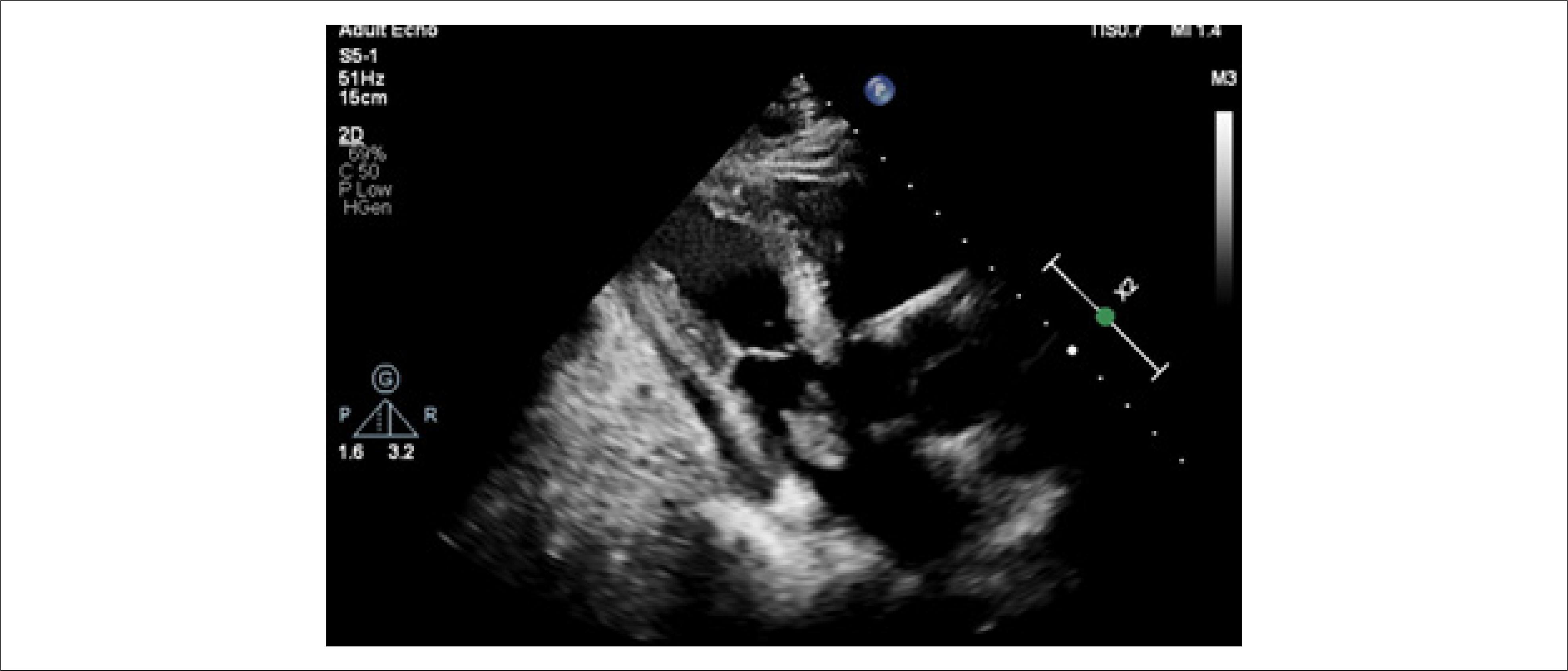
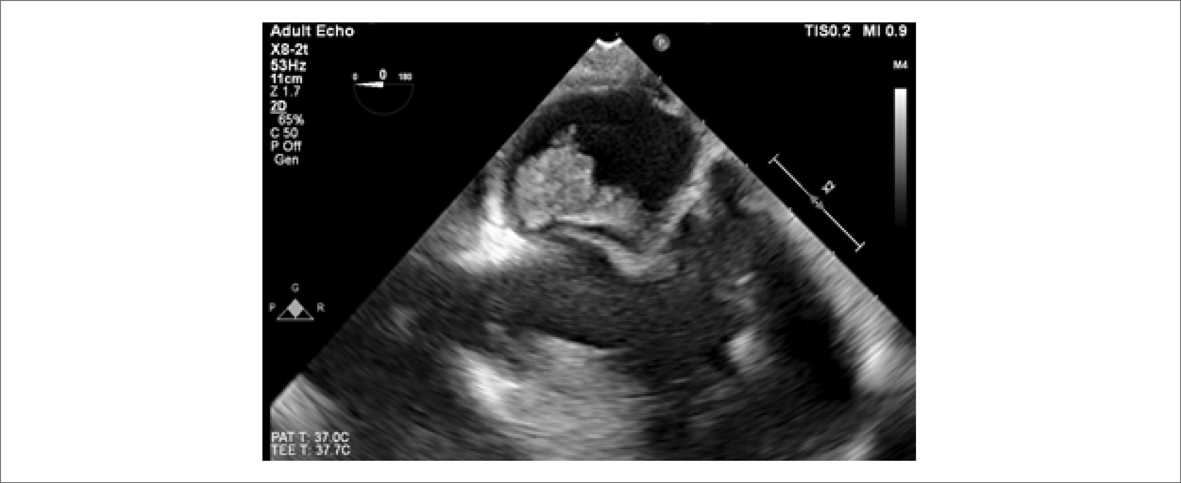
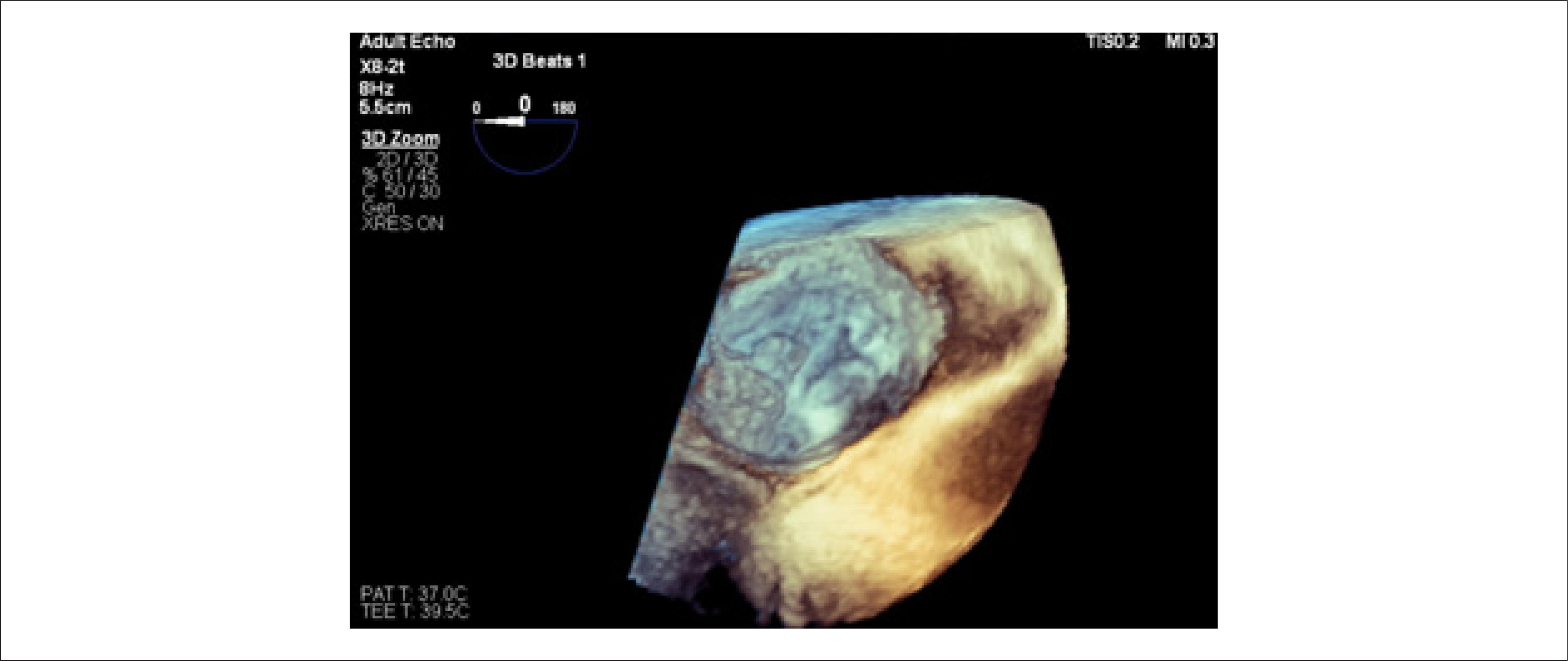
A 72-year-old male patient with a history of systemic arterial hypertension and type 2 diabetes mellitus was admitted to the emergency room three months ago with vertigo, left hemiparesis, rhyme deviation, spatial disorientation, and ataxia. He was referred for neurological evaluation with a diagnostic hypothesis of ischemic stroke ( acidente vascular encefálico isquêmico – AVEi) , of probable cardioembolic origin, confirmed by magnetic resonance imaging of the skull. The findings in the aforementioned imaging exam that describe this hypothesis of cardioembolic origin were described as a small hyperintense lesion on T2 weighting and with restriction in the diffusion study used to examine the right thalamus, suggesting acute ischemic vascular injury and small lesions with the same characteristics in the dorsal region of the midbrain and in the right cerebral peduncle. During the investigation of the neurological condition, the electrocardiogram showed sinus rhythm and nonspecific ventricular repolarization alteration, while the TTE ( Figure 1 ) showed a mobile echogenic image measuring 1.4 x 1.6 cm near the opening of the mitral valve in the region of the anterior leaflet and interatrial septum. Continuing the investigation with TEE ( Figures 2 and 3 ), an echogenic image was confirmed, attached to the septal portion of the mitral annulus, interatrial septum, and anterior leaflet of the mitral valve, measuring 22 x 13 mm, with contrast enhancement within a lesion compatible with vascularization, suggesting AM.

TEE in mid-esophagus at 0 degrees. (left interatrial myxoma visible – with largest diameters of 2.5 cm x 1.5 cm).
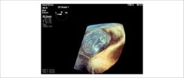
Three-dimensional (3D) TEE (3D zoom of the left atrium with visualization of myxoma attached to the membrane in the oval fossa).
The patient was referred for outpatient cardiovascular surgery, and tumor resection was recommended. During the preoperative period, a multivessel lesion was identified by cardiac catheterization, and therefore a combined procedure of intracardiac tumor resection and coronary artery bypass grafting was recommended, which was performed without complications. The material was sent for anatomopathological examination, which confirmed AM.
Following the surgery, the patient showed clinical improvement, with no new cardioembolic events. Upon hospital discharge, he was asymptomatic and was referred for outpatient follow-up with clinical cardiology.
Discussion
AM is responsible for less than 0.5% of cases of ischemic stroke.5 Recent studies report that 9% to 22% of patients with MA suffer from cardioembolic stroke. Its incidence is higher in females, at a ratio of 2:1, which most commonly appears between the third and sixth decades of life.6
AM is increasingly diagnosed in the elderly and its predominance in females disappears after the age of 65. Its most frequent location is in the left atrium (75 to 80%), inserted in the interatrial septum around the fossa ovalis.7
The main etiology of AM is unknown, with approximately 10% of the cases presenting an autosomal dominant genetic component. The classic clinical triad of AM includes obstructive symptoms, such as valve obstruction and ventricular failure; embolic manifestations in any arterial territory, with the cerebral artery being the most frequently involved; and constitutional symptoms, such as fever, asthenia, myalgias, arthralgias, and weight loss. Approximately 20% of all cases are asymptomatic, with the diagnosis being made incidentally through imaging exams. Obstructive (40% to 60%) and constitutional (30% to 90%) cardiac symptoms are the most frequent in clinical presentation, and embolic manifestations may occur in 30% to 40% of all patients.6
Patients with AM who present neurological complications are mostly young, and stroke or transient ischemic attack are the most common neurological manifestations (82%), occurring due to embolization of tumor fragments, thrombus formation, or both.8
AMs are usually solitary, with sizes ranging from 0.4 to 6.5 cm. Morphologically, they are divided into two types: type 1, with an irregular or villous surface and long pedicle, which are associated with a higher risk of embolization, and type 2, with a smooth surface and soft consistency.7
TTE is the initial examination used to detect cardiac masses, with a sensitivity of 90% and a specificity of 95%. TEE enables a more detailed visualization of the anatomical structures, which is useful in determining the size, location, adhesion, relationship with adjacent structures, and mobility of the tumor.9
Differential diagnoses of AM include vegetations, intracardiac thrombi, or artifacts.10
Surgical resection is considered curative and is recommended whenever AM is diagnosed, regardless of previous embolization. It should be performed as early as possible due to the risk of recurrence of embolism or valve obstruction. The postoperative prognosis is variable and depends on the patient's age, comorbidities, preoperative clinical status, and possible need to perform other surgical procedures associated with AM resection.7
Most AM recurrences occur in the first 4 years after tumor resection, although the risk is low. For this reason, regular follow-up with echocardiography should be performed.7
Conclusion
AM is a rare cause of AVEi , which, although it has a low population prevalence, has important clinical relevance for early surgical intervention in an attempt to reduce morbidity and mortality due to systemic embolisms caused by the tumor.
-
Sources of Funding
There were no external funding sources for this study.
-
Study Association
This study is not associated with any thesis or dissertation work.
-
Ethics Approval and Consent to Participate
This article does not contain any studies with human participants or animals performed by any of the authors.
References
-
1 Gupta A, Darbari A, Mishra A. Excision of a Left Atrial Myxoma with Mitral and Tricuspid Valve Repair. Multimed Man Cardiothorac Surg. 2020;2020. doi: 10.1510/mmcts.2020.080.
» https://doi.org/10.1510/mmcts.2020.080 -
2 Saric M, Armour AC, Arnaout MS, Chaudhry FA, Grimm RA, Kronzon I, et al. Guidelines for the Use of Echocardiography in the Evaluation of a Cardiac Source of Embolism. J Am Soc Echocardiogr. 2016;29(1):1-42. doi: 10.1016/j.echo.2015.09.011.
» https://doi.org/10.1016/j.echo.2015.09.011 -
3 Pucci A, Gagliardotto P, Zanini C, Pansini S, di Summa M, Mollo F. Histopathologic and Clinical Characterization of Cardiac Myxoma: Review of 53 Cases from a Single Institution. Am Heart J. 2000;140(1):134-8. doi: 10.1067/mhj.2000.107176.
» https://doi.org/10.1067/mhj.2000.107176 -
4 Shapiro LM. Cardiac Tumours: Diagnosis and Management. Heart. 2001;85(2):218-22. doi: 10.1136/heart.85.2.218.
» https://doi.org/10.1136/heart.85.2.218 - 5 Eira C, Mota A, Silvério R, Monteiro R, Monteiro A. Atrial Myxoma: A Rare Cause of Ischemic Stroke. Rev Soc Bras Clin Med. 2018;16(1):48-51.
-
6 Iyer P, Aung MM, Awan MU, Kososky C, Barn K. A Case of Large Atrial Myxoma Presenting as an Acute Stroke. J Community Hosp Intern Med Perspect. 2016;6(1):29604. doi: 10.3402/jchimp.v6.29604.
» https://doi.org/10.3402/jchimp.v6.29604 -
7 Bordalo AD, Alves I, Nobre AL, Silva F, Lemos A, Serpa C, et al. New Clinical Aspects of Cardiac Myxomas: A Clinical and Pathological Reappraisal. Rev Port Cardiol. 2012;31(9):567-75. doi: 10.1016/j.repc.2012.05.008.
» https://doi.org/10.1016/j.repc.2012.05.008 -
8 Baek SH, Park S, Lee NJ, Kang Y, Cho KH. Effective Mechanical Thrombectomy in a Patient with Hyperacute Ischemic Stroke Associated with Cardiac Myxoma. J Stroke Cerebrovasc Dis. 2014;23(9):e417-9. doi: 10.1016/j.jstrokecerebrovasdis.2014.05.006.
» https://doi.org/10.1016/j.jstrokecerebrovasdis.2014.05.006 -
9 Kebede S, Edmunds E, Raybould A. A Large Left Atrial Myxoma Causing Multiple Cerebral Infarcts. BMJ Case Rep. 2013;2013:bcr2013010007. doi: 10.1136/bcr-2013-010007.
» https://doi.org/10.1136/bcr-2013-010007 -
10 Ha JW, Kang WC, Chung N, Chang BC, Rim SJ, Kwon JW, et al. Echocardiographic and Morphologic Characteristics of Left Atrial Myxoma and their Relation to Systemic Embolism. Am J Cardiol. 1999;83(11):1579-82, A8. doi: 10.1016/s0002-9149(99)00156-3.
» https://doi.org/10.1016/s0002-9149(99)00156-3
Edited by
-
Editor responsible for the review:
Marcelo Tavares