
 Liver Transplantation in Alpha-1 Antitrypsin Deficiency: A Retrospective Case-Control Study in a Brazilian Center
ABSTRACT
Objectives: Alpha-1 antitrypsin deficiency (AATD) is a rare genetic metabolic disease curable with liver transplantation (LT). The aim of this study was to comparatively evaluate patients with LT due to AATD and other causes.
Methods: Observational, retrospective, analytical, case-control study evaluating 2,511 LT patients, 19 (0.75%) with AATD, from May 2002 to December 2024. Data were obtained by reviewing the medical records and stored in the REDCap program.
Results: The median age of AATD and non-AATD (NAATD) patients was similar (56.3 [Δ7-71] and 52.4 [Δ6-78]; p = 0.92, respectively). Males predominated in both groups. The pre-LT Model for End-Stage Liver Disease (MELD)-Na scores for AATD and NAATD patients were 20 (Δ8-24) and 18 (Δ6-72), respectively. Two patients with AATD had positive serologies compatible with previous hepatitis B infection. Three patients also had a clinical epidemiological diagnosis of concomitant nonalcoholic fatty liver disease. The diagnosis of AATD was made only through explant in nine patients (n = 47.4%). The mean pre-transplant alpha-1 antitrypsin dosage was 33.42 mg/dL (Δ19-59.5). All patients who underwent immunophenotyping had the PiZZ phenotype. The indication for LT was decompensated cirrhosis (DC); three patients (16.4%) had concomitant hepatocellular carcinoma (HCC), one diagnosed only in the explant. The incidence of HCC was 15.9% (AATD) and 31.3% (NAATD) (p = 0.34). None of the patients with AATD had severe lung disease. The survival of patients with AATD submitted to LT at 30 days, 1, and 5 years was 94.7, 88.2, and 64.7%, respectively, similar to the other causes (91.4, 80.1, and 68.1%; p = 0.47).
Conclusion: AATD was a rare cause of LT due to DC and HCC, predominantly in adults with piZZ phenotype without relevant pulmonary involvement, and whose diagnosis is frequently made after LT. The epidemiology, indication, prevalence of HCC, and survival were similar to those of other causes of LT.
Descriptors
Alpha-1 Antitrypsin Deficiency; Liver Transplantation; Cirrhosis; Hepatocellular Carcinoma; Retrospective Study
RESUMO
Objetivos: A deficiência de alfa-1 antitripsina (DAAT) é uma doença metabólica genética rara curável com transplante hepático (TH). O objetivo deste estudo foi avaliar comparativamente pacientes com TH por DAAT e outras causas.
Métodos: Estudo observacional, retrospectivo, analítico, caso-controle, avaliando 2.511 pacientes com TH, 19 (0,75%) com DAAT, no período de maio de 2002 a dezembro de 2024. Os dados foram obtidos por meio da revisão dos prontuários e armazenados no programa REDCap.
Resultados: A mediana de idade dos pacientes com e sem AATD (NAATD) foi semelhante [56,3 (Δ7-71) e 52,4 (Δ6-78); p = 0,92, respectivamente). O sexo masculino predominou em ambos os grupos. O modelo pré-TH para doença hepática terminal Model for End-Stage Liver Disease (MELD)-Na dos pacientes com DAAT e NDAAT foi de 20 (Δ8-24) e 18 (Δ6-72), respectivamente. Dois pacientes com DAAT apresentaram sorologias positivas compatíveis com infecção prévia por hepatite B. Três pacientes também apresentaram diagnóstico clínico e epidemiológico de doença hepática gordurosa não alcoólica concomitante. O diagnóstico de DAAT foi feito apenas por explante em nove pacientes (n = 47,4%). A dosagem média de DAAT pré-transplante foi de 33,42 mg/dL (Δ19-59,5). Todos os pacientes submetidos à imunofenotipagem apresentaram fenótipo PiZZ. A indicação para TH foi cirrose descompensada (CD); três pacientes (16,4%) apresentaram carcinoma hepatocelular (CHC) concomitante, um diagnosticado apenas no explante. A incidência de CHC foi de 15,9% (DAAT) e 31,3% (NDAAT) (p = 0,34). Nenhum dos pacientes com DAAT apresentou doença pulmonar grave. A sobrevida dos pacientes com DAAT submetidos a TH em 30 dias, 1 e 5 anos foi de 94,7; 88,2 e 64,7%, respectivamente, semelhante às outras causas (91,4, 80,1 e 68,1%; p = 0,47).
Conclusão: A DAAT foi uma causa rara de TH devido à CD e CHC, predominantemente em adultos com fenótipo piZZ sem envolvimento pulmonar relevante e cujo diagnóstico é frequentemente feito após TH. A epidemiologia, indicação, prevalência de CHC e sobrevida foram semelhantes a outras causas de TH.
Descritores
Deficiência de Alfa-1 Antitripsina; Transplante de Fígado; Cirrose; Carcinoma Hepatocelular; Estudo Retrospectivo
INTRODUCTION
Alpha-1 antitrypsin deficiency (AATD) is a rare genetic disorder primarily affecting the liver and lungs. Alpha-1 antitrypsin (AAT) is a glycoprotein that belongs to the serine protease inhibitor family and is mainly produced by hepatocytes, but it is also present in the lungs, kidneys, and intestines.1 Its primary function is to inhibit neutrophil elastase, thereby protecting the lungs from excessive proteolytic degradation of elastin and other connective tissue components, as well as from external factors such as smoking.1
AATD arises from mutations in the SERPINA1 gene, which encodes AAT and is located on the long arm of chromosome 14. The normal allele is designated as PiM, while the most common deficiency alleles are PiS and PiZ, which code for abnormal proteins that undergo polymerization in the liver. Among the variants associated with clinical disease, the Z mutation is the most prevalent.2 AATD predominantly affects individuals of European descent, with an estimated prevalence of the most severe genotype (PiZZ) ranging from one in 2,000 to 5,000 individuals in Europe, and from one in 5,000 to 7,000 individuals of European descent in countries such as Canada, the United States, Australia, and New Zealand. Based on these estimates, there are approximately 6,000 individuals with the Pi*ZZ genotype in Brazil, which has a racially diverse population, including immigrants from various European countries.2 AATD-associated liver disease is caused by the accumulation of AAT polymers inside hepatocytes, leading to inflammation, liver fibrosis, cirrhosis, and an increased risk of hepatocellular carcinoma (HCC). This manifestation is the second most common among adults with AATD and the most common in individuals under 20 years old.3 It is estimated that 85% of individuals with AATD remain undiagnosed, with many diagnosed later in life after years of symptoms and multiple medical consultations. Consequently, there is a high rate of underdiagnosis, which impedes genetic counseling and prevents proper treatment.1,3 In Brazil, reasons for underdiagnosis include limited medical knowledge about the condition, the diagnostic tests required, and the unavailability of these tests.1 To screen for the disease, it is recommended that all patients with chronic obstructive pulmonary disease (COPD), liver disease, necrotizing panniculitis, granulomatosis with polyangiitis, or bronchiectasis of unknown origin undergo serum AAT measurement.3
Despite its clinical significance, epidemiological data concerning the prevalence of AATD or the frequency of deficient alleles in Brazil and South America are limited.4 A small portion of homozygous Pi*ZZ individuals develop severe liver disease that requires liver transplantation (LT). Post-transplant survival rates are excellent. The largest burden of advanced liver disease is observed among adults rather than children. Evaluating lung function in adults prior to transplantation is essential due to the underlying risk for COPD. Although uncommon, cases of simultaneous lung and LT for AAT deficiency have been reported.5 The aim of this study was to comparatively evaluate the epidemiology and outcomes of LT in patients with AATD and other etiologies of liver disease.
METHODS
This retrospective observational cohort study utilized a case-control format, including all patients who underwent LT from May 2002 to December 2024 by the team at Hospital Universitário Walter Cantídio-Universidade Federal do Ceará (HUWC-UFC), in collaboration with São Carlos, São Camilo, and UNIMED hospitals in Fortaleza, state of Ceará. The patients were classified into two distinct groups: the first consisted of patients diagnosed with AATD, either pre- or post-transplant, while the second group included all other patients (non-AATD [NAATD]). The diagnosis of AATD was established through pre-transplant assessment of AAT concentration, confirmed by pre-transplant biopsy and/or biopsy of the transplanted organ, with a minority of cases undergoing immunophenotyping.
Data were collected from medical records, encompassing pre- and post-transplant information, and were stored in REDCap® software for analysis using R® statistical software. The variables studied included demographic data (age, gender, weight, and level of education) and clinical and laboratory variables (chronic diseases, stage of liver disease before treatment, presence of cirrhosis, liver biopsies, and AATD phenotype). Additionally, information on patient survival following transplantation was gathered. All patients were continuously monitored after transplantation for immunosuppression and complications.
The immunosuppression protocol included the use of tacrolimus (1 mg/kg) in combination with prednisone (60 mg during the 1st month, with a progressive reduction to 5 mg by the 6th month post-transplantation). Mycophenolate sodium or everolimus was added as needed in cases of renal dysfunction. The target serum level for tacrolimus was set at 8 to 10 ng/mL during the first 6 months, and 4 to 6 ng/mL after 6 months post-transplantation. The target serum level for everolimus was established at 3 to 8 ng/mL. Continuous demographic variables were presented as means ± standard deviations, while categorical variables were expressed as proportions. After confirming the normality of the data distribution, continuous variables were analyzed using the unpaired t-test.
Categorical variables were subjected to univariate analysis and, where applicable, the chi-square test or Fisher’s exact test for non-parametric variables. p-values below 0.05 were considered statistically significant.
Cases with missing data were excluded from the respective analyses (complete-case analysis). Only descriptive and univariate analyses were conducted, as the primary objective was to characterize the patient population rather than identify independent predictors. The survival curve was calculated using the Kaplan-Meier method, and differences were assessed using the log-rank test. The study protocol received approval from the Ethics Committee of the HUWC-UFC and adhered to the principles outlined in the Declaration of Helsinki.
RESULTS
From May 2002 to December 2024, the liver transplant team at HUWC-UFC performed 2,511 LTs, of which 19 (0.75%) were for patients with AATD, while the remaining 2,492 were for NAATD cases. The primary indications for LT at this center were as follows: alcoholic liver disease (32.3%), HCC (31.1%), hepatitis C (24.2%), cryptogenic cirrhosis (16.3%), hepatitis B (12.7%), hepatitis B and D (4.8%), autoimmune hepatitis (7.8%), metabolic dysfunction-associated steatotic liver disease (MASLD) (3.1%), primary sclerosing cholangitis (2.5%), Budd-Chiari syndrome (1.5%), primary biliary cirrhosis (1.3%), hemochromatosis (1.4%), secondary biliary cirrhosis (1.2%), Caroli’s disease (0.8%), Wilson’s disease (0.8%), and AATD (0.76%), among others.
With only 19 patients, AATD was one of the rarest causes of LT at our center. Pre-transplant diagnoses were confirmed in 10 patients. Nine patients (47.4%) were initially transplanted for cryptogenic cirrhosis, while the diagnosis of AATD was made only after LT based on explant analysis. Among the AATD patients, two had positive serologies indicating previous hepatitis B infection, and seven had a history of alcohol use, with only one reporting significant alcohol consumption. Three patients were clinically and epidemiologically diagnosed with concomitant nonalcoholic fatty liver disease (MASLD). Additionally, one patient was diagnosed with hepatoblastoma. The mean pre-transplant AAT level was 33.42 mg/dL (range: 19-59.5 mg/dL), whereas normal levels typically range from 200 to 400 mg/dL. Only four patients underwent immunophenotyping, all of whom were identified as PiZZ phenotypes.
The majority (61.2%) of AATD LT patients at this center originated from outside the state of Ceará. The distribution of patients’ states of residence was as follows: Ceará (8, 42.1%), Piauí (1, 5.3%), Maranhão (4, 21.1%), Rio Grande do Norte (4, 21.1%), and Pará (2, 10.5%). All AATD patients underwent LT due to decompensated cirrhosis (DC), with one patient (5.9%) also diagnosed with HCC; however, two patients with HCC were detected only through explant analysis. The indication for LT due to HCC was similar for both groups (Table 1).
Epidemiology and clinical characteristics of liver transplant patients with AATD and other causes of LT.
Although four patients were active or former smokers, only one had COPD. Two AATD patients had mild asthma diagnosed prior to LT. All patients underwent chest x-rays, and none exhibited respiratory symptoms. Five patients had normal spirometry results. The most common comorbidities among AATD patients were diabetes and hypertension, which were present in four out of 17 patients (23.5%).
Surgical complications in patients transplanted for AATD included reoperation due to bleeding (n = 1), portal vein thrombosis (PVT) (n = 1), portal vein stenosis (n = 1), biliary stenosis (n = 2), biliary fistula (n = 1), intestinal obstruction (n = 1), and intestinal subocclusion (n = 1). None of the AATD patients experienced primary graft dysfunction (PGD) or required retransplantation (ReTx). The most common non-infectious clinical complication was chronic non-dialysis renal dysfunction, observed in nine patients (50%), which was similar to findings in other patients. Other complications in the postoperative period included hypertension (n = 2), diabetes (n = 1), and dyslipidemia (n = 1). Post-transplant infections occurred in six of 17 patients (35.3%), with the following infections identified: pneumonia (n = 2), varicella-zoster (n = 1), and bloodstream infection (n = 1). Post-liver transplant renal dysfunction and infections were much more common in NAATD patients (Table 1).
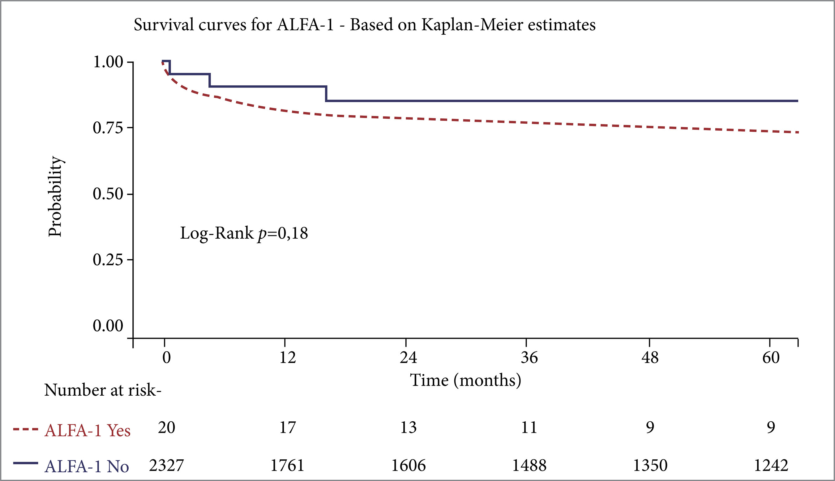
Among the 19 patients who underwent LT for AATD, three died. All patients died from infections 16 days, 4 months, and 66 months after transplant. The 30-day, 1-year, and 3-year survival rates for AATD and other causes of LT are shown in Fig. 1 and Table 2.

DISCUSSION
AATD is a rare indication for LT in Brazil, with diagnosis frequently occurring post-transplant. Similar to other LT case series involving AATD, the condition predominantly affects middle-aged men, accounting for up to 2.7% of LT indications due to cirrhosis.4 The number of AATD cases in this study is attributed to it being a reference service for LT for the entire North and Northeast regions of Brazil, which affects the understanding of how these cases develop, their epidemiological patterns, and associated mortality rates in these regions of the country.
This study is subject to certain limitations, including the small number of patients with AATD and the lack of post-liver transplant assessments, which may affect the generalizability and completeness of the findings. The PiZZ phenotype, often associated with more severe liver and lung disease and lower serum AAT levels, was the most prevalent in this study, consistent with findings from other published series.4 No population-based studies have been conducted to date in adults. The association between PI*ZZ and chronic liver disease was estimated by comparing its prevalence in cohorts with liver disease (0.8%) to the expected population frequency (0.04%). This 20-fold increase suggests a heightened risk in homozygous individuals.5
Studies show that LT normalizes AAT levels and converts the recipient’s phenotype to the normal PiMM.6 In this study, post-LT testing was not performed to confirm this. The predominance of male patients aligns with other studies, although the pediatric population was underrepresented, with only one 7-year-old child included. This may be due to pediatric cases being referred to other centers with more experience in pediatric transplantation.
The high number of LT patients without an AATD diagnosis reflects the limited access to immunophenotyping due to its cost. Given the severity and urgency of liver disease in these patients, diagnostic procedures are often deprioritized.
In a case series of 10 adults who underwent LT, aged 18 to 48 years (mean age 34), only one individual had COPD. Among 29 transplanted ZZ children, the mean age was 5 years (range: 8 months to 13 years), with reported 5-year survival rates of 60% for adults and 83% for pediatric recipients.7 Our series has a few children. A study conducted by another author showed that the risk of severe liver disease and the indication for LT in AATD is greater in adult males. Severe liver disease, as defined by the need for LT, was analyzed using three US LT databases for the period 1991-2012. It was found that 77.2% of 1,677 liver transplants with a reported diagnosis of AATD were in adults, with a peak age range of 50-64 years. Severe liver disease requiring transplantation is more than 2.5 times as likely in adults. The analysis also showed a markedly increased risk for males.8
In another study, which included 51,953 individuals diagnosed with AATD, only 2.4% required LT.9 Data from the UNOS transplant database covering 1995 to 2004 reported 406 adults with AATD, representing 1.06% of all adult LT cases during that period. Seventy-two percent of these patients were men, with a mean age of 52 years (range: 18-70). Post-LT survival in our series was similar to that of other studies, with rates of 89, 85, and 83% at 1, 3, and 5 years, respectively. Three patients had previously undergone lung transplantation.9
In the United Kingdom, AATD accounted for 2.94% of total adult LTs (10/339) and 13% of pediatric LTs (29/223). Reported 1-year survival rates were 73% for adults and 87.5% for children.10
The geographic origin of AATD patients differed from those with other LT indications. Although the state of Amazonas is the second-highest source of LT patients in this service, no AATD patients were from the Amazon Region, suggesting a lower prevalence of this mutation in Amazonian Indigenous populations.
Two AATD patients had asthma, but no pulmonary complications related to AATD were observed before or after LT in this case series. This is consistent with previous reports showing that AATD in children most often manifests as liver disease, whereas pulmonary complications such as emphysema typically emerge later in adulthood.4,11
CONCLUSION
AATD is a rare indication for LT at this Brazilian liver transplant center, with patient survival rates comparable to those of other LT etiologies. AATD was a rare cause of LT due to DC and HCC, predominantly in adults with piZZ phenotype, without relevant pulmonary involvement, and whose diagnosis is frequently made after LT. The epidemiology, indication, prevalence of HCC, and survival were similar to those of other causes of LT.
ACKNOWLEDGEMENT
Our thanks to all the health professionals at the Ceará Liver Transplant Center, especially those from the Liver Transplant Service at the HUWC-UFC and the students of the Liver Transplant and Digestive System Surgery League-UFC for their collaboration in creating the database that made this study possible.
-
FUNDING
Not applicable.
DATA AVAILABILITY STATEMENT
Data will be provided upon request.
References
-
1 American Thoracic Society; European Respiratory Society. American Thoracic Society/European Respiratory Society statement: standards for the diagnosis and management of individuals with alpha-1 antitrypsin deficiency. Am J Respir Crit Care Med, 2003;168:818-900. https://doi.org/10.1164/rccm.168.7.818
» https://doi.org/10.1164/rccm.168.7.818 -
2 Bernspang E, Sveger T, Piitulainen E. Respiratory symptoms and lung function in 30-year-old individuals with alpha-1 antitrypsin deficiency. Respir Med, 2007; 101: 1971-6. https://doi.org/10.1016/j.rmed.2007.04.003
» https://doi.org/10.1016/j.rmed.2007.04.003 -
3 Camelier AA, Souza LC, Fernandes ALG, Araújo D, Pereira MC, Mogami R. Deficiência de alfa-1 antitripsina: diagnóstico e tratamento. J Bras Pneumol. 2008; 34(7): 514-27. https://doi.org/10.1590/S1806-37132008000700012
» https://doi.org/10.1590/S1806-37132008000700012 -
4 Clark VC. Liver transplantation in alpha-1 antitrypsin deficiency. Clin Liver Dis, 2017; 21(2): 355-65. https://doi.org/10.1016/j.cld.2016.12.008
» https://doi.org/10.1016/j.cld.2016.12.008 -
5 Stoller JK, Snider GL, Brantly ML, Fallat RJ, Stockley RA, Turino GM, et al. American Thoracic Society; European Respiratory Society. Standards for the diagnosis and management of individuals with alpha-1 antitrypsin deficiency [German]. Pneumologie, 2005; 59(1): 36-8. https://doi.org/10.1055/s-2004-830176
» https://doi.org/10.1055/s-2004-830176 -
6 Putnam CW, Porter KA, Peters RL, Ashcavai M, Redeker AG, Starzl TE. Liver replacement for alpha1-antitrypsin deficiency. Surgery, 1977; 81(3): 258-61. Disponível em: https://pubmed.ncbi.nlm.nih.gov/320694/
» https://pubmed.ncbi.nlm.nih.gov/320694/ -
7 Esquivel CO, Vicente E, Van Thiel D, Gordon R, Marsh W, Makowka L, et al. Orthotopic liver transplantation for alpha-1-antitrypsin deficiency: an experience in 29 children and ten adults. Transplant Proc, 1987; 19(5): 3798-802. Disponível em: https://pmc.ncbi.nlm.nih.gov/articles/PMC2903897/
» https://pmc.ncbi.nlm.nih.gov/articles/PMC2903897/ -
8 Chu AS, Chopra KB, Perlmutter DH. Is severe progressive liver disease caused by alpha-1-antitrypsin deficiency more common in children or adults? Liver Transplantation, 2016; 22: 886-94. https://doi.org/10.1002/lt.24434
» https://doi.org/10.1002/lt.24434 -
9 Kemmer N, Kaiser T, Zacharias V, Neff GW. Alpha-1-antitrypsin deficiency: outcomes after liver transplantation. Transplantation Proceedings, 2008; 40: 1492-4. https://doi.org/10.1016/j.transproceed.2008.02.075
» https://doi.org/10.1016/j.transproceed.2008.02.075 -
10 Vennarecci G, Gunson BK, Ismail T, Hubscher SG, Kelly DA, McMaster P, et al. Transplantation for end stage liver disease related to alpha 1 antitrypsin. Transplantation. 1996; 61: 1488-95. https://doi.org/10.1097/00007890-199605270-00014
» https://doi.org/10.1097/00007890-199605270-00014 -
11 Costa MP, Silva RS, Oliveira CL, Lima MS, Almeida AF, Soares MD, et al. Clinical, laboratorial and evolutionary aspects of pediatric patients with liver disease due to alpha-1 antitrypsin deficiency. Arq Gastroenterol, 2023; 60(4): 438-49. https://doi.org/10.1590/S0004-2803.230402023-71
» https://doi.org/10.1590/S0004-2803.230402023-71
Edited by
-
Section editor:
Ilka de Fátima Santana F. Boin https://orcid.org/0000-0002-1165-2149

 Source: Elaborated by the authors.