 Survival and Prognostic Factors in Patients with Breast Cancer: Cohort Study
ABSTRACT
Introduction: Breast cancer is the most common cancer in the world and an important cause of death among women.
Objective: To analyze survival and prognostic factors in a cohort of patients with breast cancer.
Method: Retrospective cohort of 201 patients with breast cancer treated between January 2018 and December 2022 at a tertiary hospital in Caxias, Maranhão, Brazil. Data were collected between January and June 2023 and analyzed with software R, version 4.0.2. Overall survival (OS) and disease-free survival (DFS) curves were constructed using the Kaplan-Meier model. A descriptive analysis was performed by calculating absolute (n) and relative frequencies (%) and evaluating prognostic factors with Cox regression. Statistical significance was evaluated through the Wald Test, with a level of 5%.
Results: The 5-year OS and DFS were 91.3% and 72.4%, respectively. Women predominated (98.5%) over 40 years of age (86.1%) and with invasive carcinoma of no-special type (94%), G1 or G2 (79.1%), smaller than 5 cm (64.6%), without angiolymphatic (52.7%) and neural (77.6%) invasion and luminal molecular subtype (65.2%). Males had higher risk of death. Patients with angiolymphatic and neural invasion and non-expression of estrogen receptors (ER) had higher risk of recurrence.
Conclusion: The 5-year OS and DFS were 91.3% and 72.4%, respectively. Male gender, angiolymphatic and neural invasion and non-expression of ER were prognostic factors associated with higher risk of death or recurrence.
Keywords:
Prognosis; Anatomical Pathological Conditions; Health Care Outcome Assessment/statistics & numerical data; Breast Neoplasms/diagnosis
RESUMO
Introdução: O câncer de mama é o mais frequente no mundo e uma importante causa de óbito entre mulheres.
Objetivo: Analisar a sobrevida e os fatores prognósticos em uma coorte de pacientes com câncer de mama.
Método: Coorte retrospectiva de 201 pacientes com câncer de mama atendidos entre janeiro/2018 e dezembro/2022 em um hospital terciário de Caxias (MA), Brasil. A coleta de dados ocorreu entre janeiro e junho/2023. Os dados foram analisados no R, versão 4.0.2. Construíram-se as curvas de sobrevida global (SG) e sobrevida livre de doença (SLD) pelo modelo de Kaplan-Meier. Realizaram-se a análise descritiva por cálculo das frequências absolutas (n) e relativas (%) e a avaliação dos fatores prognósticos por regressão de Cox. Avaliou-se a significância estatística pelo teste de Wald, sendo o nível adotado de 5%.
Resultados: As SG e SLD em cinco anos foram de 91,3% e 72,4%, respectivamente. Predominaram mulheres (98,5%) acima de 40 anos de idade (86,1%) e com carcinoma invasivo de tipo não especial (94%), G1 ou G2 (79,1%), menores do que 5 cm (64,6%), sem invasão angiolinfática (52,7%) e neural (77,6%) e de subtipo molecular luminal (65,2%). O sexo masculino teve maior risco de óbito. Pacientes com invasão angiolinfática e neural e não expressão de receptores de estrogênio (RE) mostraram maior risco de recidiva.
Conclusão: As SG e SLD em cinco anos foram de 91,3% e 72,4%, respectivamente. Sexo masculino, invasão angiolinfática e neural e não expressão de RE foram fatores prognósticos associados a maior risco de óbito ou recidiva.
Palavras-chave:
Prognóstico; Condições Patológicas Anatômicas; Avaliação dos Resultados dos Cuidados de Saúde/estatística & dados numéricos; Neoplasias da Mama/diagnóstico
RESUMEN
Introducción: El cáncer de mama es el más común en el mundo y una importante causa de muerte entre mujeres.
Objetivo: Analizar la sobrevida y factores pronósticos en una cohorte de pacientes con cáncer de mama.
Método: Cohorte retrospectiva de 201 pacientes con cáncer de mama tratados entre enero de 2018 y diciembre de 2022 en un hospital de tercer nivel de Caxias (MA), Brasil. La recolección de datos se realizó entre enero y junio de 2023. Los datos fueron analizados en R, versión 4.0.2. Las curvas de sobrevida general (SG) y sobrevida libre de enfermedad (SSE) se construyeron utilizando el modelo de Kaplan-Meier. Se realizó un análisis descriptivo calculando frecuencias absolutas (n) y relativas (%) y evaluando los factores pronósticos mediante regresión de Cox. La significación estadística se evaluó mediante la prueba de Wald, con un nivel del 5%.
Resultados: Las SG y SSE a cinco años fueron del 91,3% y 72,4%, respectivamente. Hubo predominio de mujeres (98,5%) mayores de 40 años (86,1%) y con tumores carcinomatosos invasivos de tipo no especial (94%), G1 o G2 (79,1%), menores de 5 cm (64,6%), sin invasión linfovascular (52,7%) y neural (77,6%) y subtipo molecular luminal (65,2%). Los hombres tenían un mayor riesgo de muerte. Pacientes con invasión linfovascular y neural y ausencia de receptores de estrógenos (RE) mostraron un mayor riesgo de recurrencia.
Conclusión: Las SG y SSE a cinco años fueron del 91,3% y 72,4%, respectivamente. Sexo masculino, invasión linfovascular y neural y ausencia de ER fueron factores pronósticos asociados con mayor riesgo de muerte o recurrencia.
Palabras clave:
Pronóstico; Condiciones Patológicas Anatómicas; Evaluación de Resultado en la Atención de Salud/estadística & datos numéricos; Neoplasias de la Mama/diagnóstico
INTRODUCTION
Breast cancer is the most common among women and an important cause of death worldwide, a multifactorial disease whose incidence, mortality and survival rates depend on several factors as populational structure, lifestyle, genetic and environmental factors. Changes in risk factors have led to an increase of breast cancer for the general population along the years1.
The heterogeneity of breast cancer is attributed to molecular differences and can be categorized in three major subtypes based on the presence of expression of hormone receptors (HR), estrogen receptors (ER) and progesterone receptors (PR) and human epidermal growth factor receptor 2 (HER2): (1) HR+/HER2-; (2) HER2+; and (3) triple-negative breast cancer. Overall, subtype HR+/HER2- is the most common accounting for 70% of the cases. The subtype HER2+ (superexpression of HER2) corresponds to 15-20% of the cases and triple-negative breast cancer (ER-/PR-/HER2-) occurs in nearly 15% of the women diagnosed2.
Diagnosis of breast cancer is based on clinical exam, imaging (mammogram, ultrasound and magnetic resonance) and anatomopathological study. Treatment is based on staging and risk stratification mainly with surgery, chemotherapy, radiotherapy and molecular targeted therapy3. Currently, immunotherapy is also being adopted for some conditions as a fifth modality of treatment4.
The prognostic study of breast cancer is important to understand the course of the disease and determine therapeutic strategies, in addition to knowing the profile of mortality and relapse among institutions. The most common method utilized herein is Cox proportional hazards model to analyze several variables5,6. In a systematic review, Phung et al.6 showed that the most investigated prognostic factors for breast cancer are age at diagnosis, lymph node status, tumor size, grade of cellular differentiation, angiolymphatic invasion, HR and HER2 status, treatment, mitotic index, histological subtype, menopausal status, among others6.
The objective of the present study is to investigate the survival and prognostic factors in a cohort of patients with breast cancer.
METHOD
Retrospective, observational cohort study. Data were collected at a high complexity oncologic unit (Unacon) of a tertiary hospital (Hospital Macrorregional de Caxias Dr. Everaldo Aragão) in the city of Caxias, state of Maranhão (MA), Brazil. Beginning its activities in 2018, the service provides regional full care to patients as diagnosis, outpatient and hospital assistance, oncologic emergencies and palliative care.
Patients assisted at the unit with anatomopathological diagnosis of breast cancer performed between January 2018 and December 2021 have been enrolled. Medical procedures were funded by the National Health System (SUS). In all, the study sample consisted in 235 patients who met the inclusion criteria. 34 patients were excluded, 17 due to incomplete and/or missing data, ten for failing to complete the treatment until the end of the follow-up (December 31, 2022), five for abandonment of the treatment and two because the anatomopathology test result revealed histological subtype inconsistent with breast cancer. Eventually, the sample was formed by 201 patients.
Between January and June 2023, the following variables have been collected: sex, age at diagnosis, histological subtype, grade of cellular differentiation, tumor size, presence of neural and angiolymphatic invasion, profile of ER, PR and HER2, expression of Ki-67, molecular subtype, TNM clinical staging of the American Joint Committee on Cancer7 (AJCC) and clinical prognostic staging, surgery technique, compromise of lymph nodes at lymphadenectomy, treatment with radiotherapy, chemotherapy, hormone therapy and trastuzumab, use of zoledronic acid, occurrence of relapse and/or metastasis and outcome at the last follow-up.
The software R8 (R Core Team), version 4.0.2. was adopted to analyze the data. The Kaplan-Meier9 model was utilized to calculate the curves of overall survival (OS) and disease-free survival (DFS).
Patients can be under observation for different periods of time, some of them leave the study because the event has occurred or, for instance, sickening or death by other causes, consent withdrawal, change of address, severe adverse events forcing treatment to be terminated or end of the study.
To address these special situations, the depending variable is the time until an event in the analysis of survival and the individuals are counted as persons*time, reflecting the portion of individuals who remain under observation, for instance, they did not suffer the event and were not "censored", a term utilized for follow-up discontinuation. These analyzes can be used to estimate parameters as time to reach a percent of outcomes that occur within a time range or to compare the times for the occurrence of events in the different subgroups10.
The beginning of the time of survival for the present study was the date of the anatomopathological study. Deaths (date of death for OS) or relapses (date of relapse for DFS) occurred until the end of the follow-up were considered misses. Patients who remained alive (OS) or without relapse (DFS) until the end of the follow-up were censored at that date, respecting the maximum limit of 60 months (between 01/01/2018 and 12/ 31/2022). The cases confirmed as loss to follow-up were censored at the date of the last follow up.
Absolute (n) and relative (%) frequencies were calculated for the descriptive analysis of the variables. Bivariate and multivariate Cox proportional hazard model was applied to evaluate the prognostic factors associated with deaths, with the calculation of non-adjusted and adjusted hazard ratios (HR) and respective confidence intervals of 95% (CI 95%).
The Wald test was utilized to calculate the statistical significance and p < 0.05 was considered significant. Variables with p < 0.2 in the bivariate analysis were considered for the multivariate analysis. Cox regression analysis was utilized for the variables sex, age and those related to anatomopathological test and immunohistochemistry (histological subtype, grade of differentiation, tumor size, neural and angiolymphatic invasion, molecular subtype, expression of ER, PR, HER2 and Ki-67). Satisfactory performance is evaluated by the diagram Log-Log, which ensures the parallel scaling (homogeneity of the risk as a function of the time) between the occurrence of the events of the subgroups compared, otherwise, hazard rates vary as a function of time of follow-up10.
The Institutional Review Board (IRB) of "Universidade Estadual do Piauí (Uespi)", Teresina (PI), Brazil approved the study, report number 5,213,278 (CAAE (submission for ethical review): 54937322.4.0000.5209) in compliance with Directive 46611 of December 12, 2012 of the National Health Council. The informed consent form was waived.
RESULTS
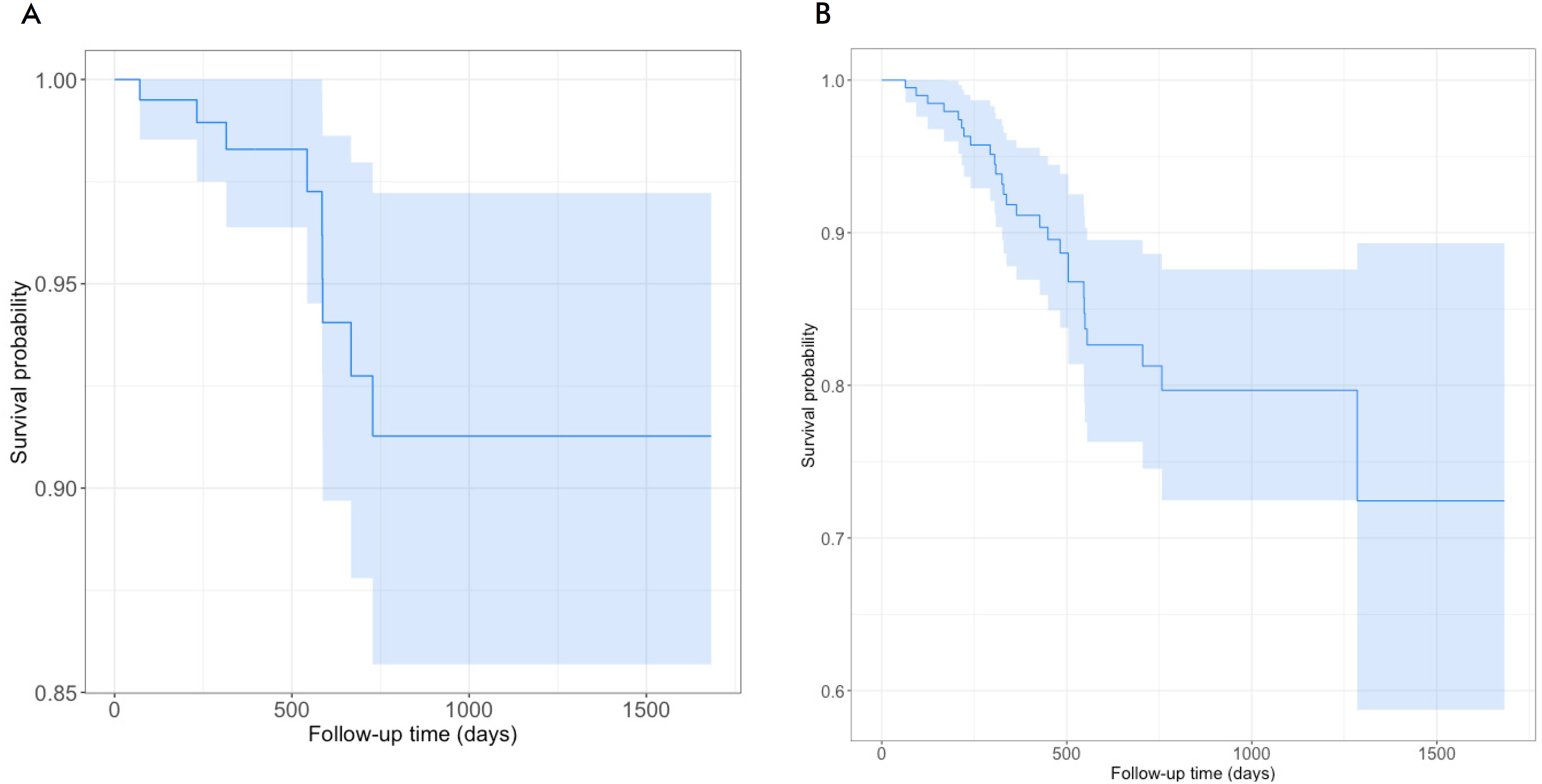
The survival curves of the patients investigated (Figure 1) show 5-year OS of 91.3% and 5-year DFS of 72.4%. OS curve stabilized after 728 days of follow-up and DFS curve, after 1,285 days of follow-up.
Survival curves of the patients investigated. (A) = overall survival; (B) = disease-free survival; Abscissa (axe x) = time of follow-up in days; Ordinate (axe y) = survival probability.
Table 1 portrays the basal data of the patients. Of the 201 patients enrolled, 198 (98.5%) were females and three (1.5%), males, 28 (13.9%) were diagnosed at younger age (less than 40 years). The most prevalent subtype was invasive carcinoma of no special type (NST) (94%), followed by ductal carcinoma in situ (DCIS) (2.5%). Other types of carcinoma were: mucinous (1%), papillary (2%), invasive lobular (1%) and neuroendocrine (0.5%); 7% were well-differentiated (G1), 72.1%, moderately differentiated (G2) and 20.9%, little differentiated (G3). The tumor of most of them (64.6%) was smaller than 5 cm at diagnosis, without angiolymphatic (52.7%) and neural (77.6%) invasion. Regarding the immunohistochemical profile, 122 (60.7%) expressed ER, 105 (52.2%), PR and 53 (26.4%), HER2. The majority (63.2%) presented low Ki-67 values ≤ 30%12; 65.2% of the tumors were subtype molecular luminal, 21.4%, triple-negative and 13.4%, HER2+. For clinical staging TNM, the most frequent were T2 (45.3%), N0 (43.8%) and M0 (85.1%); 58.2% presented clinical prognostic staging between 0 and IIB and 41.8%, between IIIA and IV.
The treatment and evolution of the patients is shown in Table 2, with 85.1% (171) who submitted to curative-intent surgery, three (1.5%), to prophylactic bilateral mastectomy and 27 (13.4%) had no indication due to very advanced stage. Of these, 83 (41.3%) had compromised lymph node at lymphadenectomy. Most of them had indication of radiotherapy (79.1%), chemotherapy (98%) and hormone therapy (57.7%). Of the 53 HER2+ patients, 29 (14.4%) were prescribed trastuzumab, 20 (10%), zoledronic acid, 27 (13.4%) relapsed, being 18 (9%) systemic, eight (4%), local and one (0.5%), regional; 48 patients (23.9%) metastasized, 30 (14.9%) already at the diagnosis and 18 (9%) as relapse of the primary tumor. Metastatic sites were bones (23 patients), lungs (23 patients), liver (11 patients), central nervous system (9 patients) and skin (5 patients). At the end of the follow-up, 147 patients (73.1%) were alive without evidences of disease, 45 (22.4%), alive with cancer and nine died (4.5%) by breast cancer.
Males had worst prognosis according to Table 3, with high risk of death in the bivariate and multivariate analysis. The bivariate analysis has also shown higher risk of death for patients with tumors larger than 5 cm and with neural and angiolymphatic invasion, but the results were not significant in the multivariate analysis.
Bivariate and multivariate Cox regression analysis of prognostic factors associated with death and relapse of the patients investigated
Tumors with angiolymphatic invasion and without expression of ER presented worst prognosis and high risk of relapse both in variate and multivariate analysis. The analysis of molecular subtypes showed high risk of relapse for triple-negative tumors, not ratified by the multivariate analysis, similar with the variable of non-expression of PR which was statistically significant only in the bivariate analysis.
The variable presence of neural invasion was not statistically significant in the bivariate analysis but was related to higher risk of relapse by the multivariate analysis. The variables age at diagnosis, grade of differentiation, tumor size, HER2 and Ki-67 were not associated with higher risk of relapse in the bivariate and multivariate analysis. For the variables sex and histological subtype, it was not possible to perform any analysis of risk of relapse, since one of the subgroups of these two variables relapsed during the follow-up period.
DISCUSSION
The program Surveillance, Epidemiology, and End Results (SEER)13 estimated for 2013-2019 a 5-year relative survival of 90.8% for patients with breast cancer. Depending on the extension of the tumor at diagnosis, this result can change to 99.3% for localized disease, 86.3% for regional disease (lymph nodes) and 31% for metastatic disease13. In Brazil, the 5-year OS of breast cancer went from 78% for the period 1995-1999 to 87% between 2005 and 200914. As earlier mentioned, the 5-year OS for the study patients was 91.3%, consistent with the literature and also related to the characteristics of the tumor extension at diagnosis13.
Breast cancer distant metastases are the most common type of relapse and the main cause of death by disease15. This type of metastasis was predominant for the study sample (66.7%). Yazdani and Haghighat analyzed DFS of 2,056 patients with breast cancer and reached a percent close to 75%, a little lower for a 5-year period15. The 5-year DFS analysis of Diniz et al. with 459 patients with non-metastatic breast cancer reached a percent of 72%16. The 5-year DFS of the present study was 72.4%, consistent with the literature.
Three male patients (1.5%) were found in the study sample. Bivariate and multivariate Cox regression analysis showed that males had statistically significant higher risk of death than females, similar to the literature17-20.
The incidence of breast cancer in young women (less than 40 years of age) is increasing, being young age a strong predictor of worst prognosis21,22. The results showed substantially higher incidence of breast cancer in women older than 40 years and bivariate and multivariate Cox regression analysis did not show impact of the variable age in the prognosis of the patients analyzed.
196 cases of the most common variables of breast carcinoma, ductal and lobular23, were found, of which 189 (94%) were invasive carcinoma NST, 5 (2.5%) ductal carcinoma in situ (DCIS) and 2 (1.0%), invasive lobular carcinoma (ILC). In addition, there were five cases of rare breast carcinoma: two cases of invasive papillary carcinoma (IPC), two cases of mucinous carcinoma and one case of neuroendocrine breast cancer (NEBC)24-26. The bivariate Cox regression analysis did not show high risk of death for patients with invasive carcinoma NST or other histological subtypes, given the high prevalence of invasive carcinoma NST (94%) of the sample, which limits the comparison.
High grade of differentiation, larger size, angiolymphatic and/or neural invasion, triple-negative molecular subtype, lack of expression of ER and PR, expression of HER2 and high Ki-67 proliferation index (> 30%)27-30 are factors classically involved in worst prognosis of breast cancer.
Most of the patients investigated had favorable prognosis, with grade of differentiation G1 or G2 (79.1%), tumors smaller than 5 cm (64.6%), lack of angiolymphatic (52.7%) and neural (77.6%) invasion, positive ER (60.7%) and PR (52.2%), negative HER2 (73.6%) and Ki-67 ≤ 30% (63.2%), the minority were subtype triple-negative (21.4%).
The bivariate and multivariate Cox regression analysis revealed that tumor larger than 5 cm and presence of neural and angiolymphatic invasion were associated with higher risk of death in the bivariate analysis. The multivariate analysis has also concluded that neural and angiolymphatic invasion were also associated with higher risk of relapse. Triple-negative subtype increased the risk of relapse, and lack of expression of ER was associated with higher risk of relapse according to the bivariate and multivariate analysis and non-expression of PR, only in the bivariate analysis.
Breast cancer staging starts with stage 0 and progresses to advanced stage IV (remote metastasis). The TNM staging is the most often system used. The most recent AJCC system, effective January 2018, has both clinical and pathologic staging systems. The pathologic stage (more accurate) is determined by examining tissue removed during a surgery. If surgery is not possible, the clinical stage based on the physical and imaging exams and biopsy is used to help plan treatment. For both systems, seven key pieces of information are evaluated: the extent of the tumor (stage T), spread to nearby lymph nodes (stage N), spread to distant sites (stage M), profile of ER, PR and HER2 and grade of differentiation (G)31.
Predominantly, the study patients presented less advanced TNM: 59.7% classified as Tis (carcinoma in situ), T1 (tumor smaller than 2 cm) or T2 (tumor between 2 and 5 cm); 67.7% were N0 (no lymph nodes affected) or N1 (1 to 3 axillary lymph nodes and/or internal mammary lymph nodes at sentinel lymph node biopsy); 85.1% had no remote metastasis at diagnosis (M0). 58.2% presented clinical prognosis strata 0, I or II.
Cancer treatment is complex and based mainly on surgery, radiotherapy, chemotherapy, hormone therapy and molecular targeted-therapy. Another possibility of treatment is zoledronic acid, reducing the risk of tumor spread to bones and other sites. Psychological support is also part of the treatment, in addition to complementary therapies as acupuncture, for instance32. The distribution of the treatment variables (surgery, lymph nodes surgical approach, radiotherapy, chemotherapy and use of trastuzumab and zoledronic acid) showed that care provided to the patients followed breast cancer international treatment standards and current SUS policies.
The methodological and sampling limitations of the study are its retrospective design conducted in a single site. In addition, sex was considered in the prognostic analysis, making less evident the knowledge of survival and prognostic factors for female patients.
CONCLUSION
The survival curves showed 5-year OS of 91.3% and 5-year DFS of 72.4%. The results of bivariate Cox regression analysis revealed that males, large tumor size and neural and angiolymphatic invasion were significantly associated with higher risk of death and by multivariate analysis, only males had worst prognosis and higher risk of death.
The presence of angiolymphatic invasion, subtype triple-negative and negativity of expression of ER and PR were significantly associated with increased risk of relapse by bivariate Cox regression analysis. The multivariate analysis concluded that neural and angiolymphatic invasion and negativity of ER had worst prognosis and higher risk of relapse.
ACKNOWLEDGMENT
To Uespi and to "Hospital Macrorregional de Caxias Dr. Everaldo Aragão" for institutional support.
References
-
1 Momenimovahed Z, Salehiniya H. Epidemiological characteristics of and risk factors for breast cancer in the world. Breast Cancer (Dove Med Press). 2019;11:151-64. doi: https://www.doi.org/10.2147/BCTT.S176070
» https://www.doi.org/10.2147/BCTT.S176070 -
2 Waks AG, Winer EP. Breast cancer treatment: a review. JAMA. 2019;321(3):288-300. doi: https://www.doi.org/10.1001/jama.2018.19323
» https://www.doi.org/10.1001/jama.2018.19323 -
3 Cardoso F, Kyriakides S, Ohno S, et al. Early breast cancer: ESMO clinical practice guidelines for diagnosis, treatment and follow-up. Ann Oncol. 2019;30(8):1194-220. doi: https://www.doi.org/10.1093/annonc/mdz173 Errata in: Ann Oncol. 2019;30(10):1674. Errata in: Ann Oncol. 2021;32(2):284.
» https://www.doi.org/10.1093/annonc/mdz173 -
4 Esteva FJ, Hubbard-Lucey VM, Tang J, et al. Immunotherapy and targeted therapy combinations in metastatic breast cancer. Lancet Oncol. 2019;20(3):e175-86. doi: https://www.doi.org/10.1016/S1470-2045(19)30026-9
» https://www.doi.org/10.1016/S1470-2045(19)30026-9 -
5 Gonçalves Junior H, Guerra MR, Duarte Cintra JR, et al. Survival study of triple-negative and non-triple-negative breast cancer in a brazilian cohort. Clin Med Insights Oncol. 2018;12:1179554918790563. doi: https://doi.org/10.1177/1179554918790563
» https://doi.org/10.1177/1179554918790563 -
6 Phung MT, Tin Tin S, Elwood JM. Prognostic models for breast cancer: a systematic review. BMC Cancer. 2019;19(1):230. doi: https://www.doi.org/10.1186/s12885-019-5442-6
» https://www.doi.org/10.1186/s12885-019-5442-6 - 7 Brierley JD, Gospodarowicz M, Wittekind Ch, editors. TNM Classification of Malignant Tumours. 8 ed. Chichester, West Sussex, UK: Wiley Blackwell; 2017.
-
8 R: The R Project for Statistical Computing [Internet]. Version 4.0.2. Viena: The R foundation. [Acesso 2024 jul 25]. Disponível em: https://www.R-project.org
» https://www.R-project.org -
9 Kaplan EL, Meier P. Nonparametric estimation from incomplete observations. J Amer Statist Assoc. 1958;53(282):457-81. doi: https://doi.org/10.2307/2281868
» https://doi.org/10.2307/2281868 -
10 Miot HA. Survival analysis in clinical and experimental studies. J Vasc Bras. 2017;16(4):267-9. doi: https://www.doi.org/10.1590/1677-5449.001604
» https://www.doi.org/10.1590/1677-5449.001604 - 11 Conselho Nacional de Saúde (BR). Resolução n° 466, de 12 de dezembro de 2012. Aprova as diretrizes e normas regulamentadoras de pesquisas envolvendo seres humanos. Diário Oficial da União, Brasília, DF. 2013 jun 13; Seção I:59.
-
12 Zhang L, Hao J, Guo J, et al. Predicting of Ki-67 expression level using diffusion-weighted and synthetic magnetic resonance imaging in invasive ductal breast cancer. Breast J. 2023;2023:6746326. doi: https://www.doi.org/10.1155/2023/6746326
» https://www.doi.org/10.1155/2023/6746326 -
13 SEER: Surveillance, Epidemiology, and End Results [Internet]. Versão 1975-2019. Washigton, DC: NCI; 2021. [Acesso 2024 jul 25]. Disponível em: https://seer.cancer.gov/data-software/documentation/seerstat/
» https://seer.cancer.gov/data-software/documentation/seerstat/ -
14 Ayala ALM, Anjos JCD, Cassol GA, et al. Sobrevida em 10 anos em mulheres com câncer de mama: coorte história de 2000-2014. Ciênc saúde coletiva. 2019;24(4):1537-50. doi: https://www.doi.org/10.1590/1413-81232018244.16722017
» https://www.doi.org/10.1590/1413-81232018244.16722017 -
15 Yazdani A, Haghighat S. Determining prognostic factors of disease-free survival in breast cancer using censored quantile regression. Breast Cancer (Auckl). 2022;16:11782234221108058. doi: https://www.doi.org/10.1177/11782234221108058
» https://www.doi.org/10.1177/11782234221108058 -
16 Diniz RW, Guerra MR, Cintra JR, et al. Disease-free survival in patients with non-metastatic breast cancer. Rev Assoc Med Bras. 2016;62(5):407-13. doi: https://www.doi.org/10.1590/1806-9282.62.05.407
» https://www.doi.org/10.1590/1806-9282.62.05.407 -
17 Chen Z, Xu L, Shi W, et al. Trends of female and male breast cancer incidence at the global, regional, and national levels, 1990-2017. Breast Cancer Res Treat. 2020;180(2):481-90. doi: https://doi.org/10.1007/s10549-020-05561-1
» https://doi.org/10.1007/s10549-020-05561-1 -
18 Gucalp A, Traina TA, Eisner JR, et al. Male breast cancer: a disease distinct from female breast cancer. Breast Cancer Res Treat. 2019;173(1):37-48. doi: https://doi.org/10.1007/s10549-018-4921-9
» https://doi.org/10.1007/s10549-018-4921-9 -
19 Hassett MJ, Somerfield MR, Baker ER, et al. Management of male breast cancer: ASCO Guideline. J Clin Oncol. 2020;38(16):1849-63. doi: https://doi.org/10.1200/JCO.19.03120
» https://doi.org/10.1200/JCO.19.03120 -
20 Fox S, Speirs V, Shaaban AM. Male breast cancer: an update. Virchows Arch. 2022;480(1):85-93. doi: https://doi.org/10.1007/s00428-021-03190-7
» https://doi.org/10.1007/s00428-021-03190-7 -
21 Fakhri N, Chad MA, Lahkim M, et al. Risk factors for breast cancer in women: an update review. Med Oncol. 2022;39(12):197. doi: https://doi.org/10.1007/s12032-022-01804-x
» https://doi.org/10.1007/s12032-022-01804-x -
22 Gao Y, Samreen N, Heller SL. Non-BRCA early-onset breast cancer in young women. Radiographics. 2022;42(1):5-22. doi: https://doi.org/10.1148/rg.210109
» https://doi.org/10.1148/rg.210109 -
23 Watkins EJ. Overview of breast cancer. JAAPA. 2019;32(10):13-7. doi: https://doi.org/10.1097/01.JAA.0000580524.95733.3d
» https://doi.org/10.1097/01.JAA.0000580524.95733.3d -
24 Ingale YP, Buch AC, Jose M, et al. Invasive papillary carcinoma of the breast – a rare case report. Med J DY Patil Vidyapeeth. 2022;15:782-4. doi: https://doi.org/10.4103/mjdrdypu.mjdrdypu_171_21
» https://doi.org/10.4103/mjdrdypu.mjdrdypu_171_21 -
25 Marrazzo E, Frusone F, Milana F, et al. Mucinous breast cancer: a narrative review of the literature and a retrospective tertiary single-centre analysis. Breast. 2020;49:87-92. doi: https://doi.org/10.1016/j.breast.2019.11.002
» https://doi.org/10.1016/j.breast.2019.11.002 -
26 Valentim MH, Monteiro V, Marques JC. Primary neuroendocrine breast carcinoma: a case report and literature review. Radiol Bras. 2014;47(2):125-7. doi: https://doi.org/10.1590/S0100-39842014000200017
» https://doi.org/10.1590/S0100-39842014000200017 -
27 Srivastava P, Wang T, Clark BZ, et al. Clinical-pathologic characteristics and response to neoadjuvant chemotherapy in triple-negative low Ki-67 proliferation (TNLP) breast cancers. NPJ Breast Cancer. 2022;8(1):51. doi: https://doi.org/10.1038/s41523-022-00415-z
» https://doi.org/10.1038/s41523-022-00415-z -
28 Donegan WL. Tumor-related prognostic factors for breast cancer. CA Cancer J Clin. 1997;47(1):28-51. doi: https://doi.org/10.3322/canjclin.47.1.28
» https://doi.org/10.3322/canjclin.47.1.28 -
29 Roses DF, Bell DA, Flotte TJ, et al. Pathologic predictors of recurrence in stage 1 (TINOMO) breast cancer. Am J Clin Pathol. 1982;78(6):817-20. doi: https://www.doi.org/10.1093/ajcp/78.6.817
» https://www.doi.org/10.1093/ajcp/78.6.817 -
30 Zagami P, Carey LA. Triple negative breast cancer: pitfalls and progress. NPJ Breast Cancer. 2022;8(1):95. doi: https://www.doi.org/10.1038/s41523-022-00468-0
» https://www.doi.org/10.1038/s41523-022-00468-0 -
31 American Cancer Society [Internet]. Atlanta: ACS; [sem data]. Breast Cancer Stages. [acesso 2023 set 9]. Disponível em: https://cancer.org/cancer/types/breast-cancer/understanding-a-breast-cancer-diagnosis/stages-of-breast-cancer.html
» https://cancer.org/cancer/types/breast-cancer/understanding-a-breast-cancer-diagnosis/stages-of-breast-cancer.html -
32 National Health Service [Internet]. Londres: NHS; [sem data]. Treatment: Breast cancer in women. [acesso 2023 set 9]. Disponível em: https://nhs.uk/conditions/breast-cancer/treatment/
» https://nhs.uk/conditions/breast-cancer/treatment/
Edited by
-
Scientific-editor:
Anke Bergmann. Orcid iD: https://orcid.org/0000-0002-1972-8777